Uterine prolapse is a form of pelvic organ prolapse in which the uterus and a portion of the upper vagina protrude into the vaginal canal and, in severe cases, through the opening of the vagina.[4] It is most often caused by injury or damage to structures that hold the uterus in place within the pelvic cavity.[2] Symptoms may include vaginal fullness, pain with sexual intercourse, difficulty urinating, and urinary incontinence.[4][1] Risk factors include older age, pregnancy, vaginal childbirth, obesity, chronic constipation, and chronic cough.[1] Prevalence, based on physical exam alone, is estimated to be approximately 14%.
Diagnosis is based on a symptom history and physical examination, including pelvic examination.[4] Preventive efforts include managing medical risk factors, such as chronic lung conditions, smoking cessation, and maintaining a healthy weight.[1] Management of mild cases of uterine prolapse include pelvic floor therapy and pessaries. More severe cases may require surgical intervention, including removal of the uterus or surgical fixation of the upper portion of the vagina to a nearby pelvic structure.[4] Outcomes following management are generally positive with reported improvement in quality of life.[5]
Signs and symptoms
Uterine prolapse in a 71 year old woman, with the cervix visible in the vaginal orifice.
While uterine prolapse is rarely life-threatening, the symptoms associated with uterine prolapse can have a significant impact on quality of life.[2] The severity of prolapse symptoms does not necessarily correlate with the degree of prolapse, and one may experience little to no bothersome symptoms with even advanced prolapse.[2][3] Additionally, different forms of pelvic organ prolapse often present with similar symptoms.[2]
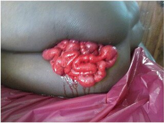
Most women who experience pelvic organ prolapse do not have symptoms.[2] When symptoms are present, the most common and most specific symptoms for uterine prolapse—and organ prolapse in general—into the vagina are bulge symptoms, such as pelvic pressure, vaginal fullness, or a palpable vaginal bulge, and these symptoms are often more common and more severe if the prolapse reaches the vaginal hymen.[2][3] Urinary symptoms, such as uncontrollable loss of urine or difficulty urinating, may also be present.[3] Complete uterine prolapse in which the uterus protrudes through the vaginal hymen is known as procidentia.[6] In the absence of treatment, symptoms of procidentia may include purulent vaginal discharge, ulceration, and bleeding.[1][6] Complications of procidentia include urinary obstruction.[6]
People may also report sexual dysfunction symptoms, such as pain with sexual intercourse and decreased libido.[4][2] There is conflicting data concerning the effect of pelvic organ prolapse on sexual function.[2][3] The severity of the symptoms associated with prolapse seems to have a negative effect on sexual activity and reported satisfaction. Mild or asymptomatic prolapse does not seem to be associated with sexual complaints while more symptomatic prolapse is associated with more negative sexual symptoms.[3]
Causes
The most common risk factor associated with uterine prolapse is vaginal childbirth.[2][6][7] The risk of prolapse increased with each vaginal delivery, and people who have had multiple vaginal deliveries are more likely to develop prolapse compared with those who have had a single vaginal delivery.[2] Operative vaginal delivery, especially when obstetrical forceps are used, has been found to increase the odds of pelvic organ prolapse when compared to non-operative vaginal delivery.[2][7]
Age also plays a significant role in uterine prolapse, with prevalence increasing with each decade of life due in part to age-related changes to pelvic support structures and menopause-related reduction in estrogen levels.[2] Additionally, conditions that chronically increase the pressure within the abdomen can also predispose people to uterine prolapse.[2][1][7] This includes chronic obstructive pulmonary disease (COPD), obesity, chronic cough, straining due to chronic constipation, and repetitive heavy lifting.[2][7] Tobacco smoking has been found to be correlated to pelvic organ prolapse both due to the risk of developing lung conditions that lead to chronic cough or COPD as well as the negative effects of tobacco chemicals on connective tissue.[2]
Pathophysiology
The uterosacral ligaments connect the base of the uterus to the sacrum
The uterus is normally held in place by the combined effort of pelvic floor muscles, various ligaments, pelvic fascia, and the vaginal wall.[2][6] The levator ani muscle plays the most significant role in pelvic organ support by acting as a basket that keeps the pelvic organs suspended.[2] The uterosacral ligaments are especially important in providing support to the uterus by attaching and holding the uterus, cervix, and upper vagina to the sacrum.[2][3]
Schematic of the female reproductive system with a frontal view. 43 depicts the pelvic floor muscles that support the uterus in the pelvic cavity.
Uterine prolapse occurs when there is a disruption to any of the structures mentioned above that help hold the uterus in place.[2][6] Weakening of the levator ani muscles can occur during vaginal childbirth, in which portions of the muscle can detach from the bony pelvis, or through age-related changes to musculature, and this can lead to a loss of support for the uterus.[2] Pregnancy, vaginal childbirth, or injury can also stretch and weaken the uterosacral ligaments, leading to poor suspension or positioning of the uterus so that it is no longer supported by pelvic floor muscles.[3] Problems with the vaginal wall, such as trauma or loss of smooth muscle support in the wall, can lead to the uterus collapsing downward due to a loss of support.[2] When the uterus prolapses, it also drags the upper portion of the vagina (the apical vagina) along with it due to its anatomic relationship with the apical vagina.[6]
Additionally, the pelvic musculature and connective tissues are estrogen sensitive and respond to changes in estrogen level.[2] Estrogen deficiency, which can occur during menopause, can affect the production of collagen that is needed to build connective tissue that makes up ligaments and fascia, which can contribute to uterine prolapse.[2] This is also a reason that connective tissue disorders can predispose certain people to uterine prolapse.[2]
Diagnosis and management
Diagnosis
Diagnosis of uterine prolapse is based on a history of symptoms, which may include symptom questionnaires, and a physical exam.[1][2] Usually, the physical exam involves a vaginal exam, often with a speculum, and a pelvic exam.[2][6] The extent and severity of prolapse is commonly documented using the Pelvic Organ Prolapse Quantification (POP-Q) system.[1][2]
Management
The management of uterine prolapse may be conservative or surgical, depending on factors such as personal preference, symptom severity, and extent of prolapse.[2] Additionally, management of existing medical conditions that can contribute to prolapse, such as chronic lung conditions or obesity, are important to prevent progression of uterine prolapse and reduce symptom burden.[6]
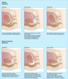
Conservative
Different types of pessaries may be inserted into the vaginal canal for the conservative management of uterine prolapse
Conservative options include pelvic floor muscle strengthening exercises and pessaries.[6][2]Pelvic floor muscle training (PFMT), also known as Kegel exercise, has been found to improve the bulk and urinary symptoms associated with pelvic organ prolapse and improve quality of life when performed consistently and correctly.[2][8]Pessaries are a mechanical treatment that supports the vagina and elevates the prolapsed uterus to its anatomically correct position.[9] Pessaries are frequently offered as a first-line management option for uterine prolapse, especially amongst people who cannot or do not wish to undergo surgery, due to their affordability and low-risk profile compared to more invasive procedures.[6] When properly fitted, pessaries have been found to improve bulk and pressure symptoms associated with prolapse and improve quality of life measures.[3][5]
Surgical
There are many surgical options available for the treatment of uterine prolapse, which may be performed through a vaginal procedure or through the abdomen.[2][10] Generally, vaginal procedures are considered to be less invasive, offer a quicker recovery, and have a shorter operative time compared to abdominal procedures, but abdominal procedures offer longer-term results and potentially reduce risk of postoperative vaginal pain with intercourse.[2]Laparoscopic and robotic approaches to abdominal procedures in prolapse surgery have become more common as they require smaller incision sites, result in less blood loss, and have shorter hospital stays. [2][10]
Vaginal vault suspension (known as colpopexy), in which the upper portion of the vagina is surgically connected to another structure in the pelvis, is commonly performed in the treatment of uterine prolapse.[2][10] Forms of colpopexy include sacrocolpopexy, in which the vaginal vault is attached to the sacrum using a surgical mesh; sacrospinous ligament fixation, in which the upper vagina is attached to the sacrospinous ligaments; and uterosacral ligament vaginal vault suspension, in which the upper vagina is attached to the uterosacral ligaments.[2] Colpopexy can be performed with or without a hysterectomy. If performed without a hysterectomy, the procedure is known as a hysteropexy. Hysteropexy procedures include sacrohysteropexy and sacrospinous hysteropexy.[2]
In severe cases of prolapse where the person no longer desires vaginal intercourse and has contraindications to more invasive surgery, vaginal closure procedures may be offered.[10] These include LeFort partial colpocleisis and complete colpocleisis, in which the vagina is sutured closed.[10]
Also taken into consideration prior to surgery is use of native, or one's own, tissue versus a synthetic mesh. Generally, mesh may be considered in instances where the connective tissue is weak or absent, if there is an empty space at the surgical site that needs to be bridged, or if there is a high risk of prolapse recurrence.[2] Synthetic mesh is indicated and used for sacrocolpopexy and sacrohysteropexy procedures.[2] However, the use of synthetic mesh transvaginally, or within the vaginal tissue itself, is not indicated and is not routinely used for apical vaginal or uterine prolapse due to a lack of safety and effectiveness data, higher rate of mesh exposure compared with native tissue repair, and lack of data regarding long-term outcomes and complication rates.[3][2][10]
Outcomes
Overall, it appears that quality of life was found to be significantly improved for people with pelvic organ prolapse after surgical or pessary management.[5]
It can be difficult to determine success when discussing the outcomes of surgical intervention for pelvic organ prolapse due to multiple factors that can define success, such as anatomic success versus patient-reported outcome measures.[10] Improvement of vaginal bulge symptoms after surgery appears to be more of a measure of success for patients themselves than does anatomic success alone.[3]
The rate of pelvic organ prolapse recurrence following surgery depends on several factors, the most significant being patient age (patients younger than 60 years have higher likelihood of recurrence), POP-Q stage (POP-Q greater than 3 has higher likelihood of recurrence), surgeon's experience performing the procedure, and prior history of pelvic surgery.[11][12] Additionally, the type of surgery, for instance vaginal versus abdominal, also affects recurrence rate.[3][13] The rates of reoperation following pelvic organ prolapse surgery ranges from 3.4% to 9.7%.[3] Reoperation rates appear to be higher with transvaginal mesh repair compared to other procedures, due in part to complications such as mesh exposure.[3]
Epidemiology
Numerical values regarding prevalence of uterine prolapse differ based on whether the epidemiologic study in question uses a physical exam or a symptom questionnaire to determine the presence of prolapse.[3] Prevalence of pelvic organ prolapse was found to be consistently higher when physical exam was used (for uterine prolapse, this was 14.2%[14] in one study and 3.8% in another[3]) compared to a symptom-based determination in which the prevalence of any type of prolapse, including uterine prolapse, was 2.9% to 8% in the U.S.[3] Using Women's Health Initiative data, the incidence of grades 1 to 3 uterine prolapse was approximately 1.5/100 women-years and progression of uterine prolapse was found to be about 1.9%.[3]
History
The first mention of uterine prolapse in medical literature was in the Kahun papyrus, circa 1835 B.C.E, which read, "of a woman whose posterior, belly, and branching of her thighs are painful, say thou as to it, it is the falling of the womb."[15] The treatment at the time, documented on the Ebers papyrus, was to rub the afflicted person with a mixture of "oil of the earth [and] fedder",[15] or petroleum and manure.
Throughout Western history, advancements in the management of uterine prolapse have been hampered by a poor understanding of female pelvic anatomy.[15] During the Hippocratic era, approximately 460 B.C.E., it was thought that the uterus was akin to an animal.[15] Therefore, common treatments included fumigation, placing a foul-smelling object near the uterus to convince it to move into the vagina; the use of topical astringents, such as vinegar; and succussion, in which a woman was tied upside-down and shaken until the prolapse reduced.[15]
During the first century C.E., the Greek physician Soranus would disagree with many of these practices and recommended the use of wool, dipped in vinegar or wine and inserted into the vagina, to lift the uterus back into place.[15] He would also go on to recommend surgical removal of gangrenous portions of a prolapsed uterus.[15] However, these ideas did not become commonly accepted practices during that era, and the Middle Ages brought about a return to previous beliefs and practices for uterine prolapse.[15] In 1603, for instance, it was recommended that burning the prolapsed uterus with a hot iron would frighten it back into the vagina.[15]
In 1543, Andreus Vesalius, professor of anatomy at Padua, published De Humani Corporis Fabrica Libri Septem, which included an accurate depiction of the entire female genital tract, including the uterus
Towards the end of the 16th century, pessaries became more common in the management of uterine prolapse, due in part to advances in anatomic knowledge of the female genitourinary tract earlier in the century.[15] Pessaries were usually made out of wax, metal, glass, or wood. Charles Goodyear's invention of volcanized rubber in the mid 1800s made it possible to produce pessaries that would not decompose.[15] However, even into the 1800s, alternative practices were still used, such as the use of sea-water douches, postural exercises, and leeching.[15]
Although the use of surgery in the treatment of uterine prolapse had been described previously, the 19th century saw advances in surgical techniques.[15] During the mid to late 1800s, surgical attempts to manage uterine prolapse included narrowing the vaginal vault, suturing the perineum, and amputating the cervix.[15] In 1877, LeFort described the process of a partial colpocleisis.[15] In 1861, Choppin in New Orleans reported the first instance in which vaginal hysterectomy was performed for uterine prolapse. Prior to that, vaginal hysterectomies were mainly performed for malignancies.[15]
Following Alwin Mackenrodt's 1895 publication of a comprehensive description of the female pelvic floor connective tissue, Fothergill began working on the Manchester-Fothergill surgery with the belief that the cardinal and uterosacral ligaments were key support structures for the uterus.[15] In 1907, Josef Haban and Julius Tandler theorized that the levator ani muscles were also very important for uterine support.[15] Combined with a better understanding of female pelvic floor connective tissue, these ideas would go on to influence surgical approaches for the treatment of uterine prolapse.[15]
By the early 20th century, different techniques for vaginal hysterectomies had been described and performed. As a result, post-hysterectomy vaginal vault prolapse became more common and a growing concern for some surgeons, and new techniques to correct this complication were attempted.[15] In 1957, Arthure and Savage of London's Charing Cross Hospital, suspecting that uterine prolapse could not be cured with hysterectomy alone, published their surgical technique of sacral hysteropexy.[15] Their technique is still used in modern practice with the addition of a graft.[15]
Society and culture
Vaginal mesh kits were introduced to the U.S. market in 2004 through the U.S. Food and Drug Administration (FDA) pathway that did not require companies to demonstrate both safety and efficacy of the product if they were able to demonstrate that their product was similar to previous products already in the market.[3][18] However, there was concern over reports of increased rates of postoperative complications over the next several years. That, in addition to the lack of available data that transvaginal mesh products were superior to other forms of surgical intervention[18] and the expedited process which the vaginal mesh kits were introduced to the market, the FDA released a Safety Communication in 2011 that described serious complications associated with transvaginal mesh as "not rare".[3] In 2019, the FDA ordered manufacturers to halt sales transvaginal mesh intended for repair of pelvic organ prolapse.[3][19] This does not include surgical mesh used during sacrocolpopexy, sacrohysteropexy, or transurethral sling procedures.[19]
Since 2008, a number of class actionlawsuits have been filed and settled against several manufacturers of transvaginal mesh after people reported complications following surgery.[20]
Related Research Articles
In medicine, prolapse is a condition in which organs fall down or slip out of place. It is used for organs protruding through the vagina, rectum, or for the misalignment of the valves of the heart. A spinal disc herniation is also sometimes called "disc prolapse". Prolapse means "to fall out of place", from the Latin prolabi meaning "to fall out".
Hysterectomy is the surgical removal of the uterus and cervix. Supracervical hysterectomy refers to removal of the uterus while the cervix is spared. These procedures may also involve removal of the ovaries (oophorectomy), fallopian tubes (salpingectomy), and other surrounding structures. The term “partial” or “total” hysterectomy are lay-terms that incorrectly describe the addition or omission of oophorectomy at the time of hysterectomy. These procedures are usually performed by a gynecologist. Removal of the uterus renders the patient unable to bear children and has surgical risks as well as long-term effects, so the surgery is normally recommended only when other treatment options are not available or have failed. It is the second most commonly performed gynecological surgical procedure, after cesarean section, in the United States. Nearly 68 percent were performed for conditions such as endometriosis, irregular bleeding, and uterine fibroids. It is expected that the frequency of hysterectomies for non-malignant indications will continue to fall given the development of alternative treatment options.
A pessary is a prosthetic device inserted into the vagina for structural and pharmaceutical purposes. It is most commonly used to treat stress urinary incontinence to stop urinary leakage and to treat pelvic organ prolapse to maintain the location of organs in the pelvic region. It can also be used to administer medications locally in the vagina or as a method of contraception.
The pelvic floor or pelvic diaphragm is an anatomical location in the human body, which has an important role in urinary and anal continence, sexual function and support of the pelvic organs. The pelvic floor includes muscles, both skeletal and smooth, ligaments and fascia. and separates between the pelvic cavity from above, and the perineum from below. It is formed by the levator ani muscle and coccygeus muscle, and associated connective tissue.
In gynecology, a rectocele or posterior vaginal wall prolapse results when the rectum bulges (herniates) into the vagina. Two common causes of this defect are childbirth and hysterectomy. Rectocele also tends to occur with other forms of pelvic organ prolapse, such as enterocele, sigmoidocele and cystocele.
Pelvic floor dysfunction is a term used for a variety of disorders that occur when pelvic floor muscles and ligaments are impaired. The condition affects up to 50 percent of women who have given birth. Although this condition predominantly affects women, up to 16 percent of men are affected as well. Symptoms can include pelvic pain, pressure, pain during sex, urinary incontinence (UI), overactive bladder, bowel incontinence, incomplete emptying of feces, constipation, myofascial pelvic pain and pelvic organ prolapse. When pelvic organ prolapse occurs, there may be visible organ protrusion or a lump felt in the vagina or anus. Research carried out in the UK has shown that symptoms can restrict everyday life for women. However, many people found it difficult to talk about it and to seek care, as they experienced embarrassment and stigma.
Adenomyosis is a medical condition characterized by the growth of cells that proliferate on the inside of the uterus (endometrium) atypically located among the cells of the uterine wall (myometrium), as a result, thickening of the uterus occurs. As well as being misplaced in patients with this condition, endometrial tissue is completely functional. The tissue thickens, sheds and bleeds during every menstrual cycle.
The cystocele, also known as a prolapsed bladder, is a medical condition in which a woman's bladder bulges into her vagina. Some may have no symptoms. Others may have trouble starting urination, urinary incontinence, or frequent urination. Complications may include recurrent urinary tract infections and urinary retention. Cystocele and a prolapsed urethra often occur together and is called a cystourethrocele. Cystocele can negatively affect quality of life.
Pelvic organ prolapse (POP) is characterized by descent of pelvic organs from their normal positions into the vagina. In women, the condition usually occurs when the pelvic floor collapses after gynecological cancer treatment, childbirth or heavy lifting. Injury incurred to fascia membranes and other connective structures can result in cystocele, rectocele or both. Treatment can involve dietary and lifestyle changes, physical therapy, or surgery.
Adenomyoma is a tumor (-oma) including components derived from glands (adeno-) and muscle (-my-). It is a type of complex and mixed tumor, and several variants have been described in the medical literature. Uterine adenomyoma, the localized form of uterine adenomyosis, is a tumor composed of endometrial gland tissue and smooth muscle in the myometrium. Adenomyomas containing endometrial glands are also found outside of the uterus, most commonly on the uterine adnexa but can also develop at distant sites outside of the pelvis. Gallbladder adenomyoma, the localized form of adenomyomatosis, is a polypoid tumor in the gallbladder composed of hyperplastic mucosal epithelium and muscularis propria.
Vaginectomy is a surgery to remove all or part of the vagina. It is one form of treatment for individuals with vaginal cancer or rectal cancer that is used to remove tissue with cancerous cells. It can also be used in gender-affirming surgery. Some people born with a vagina who identify as trans men or as nonbinary may choose vaginectomy in conjunction with other surgeries to make the clitoris more penis-like (metoidioplasty), construct of a full-size penis (phalloplasty), or create a relatively smooth, featureless genital area.
The vaginal cuff is the upper portion of the vagina that opens up into the peritoneum and is sutured shut after the removal of the cervix and uterus during a hysterectomy.
Female genital disease is a disorder of the structure or function of the female reproductive system that has a known cause and a distinctive group of symptoms, signs, or anatomical changes. The female reproductive system consists of the ovaries, fallopian tubes, uterus, vagina, and vulva. Female genital diseases can be classified by affected location or by type of disease, such as malformation, inflammation, or infection.
Surgical mesh is a medical implant made of loosely woven mesh, which is used in surgery as either a permanent or temporary structural support for organs and other tissues. Surgical mesh can be made from both inorganic and biological materials and is used in a variety of surgeries, although hernia repair is the most common application. It can also be used for reconstructive work, such as in pelvic organ prolapse or to repair physical defects created by extensive resections or traumatic tissue loss.
Vaginal evisceration is an evisceration of the small intestine that occurs through the vagina, typically subsequent to vaginal hysterectomy, and following sexual intercourse after the surgery. It is a surgical emergency.
A urogenital fistula is an abnormal tract that exists between the urinary tract and bladder, ureters, or urethra. A urogenital fistula can occur between any of the organs and structures of the pelvic region. A fistula allows urine to continually exit through and out the urogenital tract. This can result in significant disability, interference with sexual activity, and other physical health issues, the effects of which may in turn have a negative impact on mental or emotional state, including an increase in social isolation. Urogenital fistulas vary in etiology. Fistulas are usually caused by injury or surgery, but they can also result from malignancy, infection, prolonged and obstructed labor and deliver in childbirth, hysterectomy, radiation therapy or inflammation. Of the fistulas that develop from difficult childbirth, 97 percent occur in developing countries. Congenital urogenital fistulas are rare; only ten cases have been documented. Abnormal passageways can also exist between the vagina and the organs of the gastrointestinal system, and these may also be termed fistulas.
Vaginal anomalies are abnormal structures that are formed during the prenatal development of the female reproductive system and are rare congenital defects that result in an abnormal or absent vagina.
The vaginal support structures are those muscles, bones, ligaments, tendons, membranes and fascia, of the pelvic floor that maintain the position of the vagina within the pelvic cavity and allow the normal functioning of the vagina and other reproductive structures in the female. Defects or injuries to these support structures in the pelvic floor leads to pelvic organ prolapse. Anatomical and congenital variations of vaginal support structures can predispose a woman to further dysfunction and prolapse later in life. The urethra is part of the anterior wall of the vagina and damage to the support structures there can lead to incontinence and urinary retention.
Squamous cell carcinoma of the vagina is a potentially invasive type of cancer that forms in the tissues of the vagina. Though uncommonly diagnosed, squamous cell cancer of the vagina (SCCV) is the most common type of vaginal cancer, accounting for 80-90% of cases as well as 2% of all gynecological cancers. SCCV forms in squamous cells, which are the thin, flat cells lining the vagina. SCCV initially spreads superficially within the vaginal wall and can slowly spread to invade other vaginal tissues. Because of its slow growth, this cancer may cause no symptoms, or it may present with signs like irregular bleeding, pain, or a vaginal mass. This carcinoma can metastasize to the lungs or less frequently to the liver, bone, or other sites. SCCV has many risk factors in common with cervical cancer and is similarly strongly associated with infection with oncogenic strains of human papillomavirus (HPV). Diagnosis of SCCV is done by pelvic exam and biopsy of the tissue. Treatment and prognosis will depend on the stage, location, and characteristics of the cancer.
Transvaginal mesh, also known as vaginal mesh implant, is a net-like surgical tool that is used to treat pelvic organ prolapse (POP) and stress urinary incontinence (SUI) among female patients. The surgical mesh is placed transvaginally to reconstruct weakened pelvic muscle walls and to support the urethra or bladder.
↑ Schulten SF, Claas-Quax MJ, Weemhoff M, van Eijndhoven HW, van Leijsen SA, Vergeldt TF, etal. (August 2022). "Risk factors for primary pelvic organ prolapse and prolapse recurrence: an updated systematic review and meta-analysis". American Journal of Obstetrics and Gynecology. 227 (2): 192–208. doi:10.1016/j.ajog.2022.04.046. hdl:2066/282895. PMID35500611. S2CID248487990.
↑ Maher C, Yeung E, Haya N, Christmann-Schmid C, Mowat A, Chen Z, Baessler K (July 2023). "Surgery for women with apical vaginal prolapse". The Cochrane Database of Systematic Reviews. 2023 (7): CD012376. doi:10.1002/14651858.CD012376.pub2. PMC10370901. PMID37493538.
This page is based on this Wikipedia article Text is available under the CC BY-SA 4.0 license; additional terms may apply. Images, videos and audio are available under their respective licenses.