Viral envelope

Last updated
Schematic of a Cytomegalovirus, coat = envelope CMVschema.svg
Schematic of a Cytomegalovirus, coat = envelope

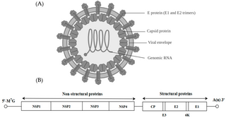
A viral envelope is the outermost layer of many types of viruses. [1] It protects the genetic material in their life cycle when traveling between host cells. Not all viruses have envelopes. A viral envelope protein or E protein is a protein in the envelope, which may be acquired by the capsid from an infected host cell.

Contents

Numerous human pathogenic viruses in circulation are encased in lipid bilayers, and they infect their target cells by causing the viral envelope and cell membrane to fuse. Although there are effective vaccines against some of these viruses, there is no preventative or curative medicine for the majority of them. In most cases, the known vaccines operate by inducing antibodies that prevent the pathogen from entering cells. This happens in the case of enveloped viruses when the antibodies bind to the viral envelope proteins.

The membrane fusion event that triggers viral entrance is caused by the viral fusion protein. Many enveloped viruses only have one protein visible on the surface of the particle, which is required for both mediating adhesion to the cell surface and for the subsequent membrane fusion process. To create potentially protective vaccines for human pathogenic enveloped viruses for which there is currently no vaccine, it is essential to comprehend how antibodies interact with viral envelope proteins, particularly with the fusion protein, and how antibodies neutralize viruses.

Enveloped viruses enter cells by joining a cellular membrane to their lipid bilayer membrane. Priming by proteolytic processing, either of the fusion protein or of a companion protein, is necessary for the majority of viral fusion proteins. The priming stage then gets the fusion protein ready for triggering by the processes that go along with attachment and uptake, which frequently happens during transport of the fusion protein to the cell surface but may also happen extracellularly. So far, structural studies have revealed two kinds of viral fusion proteins. These proteins are believed to catalyze the same mechanism in both situations, resulting in the fusing of two bilayers. In other words, these proteins operate as enzymes, which while having various structural variations catalyze the same chemical reaction. [2]

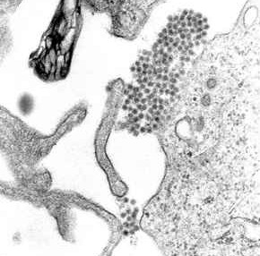
The envelopes are typically derived from portions of the host cell membranes (phospholipids and proteins), but include some viral glycoproteins. One of the main parts of human pathogenic viruses is glycoprotein. They have been shown to play significant roles in immunity and infection. [3] Viral glycoproteins, a new class of cellular inhibitory proteins has been discovered. These include the E3 ubiquitin ligases of the membrane-associated RING-CH (MARCH) family, which among other things, inhibits the expression of cell surface proteins implicated in adaptive immunity. [4] Being made up mostly of host membrane, the viral envelope can also have the proteins associated with the host cell within their membrane after budding. [5] Many enveloped viruses mature by budding at the plasma membrane, which allows them to be discharged from infected cells. During this procedure, viral transmembrane proteins, also known as spike proteins, are integrated into membrane vesicles containing components of the viral core (capsid).

For a very long time, it was thought that the spike proteins, which are necessary for infectivity, were directly incorporated into the viral core through their cytoplasmic domains. Recent research suggests that while such direct interactions may be what causes the budding of alphaviruses, this may not be the case for retroviruses and negative strand RNA viruses. These viruses can form bud particles even in the absence of spike proteins by relying only on viral core components. The spike proteins can occasionally be produced as virus-like particles without the viral core. Therefore, optimal budding and release may be dependent on a coordinated "push-and-pull" action between core and spike, where oligomerization of both components is essential. [6]

They may help viruses avoid the host immune system. TAM receptor tyrosine kinases increase phagocytic clearance of apoptotic cells and inhibit immunological responses brought on by Toll-like receptors and type I interferons (IFNs) when they are activated by the ligands Gas6 and Protein S. The phospholipid phosphatidylserine may be seen on the membranes of several enveloped viruses, which they employ to bind Gas6 and Protein S to activate TAM receptors.

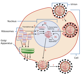
Ligand-coated viruses stimulate type I IFN signaling, activate TAM receptors on dendritic cells (DCs), and suppress type II interferon signaling to circumvent host defenses and advance infection.TAM-deficient DCs exhibit type I IFN responses that are more pronounced than those of wild-type cells in response to viral exposure. As a result, flaviviruses and pseudo typed retroviruses have a harder time infecting TAM-deficient DCs, albeit infection can be brought back by type I IFN antibodies. A TAM kinase inhibitor, meanwhile, prevents infection of wild-type DCs. TAM receptors, which are potential targets for therapy, are thereby activated by viruses to reduce type I IFN signaling. [7] Glycoproteins on the surface of the envelope serve to identify and bind to receptor sites on the host's membrane. The particular set of viral proteins are engaged in a series of structural changes. When these changes are set/finished, there is then and only then, fusion with the host membrane. [8] These glycoproteins mediate the interaction between virion and host cell, typically initiating the fusion between the viral envelope and the host's cellular membrane. [9] In some cases, the virus with an envelope will form an endosome within the host cell. [10] There are three main types of viral glycoproteins: Envelope proteins, membrane proteins, and spike proteins (E, M, and S). [11] The viral envelope then fuses with the host's membrane, allowing the capsid and viral genome to enter and infect the host.[ citation needed ]

All enveloped viruses also have a capsid, another protein layer, between the envelope and the genome. [1] The virus wraps its delicate nucleic acid with a protein shell known as the capsid, from the Latin capsa, meaning "box," in order to shield it from this hostile environment. Similar to how numerous bricks come together to form a wall, the capsid is made up of one or more distinct protein types that repeatedly repeat to form the whole capsid. This repetitive pattern creates a robust but rather flexible capsid. The nucleic acid inside the capsid is appropriately protected by its modest size and physical difficulty in opening it. The nucleocapsid of the virion is made up of the nucleic acid and the capsid. Remember that the genomes of most viruses are very small. Genes code for instructions to make proteins, so small genomes cannot code for many proteins. Therefore, the virion capsid consists of one or only a few proteins that repeat over and over  to form the structure. The viral nucleic acid  would be physically too large to fit inside the capsid if it consisted of more than  a few proteins. [12] The capsid, having a focused role of protecting the genome in addition to immune recognition evasion. [13] The viral capsid is known for its protection of RNA before it is inserted into the host cell, unlike the viral envelope which protects the protein capsid. [14]

The cell from which a virus buds often dies or is weakened, and sheds more viral particles for an extended period. The lipid bilayer envelope of these viruses is relatively sensitive to desiccation, heat, and amphiphiles such as soap and detergents, therefore these viruses are easier to sterilize than non-enveloped viruses, have limited survival outside host environments, and typically must transfer directly from host to host. Viral envelope persistence, whether it be enveloped or naked, are a factor in determining longevity of a virus on inanimate surfaces. [15] Enveloped viruses possess great adaptability and can change in a short time in order to evade the immune system. Enveloped viruses can cause persistent infections.[ citation needed ]

Vaccination against enveloped viruses can function by neutralizing the glycoprotein activity with antibodies. [16]

Examples of enveloped viruses

The following are some examples of enveloped virus species:

Examples of non-enveloped viruses

The following are some examples of viruses without envelopes:

See also

Related Research Articles

<i>Dengue virus</i> Species of virus

Dengue virus (DENV) is the cause of dengue fever. It is a mosquito-borne, single positive-stranded RNA virus of the family Flaviviridae; genus Flavivirus. Four serotypes of the virus have been found, and a reported fifth has yet to be confirmed, all of which can cause the full spectrum of disease. Nevertheless, scientists' understanding of dengue virus may be simplistic as, rather than distinct antigenic groups, a continuum appears to exist. This same study identified 47 strains of dengue virus. Additionally, coinfection with and lack of rapid tests for Zika virus and chikungunya complicate matters in real-world infections.

<i>Measles morbillivirus</i> Species of virus

Measles morbillivirus(MeV), also called measles virus (MV), is a single-stranded, negative-sense, enveloped, non-segmented RNA virus of the genus Morbillivirus within the family Paramyxoviridae. It is the cause of measles. Humans are the natural hosts of the virus; no animal reservoirs are known to exist.

<span class="mw-page-title-main">Viral protein</span>

The term viral protein refers to both the products of the genome of a virus and any host proteins incorporated into the viral particle. Viral proteins are grouped according to their functions, and groups of viral proteins include structural proteins, nonstructural proteins, regulatory proteins, and accessory proteins. Viruses are non-living and do not have the means to reproduce on their own, instead depending on their host cell's machinery to do this. Thus, viruses do not code for most of the proteins required for their replication and the translation of their mRNA into viral proteins, but use proteins encoded by the host cell for this purpose.

<i>Gammaretrovirus</i> Genus of viruses

Gammaretrovirus is a genus in the Retroviridae family. Example species are the murine leukemia virus and the feline leukemia virus. They cause various sarcomas, leukemias and immune deficiencies in mammals, reptiles and birds.

<i>Alphavirus</i> Genus of viruses

Alphavirus is a genus of RNA viruses, the sole genus in the Togaviridae family. Alphaviruses belong to group IV of the Baltimore classification of viruses, with a positive-sense, single-stranded RNA genome. There are 32 alphaviruses, which infect various vertebrates such as humans, rodents, fish, birds, and larger mammals such as horses, as well as invertebrates. Alphaviruses that could infect both vertebrates and arthropods are referred dual-host alphaviruses, while insect-specific alphaviruses such as Eilat virus and Yada yada virus are restricted to their competent arthropod vector. Transmission between species and individuals occurs mainly via mosquitoes, making the alphaviruses a member of the collection of arboviruses – or arthropod-borne viruses. Alphavirus particles are enveloped, have a 70 nm diameter, tend to be spherical, and have a 40 nm isometric nucleocapsid.

The genome and proteins of HIV (human immunodeficiency virus) have been the subject of extensive research since the discovery of the virus in 1983. "In the search for the causative agent, it was initially believed that the virus was a form of the Human T-cell leukemia virus (HTLV), which was known at the time to affect the human immune system and cause certain leukemias. However, researchers at the Pasteur Institute in Paris isolated a previously unknown and genetically distinct retrovirus in patients with AIDS which was later named HIV." Each virion comprises a viral envelope and associated matrix enclosing a capsid, which itself encloses two copies of the single-stranded RNA genome and several enzymes. The discovery of the virus itself occurred two years following the report of the first major cases of AIDS-associated illnesses.

<i>Pestivirus</i> Genus of viruses

Pestivirus is a genus of viruses, in the family Flaviviridae. Viruses in the genus Pestivirus infect mammals, including members of the family Bovidae and the family Suidae. There are 11 species in this genus. Diseases associated with this genus include: hemorrhagic syndromes, abortion, and fatal mucosal disease.

The murine leukemia viruses are retroviruses named for their ability to cause cancer in murine (mouse) hosts. Some MLVs may infect other vertebrates. MLVs include both exogenous and endogenous viruses. Replicating MLVs have a positive sense, single-stranded RNA (ssRNA) genome that replicates through a DNA intermediate via the process of reverse transcription.

<span class="mw-page-title-main">Herpes simplex virus</span> Species of virus

Herpes simplex virus1 and 2, also known by their taxonomic names Human alphaherpesvirus 1 and Human alphaherpesvirus 2, are two members of the human Herpesviridae family, a set of viruses that produce viral infections in the majority of humans. Both HSV-1 and HSV-2 are very common and contagious. They can be spread when an infected person begins shedding the virus.

Simian foamy virus (SFV) is a species of the genus Spumavirus that belongs to the family of Retroviridae. It has been identified in a wide variety of primates, including prosimians, New World and Old World monkeys, as well as apes, and each species has been shown to harbor a unique (species-specific) strain of SFV, including African green monkeys, baboons, macaques, and chimpanzees. As it is related to the more well-known retrovirus human immunodeficiency virus (HIV), its discovery in primates has led to some speculation that HIV may have been spread to the human species in Africa through contact with blood from apes, monkeys, and other primates, most likely through bushmeat-hunting practices.

<i>Murine respirovirus</i> Sendai virus, virus of rodents

Murine respirovirus, formerly Sendai virus (SeV) and previously also known as murine parainfluenza virus type 1 or hemagglutinating virus of Japan (HVJ), is an enveloped, 150-200 nm–diameter, negative sense, single-stranded RNA virus of the family Paramyxoviridae. It typically infects rodents and it is not pathogenic for humans or domestic animals.

<span class="mw-page-title-main">Viral entry</span> Earliest stage of infection in the viral life cycle

Viral entry is the earliest stage of infection in the viral life cycle, as the virus comes into contact with the host cell and introduces viral material into the cell. The major steps involved in viral entry are shown below. Despite the variation among viruses, there are several shared generalities concerning viral entry.

<span class="mw-page-title-main">Spike protein</span> Glycoprotein spike on a viral capsid or viral envelope

In virology, a spike protein or peplomer protein is a protein that forms a large structure known as a spike or peplomer projecting from the surface of an enveloped virus. The proteins are usually glycoproteins that form dimers or trimers.

Env is a viral gene that encodes the protein forming the viral envelope. The expression of the env gene enables retroviruses to target and attach to specific cell types, and to infiltrate the target cell membrane.

<span class="mw-page-title-main">Viral shedding</span> Dissemination of mature virions from host cell

Viral shedding is the expulsion and release of virus progeny following successful reproduction during a host cell infection. Once replication has been completed and the host cell is exhausted of all resources in making viral progeny, the viruses may begin to leave the cell by several methods.

Avian sarcoma leukosis virus (ASLV) is an endogenous retrovirus that infects and can lead to cancer in chickens; experimentally it can infect other species of birds and mammals. ASLV replicates in chicken embryo fibroblasts, the cells that contribute to the formation of connective tissues. Different forms of the disease exist, including lymphoblastic, erythroblastic, and osteopetrotic.

Mason-Pfizer monkey virus (M-PMV), formerly Simian retrovirus (SRV), is a species of retroviruses that usually infect and cause a fatal immune deficiency in Asian macaques. The ssRNA virus appears sporadically in mammary carcinoma of captive macaques at breeding facilities which expected as the natural host, but the prevalence of this virus in feral macaques remains unknown. M-PMV was transmitted naturally by virus-containing body fluids, via biting, scratching, grooming, and fighting. Cross contaminated instruments or equipment (fomite) can also spread this virus among animals.

Bovine foamy virus (BFV) is a ss(+)RNA retrovirus that belongs to the genus spumaviridae. Spumaviruses differ from the other six members of family retroviridae, both structurally and in pathogenic nature. Spumaviruses derive their name from spuma the latin for "foam". The 'foam' aspect of 'foamy virus' comes from syncytium formation and the rapid vacuolization of infected cells, creating a 'foamy' appearance.

Rio Negro virus is an alphavirus that was first isolated in Argentina in 1980. The virus was first called Ag80-663 but was renamed to Rio Negro virus in 2005. It is a former member of the Venezuelan equine encephalitis complex (VEEC), which are a group of alphaviruses in the Americas that have the potential to emerge and cause disease. Río Negro virus was recently reclassified as a distinct species. Closely related viruses include Mucambo virus and Everglades virus.

<span class="mw-page-title-main">Coronavirus spike protein</span> Glycoprotein spike on a viral capsid or viral envelope

Spike (S) glycoprotein is the largest of the four major structural proteins found in coronaviruses. The spike protein assembles into trimers that form large structures, called spikes or peplomers, that project from the surface of the virion. The distinctive appearance of these spikes when visualized using negative stain transmission electron microscopy, "recalling the solar corona", gives the virus family its main name.

References

  1. 1 2 HURLBERT, RONALD E. Fundamentals of Microbiology 102. Chapter #11: Viruses. Archived from the original on 2008-11-10. Retrieved 2008-11-07.
  2. Rey, Felix A.; Lok, Shee-Mei (2018-03-08). "Common Features of Enveloped Viruses and Implications for Immunogen Design for Next-Generation Vaccines". Cell. 172 (6): 1319–1334. doi:10.1016/j.cell.2018.02.054. ISSN   0092-8674. PMC   7112304 . PMID   29522750. S2CID   3775608.
  3. Banerjee, Nilotpal; Mukhopadhyay, Sumi (March 2016). "Viral glycoproteins: biological role and application in diagnosis". Virusdisease. 27 (1): 1–11. doi:10.1007/s13337-015-0293-5. ISSN   2347-3584. PMC   4758313 . PMID   26925438.
  4. Lun, Cheng Man; Waheed, Abdul A.; Majadly, Ahlam; Powell, Nicole; Freed, Eric O. (2021-03-16). "Mechanism of Viral Glycoprotein Targeting by Membrane-Associated RING-CH Proteins". mBio. 12 (2): e00219–21. doi:10.1128/mBio.00219-21. ISSN   2150-7511. PMC   8092221 . PMID   33727347.
  5. Gelderblom HR. Structure and Classification of Viruses. In: Baron S, editor. Medical Microbiology. 4th edition. Galveston (TX): University of Texas Medical Branch at Galveston; 1996. Chapter 41. Available from: https://www.ncbi.nlm.nih.gov/books/NBK8174/
  6. Cadd, T. L.; Skoging, U.; Liljeström, P. (November 1997). "Budding of enveloped viruses from the plasma membrane". BioEssays: News and Reviews in Molecular, Cellular and Developmental Biology. 19 (11): 993–1000. doi:10.1002/bies.950191109. ISSN   0265-9247. PMC   7161837 . PMID   9394621.
  7. Bhattacharyya, Suchita; Zagórska, Anna; Lew, Erin D.; Shrestha, Bimmi; Rothlin, Carla V.; Naughton, John; Diamond, Michael S.; Lemke, Greg; Young, John A.T. (2013-08-14). "Enveloped Viruses Disable Innate Immune Responses in Dendritic Cells by Direct Activation of TAM Receptors". Cell Host & Microbe. 14 (2): 136–147. doi:10.1016/j.chom.2013.07.005. ISSN   1931-3128. PMC   3779433 . PMID   23954153.
  8. Benhaim, Mark A.; Lee, Kelly K. (2020-04-08). "New Biophysical Approaches Reveal the Dynamics and Mechanics of Type I Viral Fusion Machinery and Their Interplay with Membranes". Viruses. 12 (4): E413. doi: 10.3390/v12040413 . ISSN   1999-4915. PMC   7232462 . PMID   32276357.
  9. Navaratnarajah, C.K. et al. “Assembly of Viruses: Enveloped Particles.” Encyclopedia of Virology (2008): 193–200. doi : 10.1016/B978-012374410-4.00667-1
  10. White, Judith M, and Gary R Whittaker. “Fusion of Enveloped Viruses in Endosomes.” Traffic vol. 17,6 (2016): 593-614. doi : 10.1111/tra.12389
  11. Banerjee, Nilotpal, and Sumi Mukhopadhyay. “Viral glycoproteins: biological role and application in diagnosis.” Virusdisease vol. 27,1 (2016): 1-11. doi : 10.1007/s13337-015-0293-5
  12. Louten, Jennifer (2016). "Virus Structure and Classification". Essential Human Virology: 19–29. doi:10.1016/B978-0-12-800947-5.00002-8. ISBN   9780128009475. PMC   7150055 .
  13. Stuart, David I.; Ren, Jingshan; Wang, Xiangxi; Rao, Zihe; Fry, Elizabeth E. (May 2019). "Hepatitis A Virus Capsid Structure". Cold Spring Harbor Perspectives in Medicine. 9 (5): a031807. doi:10.1101/cshperspect.a031807. ISSN   2157-1422. PMC   6496327 . PMID   30037986.
  14. Cliver, Dean O. (2009). "Capsid and Infectivity in Virus Detection". Food and Environmental Virology. 1 (3): 123–128. doi:10.1007/s12560-009-9020-y. ISSN   1867-0334. PMC   2837222 . PMID   20234879.
  15. Firquet, Swan; Beaujard, Sophie; Lobert, Pierre-Emmanuel; Sané, Famara; Caloone, Delphine; Izard, Daniel; Hober, Didier (June 2015). "Survival of Enveloped and Non-Enveloped Viruses on Inanimate Surfaces". Microbes and Environments. 30 (2): 140–144. doi:10.1264/jsme2.ME14145. ISSN   1342-6311. PMC   4462923 . PMID   25843687.
  16. Rey, Felix A.; Lok, Shee-Mei (8 March 2018). "Common Features of Enveloped Viruses and Implications for Immunogen Design for Next-Generation Vaccines". Cell. 172 (6): 1319–1334. doi:10.1016/j.cell.2018.02.054. PMC   7112304 . PMID   29522750.
  17. "The Rabies Virus". CDC. Archived from the original on 2017-01-18. Retrieved 2008-11-07.