Human parainfluenza viruses

Last updated
Human parainfluenza viruses
Parainfluenza virus TEM PHIL 271 lores.jpg
Transmission electron micrograph of a parainfluenza virus. Two intact particles and free filamentous nucleocapsid
Scientific classification OOjs UI icon edit-ltr.svg
(unranked): Virus
Realm: Riboviria
Kingdom: Orthornavirae
Phylum: Negarnaviricota
Class: Monjiviricetes
Order: Mononegavirales
Family: Paramyxoviridae
Groups included
Cladistically included but traditionally excluded taxa

Human parainfluenza viruses (HPIVs) are the viruses that cause human parainfluenza. HPIVs are a paraphyletic group of four distinct single-stranded RNA viruses belonging to the Paramyxoviridae family. These viruses are closely associated with both human and veterinary disease. [2] Virions are approximately 150–250 nm in size and contain negative sense RNA with a genome encompassing about 15,000 nucleotides. [3]

Contents

Fusion glycoprotein trimer, Human parainfluenza virus 3 (HPIV3). 1ztm.jpg
Fusion glycoprotein trimer, Human parainfluenza virus 3 (HPIV3).

The viruses can be detected via cell culture, immunofluorescent microscopy, and PCR. [4] HPIVs remain the second main cause of hospitalisation in children under 5 years of age for a respiratory illness (only respiratory syncytial virus (RSV) causes more respiratory hospitalisations for this age group). [5]

Classification

The first HPIV was discovered in the late 1950s. The taxonomic division is broadly based on antigenic and genetic characteristics, forming four major serotypes or clades, which today are considered distinct viruses. [6] These include:

VirusGenBank acronymNCBI taxonomyNotes
Human parainfluenza virus type 1HPIV-1 12730 Most common cause of croup
Human parainfluenza virus type 2HPIV-2 11212 Causes croup and other upper and lower respiratory tract illnesses
Human parainfluenza virus type 3HPIV-3 11216 Associated with bronchiolitis and pneumonia
Human parainfluenza virus type 4HPIV-4 11203 Includes subtypes 4a and 4b

HPIVs belong to two genera: Respirovirus (HPIV-1 & HPIV-3) and Rubulavirus (HPIV-2 & HPIV-4). [3]

Viral structure and organisation

HPIVs are characterised by producing enveloped virions and containing single stranded negative sense RNA. [3] Non-infectious virions have also been reported to contain RNA with positive polarity. [3] HPIV genomes are about 15,000 nucleotides in length and encode six key structural proteins. [3]

The structural gene sequence of HPIVs is as follows: 3′-NP-P-M-F-HN-L-5′ (the protein prefixes and further details are outlined in the table below). [7]

Structural proteinLocationFunction
Hemagglutinin-neuraminidase (HN)EnvelopeAttachment and cell entry
Fusion Protein (F)EnvelopeFusion and cell entry
Matrix Protein (M)Within the envelopeAssembly
Nucleoprotein (NP) Nucleocapsid Forms a complex with the RNA genome
Phosphoprotein (P) Nucleocapsid Forms as part of RNA polymerase complex
Large Protein (L) Nucleocapsid Forms as part of RNA polymerase complex

With the advent of reverse genetics, it has been found that the most efficient human parainfluenza viruses (in terms of replication and transcription) have a genome nucleotide total that is divisible by the number 6. This has led to the "rule of six" being coined. Exceptions to the rule have been found, and its exact advantages are not fully understood. [8]

Electrophoresis has shown that the molecular weight of the proteins for the four HPIVs are similar (with the exception of the phosphoprotein, which shows significant variation). [3] [9]

Viral entry and replication

Viral replication is initiated only after successful entry into a cell by attachment and fusion between the virus and the host cell lipid membrane. Viral RNA (vRNA) is initially associated with nucleoprotein (NP), phosphoprotein (P) and the large protein (L). The hemagglutininneuraminidase (HN) is involved with viral attachment and thus hemadsorption and hemagglutination. Furthermore, the fusion (F) protein is important in aiding the fusion of the host and viral cellular membranes, eventually forming syncytia. [10]

Initially the F protein is in an inactive form (F0) but can be cleaved by proteolysis to form its active form, F1 and F2, linked by di-sulphide bonds. Once complete, this is followed by the HPIV nucleocapsid entering the cytoplasm of the cell. Subsequently, genomic transcription occurs using the viruses own 'viral RNA-dependent RNA polymerase' (L protein). The cell's own ribosomes are then tasked with translation, forming the viral proteins from the viral mRNA. [10]

Towards the end of the process, (after the formation of the viral proteins) the replication of the viral genome occurs. Initially, this occurs with the formation of a positive-sense RNA (intermediate step, necessary for producing progeny), and finally, negative-sense RNA is formed which is then associated with the nucleoprotein. This may then be either packaged and released from the cell by budding or used for subsequent rounds of transcription and replication. [11]

The observable and morphological changes that can be seen in infected cells include the enlargement of the cytoplasm, decreased mitotic activity and 'focal rounding', with the potential formation of multi-nucleate cells (syncytia). [12]

The pathogenicity of HPIVs is mutually dependent on the viruses having the correct accessory proteins that are able to elicit anti-interferon properties. This is a major factor in the clinical significance of disease. [11]

Host range

The main host remains the human. However, infections have been induced in other animals (both under natural and experimental situations), although these were always asymptomatic. [13]

Clinical significance

It is estimated that there are 5 million children with lower respiratory infections (LRI) each year in the United States alone. [14] HPIV-1, HPIV-2 and HPIV-3 have been linked with up to a third of these infections. [15] Upper respiratory infections (URI) are also important in the context of HPIV, however, they are caused to a lesser extent by the virus. [16] The highest rates of serious HPIV illnesses occur among young children, and surveys have shown that about 75% of children aged 5 or older have antibodies to HPIV-1.[ citation needed ]

For infants and young children, it has been estimated that about 25% will develop "clinically significant disease". [17]

Repeated infection throughout the life of the host is not uncommon and symptoms of later breakouts include upper respiratory tract illness, such as cold and a sore throat. [3] The incubation period for all four serotypes is 1 to 7 days. [18] In immunosuppressed people, parainfluenza virus infections can cause severe pneumonia, which can be fatal. [19]

HPIV-1 and HPIV-2 have been demonstrated to be the principal causative agent behind croup (laryngotracheobronchitis), which is a viral disease of the upper airway and is mainly problematic in children aged 6–48 months of age. [20] [21] Biennial epidemics starting in autumn are associated with both HPIV-1 and -2; however, HPIV-2 can also have yearly outbreaks. [14] Additionally, HPIV-1 tends to cause biennial outbreaks of croup in the fall. In the United States, large peaks have presently been occurring during odd-numbered years.[ citation needed ]

HPIV-3 has been closely associated with bronchiolitis and pneumonia, and principally targets those aged <1 year. [22]

HPIV-4 remains infrequently detected. It is now believed to be more common than previously thought but less likely to cause severe disease. By the age of 10, the majority of children are seropositive for HPIV-4 infectionthis may be indicative of a large proportion of asymptomatic or mild infections. [3]

Those with compromised immunity have a higher risk of infection and mortality and may fall ill with more extreme forms of LRI. [13] Associations between HPIVs and neurologic disease are known. For example, hospitalisation with certain HPIVs has a strong association with febrile seizures. [23] HPIV-4b has the strongest association (up to 62%)[ vague ] followed by HPIV-3 and -1. [3]

HPIVs have also been linked with rare cases of viral meningitis [24] and Guillain–Barré syndrome. [12]

HPIVs are spread from person to person (i.e., horizontal transmission) by contact with infected secretions in respiratory droplets or contaminated surfaces or objects. Infection can occur when infectious material contacts the mucous membranes of the eyes, mouth, or nose, and possibly through the inhalation of droplets generated by a sneeze or cough. HPIVs can remain infectious in airborne droplets for over an hour.[ citation needed ]

Airway inflammation

The inflammation of the airway is a common attribute of HPIV infection. It is believed to occur due to the large scale upregulation of inflammatory. Common cytokines observed to be upregulated include IFN–α, various interleukins (i.e., IL–2, IL-6), and TNF–α. Various chemokines and inflammatory proteins are also believed to be associated with the common symptoms of HPIV infection. [12]

Recent evidence suggests that the virus-specific antibody immunoglobulin E may be responsible for mediating the large-scale releases of histamine in the trachea that are believed to cause croup. [12] [25]

Immunology

The body's primary defense against HPIV infection is adaptive immunity involving both humoral and cellular immunity. With humoral immunity, antibodies that bind to the surface viral proteins HN and F protect against later infection. [26] Patients with defective cell-mediated immunity also experience more severe infection, suggesting that T cells are important in clearing infection. [12]

Diagnosis

Diagnosis can be made in several ways, encompassing a range of multi-faceted techniques: [4]

Because of the similarity in terms of the antigenic profile between the viruses, hemagglutination assay (HA) or hemadsorption inhibition (HAdI) processes are often used. Both complement fixation, neutralisation, and enzyme linked immunosorbent assays – ELISA, can also be used to aid in the process of distinguishing between viral serotypes. [3]

Morbidity and mortality

Mortality caused by HPIVs in developed regions of the world remains rare. Where mortality has occurred, it is principally in the three core risk groups (very young, elderly and immuno-compromised). Long-term changes can however be associated with airway remodeling and are believed to be a significant cause of morbidity. [27] The exact associations between HPIVs and diseases such as chronic obstructive pulmonary disease (COPD) are still being investigated. [28]

In developing regions of the world, preschool children remain the highest mortality risk group. Mortality may be a consequence of primary viral infection or secondary problems, such as bacterial infection. Predispositions, such as malnutrition and other deficiencies, may further elevate the chances of mortality associated with infection. [12]

Overall, LRIs cause approximately 25–30% of total deaths in preschool children in the developing world. HPIVs are believed to be associated with 10% of all LRI cases, thus remaining a significant cause of mortality. [12]

Risk factors

Numerous factors have been suggested and linked to a higher risk of acquiring the infection, inclusive of malnutrition, vitamin A deficiency, absence of breastfeeding during the early stages of life, environmental pollution and overcrowding. [29]

Prevention

Despite decades of research, no vaccines currently exist. [30]

Recombinant technology has however been used to target the formation of vaccines for HPIV-1, -2 and -3 and has taken the form of several live-attenuated intranasal vaccines. Two vaccines in particular were found to be immunogenic and well tolerated against HPIV-3 in phase I trials. HPIV-1 and -2 vaccine candidates remain less advanced. [17]

Vaccine techniques which have been used against HPIVs are not limited to intranasal forms, but also viruses attenuated by cold passage, host range attenuation, chimeric construct vaccines and also introducing mutations with the help of reverse genetics to achieve attenuation. [31]

Maternal antibodies may offer some degree of protection against HPIVs during the early stages of life via the colostrum in breast milk. [32]

Medication

Ribavirin is one medication which has shown good potential for the treatment of HPIV-3 given recent in-vitro tests (in-vivo tests show mixed results). [12] Ribavirin is a broad-spectrum antiviral, and as of 2012, was being administered to those who are severely immuno-compromised, despite the lack of conclusive evidence for its benefit. [12] Protein inhibitors and novel forms of medication have also been proposed to relieve the symptoms of infection. [13]

Furthermore, antibiotics may be used if a secondary bacterial infection develops. Corticosteroid treatment and nebulizers are also a first line choice against croup if breathing difficulties ensue. [12]

Interactions with the environment

Parainfluenza viruses last only a few hours in the environment and are inactivated by soap and water. Furthermore, the virus can also be easily destroyed using common hygiene techniques and detergents, disinfectants and antiseptics. [4]

Environmental factors which are important for HPIV survival are pH, humidity, temperature and the medium within which the virus is found. The optimal pH is around the physiologic pH values (7.4 to 8.0), whilst at high temperatures (above 37 °C) and low humidity, infectivity reduces. [33]

The majority of transmission has been linked to close contact, especially in nosocomial infections. Chronic care facilities and doctors' surgeries are also known to be transmission 'hotspots' with transmission occurring via aerosols, large droplets and also fomites (contaminated surfaces). [34]

The exact infectious dose remains unknown. [13]

Economic burden

In economically disadvantaged regions of the world, HPIV infection can be measured in terms of mortality. In the developed world where mortality remains rare, the economic costs of the infection can be estimated. Estimates from the US are suggestive of a cost (based on extrapolation) in the region of $200 million per annum. [3]

Related Research Articles

<span class="mw-page-title-main">Rhinovirus</span> Genus of viruses (Enterovirus)

The rhinovirus is a positive-sense, single-stranded RNA virus belonging to the genus Enterovirus in the family Picornaviridae. Rhinovirus is the most common viral infectious agent in humans and is the predominant cause of the common cold.

<i>Rotavirus</i> Specific genus of RNA viruses

Rotaviruses are the most common cause of diarrhoeal disease among infants and young children. Nearly every child in the world is infected with a rotavirus at least once by the age of five. Immunity develops with each infection, so subsequent infections are less severe. Adults are rarely affected. Rotavirus is a genus of double-stranded RNA viruses in the family Reoviridae. There are nine species of the genus, referred to as A, B, C, D, F, G, H, I and J. Rotavirus A is the most common species, and these rotaviruses cause more than 90% of rotavirus infections in humans.

<i>Paramyxoviridae</i> Family of viruses

Paramyxoviridae is a family of negative-strand RNA viruses in the order Mononegavirales. Vertebrates serve as natural hosts. Diseases associated with this family include measles, mumps, and respiratory tract infections. The family has four subfamilies, 17 genera, three of which are unassigned to a subfamily, and 78 species.

<span class="mw-page-title-main">Croup</span> Respiratory infection often caused by a virus

Croup, also known as laryngotracheobronchitis, is a type of respiratory infection that is usually caused by a virus. The infection leads to swelling inside the trachea, which interferes with normal breathing and produces the classic symptoms of "barking/brassy" cough, inspiratory stridor and a hoarse voice. Fever and runny nose may also be present. These symptoms may be mild, moderate, or severe. Often it starts or is worse at night and normally lasts one to two days.

<i>Adenoviridae</i> Family of viruses

Adenoviruses are medium-sized, nonenveloped viruses with an icosahedral nucleocapsid containing a double-stranded DNA genome. Their name derives from their initial isolation from human adenoids in 1953.

<span class="mw-page-title-main">Respiratory syncytial virus</span> Species of virus

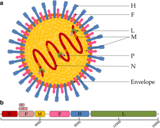
Respiratory syncytial virus (RSV), also called human respiratory syncytial virus (hRSV) and human orthopneumovirus, is a contagious virus that causes infections of the respiratory tract. It is a negative-sense, single-stranded RNA virus. Its name is derived from the large cells known as syncytia that form when infected cells fuse.

<i>Human metapneumovirus</i> Species of virus

Human metapneumovirus is a negative-sense single-stranded RNA virus of the family Pneumoviridae and is closely related to the Avian metapneumovirus (AMPV) subgroup C. It was isolated for the first time in 2001 in the Netherlands by using the RAP-PCR technique for identification of unknown viruses growing in cultured cells. As of 2016, it was the second most common cause of acute respiratory tract illness in otherwise-healthy children under the age of 5 in a large US outpatient clinic.

Coinfection is the simultaneous infection of a host by multiple pathogen species. In virology, coinfection includes simultaneous infection of a single cell by two or more virus particles. An example is the coinfection of liver cells with hepatitis B virus and hepatitis D virus, which can arise incrementally by initial infection followed by superinfection.

Viral pneumonia is a pneumonia caused by a virus. Pneumonia is an infection that causes inflammation in one or both of the lungs. The pulmonary alveoli fill with fluid or pus making it difficult to breathe. Pneumonia can be caused by bacteria, viruses, fungi or parasites. Viruses are the most common cause of pneumonia in children, while in adults bacteria are a more common cause.

<span class="mw-page-title-main">Respiratory tract infection</span> Infectious disease affecting nose, throat and lungs

Respiratory tract infections (RTIs) are infectious diseases involving the lower or upper respiratory tract. An infection of this type usually is further classified as an upper respiratory tract infection or a lower respiratory tract infection. Lower respiratory infections, such as pneumonia, tend to be far more severe than upper respiratory infections, such as the common cold.

<i>Human coronavirus NL63</i> Species of virus

Human coronavirus NL63 (HCoV-NL63) is a species of coronavirus, specifically a Setracovirus from among the Alphacoronavirus genus. It was identified in late 2004 in patients in the Netherlands by Lia van der Hoek and Krzysztof Pyrc using a novel virus discovery method VIDISCA. Later on the discovery was confirmed by the researchers from the Rotterdam, the Netherlands The virus is an enveloped, positive-sense, single-stranded RNA virus which enters its host cell by binding to ACE2. Infection with the virus has been confirmed worldwide, and has an association with many common symptoms and diseases. Associated diseases include mild to moderate upper respiratory tract infections, severe lower respiratory tract infection, croup and bronchiolitis.

<i>Murine respirovirus</i> Sendai virus, virus of rodents

Murine respirovirus, formerly Sendai virus (SeV) and previously also known as murine parainfluenza virus type 1 or hemagglutinating virus of Japan (HVJ), is an enveloped, 150-200 nm–diameter, negative sense, single-stranded RNA virus of the family Paramyxoviridae. It typically infects rodents and it is not pathogenic for humans or domestic animals.

Palivizumab, sold under the brand name Synagis, is a monoclonal antibody produced by recombinant DNA technology used to prevent severe disease caused by respiratory syncytial virus (RSV) infections. It is recommended for infants at high-risk for RSV due to conditions such as prematurity or other medical problems including heart or lung diseases.

<span class="mw-page-title-main">Antibody-dependent enhancement</span> Antibodies rarely making an infection worse instead of better

Antibody-dependent enhancement (ADE), sometimes less precisely called immune enhancement or disease enhancement, is a phenomenon in which binding of a virus to suboptimal antibodies enhances its entry into host cells, followed by its replication. The suboptimal antibodies can result from natural infection or from vaccination. ADE may cause enhanced respiratory disease, but is not limited to respiratory disease. It has been observed in HIV, RSV virus and Dengue virus and is monitored for in vaccine development.

<span class="mw-page-title-main">Virus</span> Infectious agent that replicates in cells

A virus is a submicroscopic infectious agent that replicates only inside the living cells of an organism. Viruses infect all life forms, from animals and plants to microorganisms, including bacteria and archaea. Viruses are found in almost every ecosystem on Earth and are the most numerous type of biological entity. Since Dmitri Ivanovsky's 1892 article describing a non-bacterial pathogen infecting tobacco plants and the discovery of the tobacco mosaic virus by Martinus Beijerinck in 1898, more than 11,000 of the millions of virus species have been described in detail. The study of viruses is known as virology, a subspeciality of microbiology.

<span class="mw-page-title-main">Influenza</span> Infectious disease

Influenza, commonly known as "the flu" or just "flu", is an infectious disease caused by influenza viruses. Symptoms range from mild to severe and often include fever, runny nose, sore throat, muscle pain, headache, coughing, and fatigue. These symptoms begin one to four days after exposure to the virus and last for about two to eight days. Diarrhea and vomiting can occur, particularly in children. Influenza may progress to pneumonia from the virus or a subsequent bacterial infection. Other complications include acute respiratory distress syndrome, meningitis, encephalitis, and worsening of pre-existing health problems such as asthma and cardiovascular disease.

<i>Pneumoviridae</i> Family of viruses

Pneumoviridae is a family of negative-strand RNA viruses in the order Mononegavirales. Humans, cattle, and rodents serve as natural hosts. Respiratory tract infections are associated with member viruses such as human respiratory syncytial virus. There are five species in the family which are divided between the genera Metapneumovirus and Orthopneumovirus. The family used to be considered as a sub-family of Paramyxoviridae, but has been reclassified as of 2016.

A respiratory syncytial virus vaccine, or RSV vaccine, is a vaccine that protects against respiratory syncytial virus. RSV affects an estimated 64 million people and causes 160,000 deaths worldwide each year.

<span class="mw-page-title-main">Jason McLellan</span> American structural biologist

Jason S. McLellan is a structural biologist, professor in the Department of Molecular Biosciences and Robert A. Welch Chair in Chemistry at The University of Texas at Austin who specializes in understanding the structure and function of viral proteins, including those of coronaviruses. His research focuses on applying structural information to the rational design of vaccines and other therapies for viruses, including SARS-CoV-2, the novel coronavirus that causes COVID-19, and respiratory syncytial virus (RSV). McLellan and his team collaborated with researchers at the National Institute of Allergy and Infectious Diseases’ Vaccine Research Center to design a stabilized version of the SARS-CoV-2 spike protein, which biotechnology company Moderna used as the basis for the vaccine mRNA-1273, the first COVID-19 vaccine candidate to enter phase I clinical trials in the U.S. At least three other vaccines use this modified spike protein: those from Pfizer and BioNTech; Johnson & Johnson and Janssen Pharmaceuticals; and Novavax.

Bovine respiratory syncytial virus (BRSV) is pneumovirus closely related to human respiratory syncytial virus (RSV) that is a common cause of respiratory disease in cattle, particularly calves. It is a negative-sense, single-stranded RNA virus that replicates in the cytoplasm of the cell. Similarly to other single-stranded RNA viruses, the genome of BRSV has a high mutation rate, which results in great antigenetic variation. Thus, BRSV can be split into four different subgroups based on antigen expression.

References

  1. "Virus Taxonomy: 2018 Release". International Committee on Taxonomy of Viruses (ICTV). October 2018. Retrieved 25 January 2019.
  2. Vainionpää R, Hyypiä T (April 1994). "Biology of parainfluenza viruses". Clin. Microbiol. Rev. 7 (2): 265–275. doi:10.1128/CMR.7.2.265. PMC   358320 . PMID   8055470.
  3. 1 2 3 4 5 6 7 8 9 10 11 Henrickson, KJ (April 2003). "Parainfluenza viruses". Clinical Microbiology Reviews. 16 (2): 242–264. doi:10.1128/CMR.16.2.242-264.2003. PMC   153148 . PMID   12692097.
  4. 1 2 3 "Human Parainfluenza Viruses". Centers for Disease Control and Prevention (2011). Archived from the original on 20 March 2012. Retrieved 21 March 2012.
  5. Schmidt, Alexander; Anne Schaap-Nutt; Emmalene J Bartlett; Henrick Schomacker; Jim Boonyaratanakornkit; Ruth A Karron; Peter L Collins (1 February 2011). "Progress in the development of human parainfluenza virus vaccines". Expert Review of Respiratory Medicine. 5 (4): 515–526. doi:10.1586/ers.11.32. PMC   3503243 . PMID   21859271.
  6. Baron, S.; Enders, G. (1996). "Paramyxoviruses". Parainfluenza Viruses. University of Texas Medical Branch at Galveston. ISBN   9780963117212. PMID   21413341 . Retrieved 2009-03-15.
  7. Hunt, Dr. Margaret. "PARAINFLUENZA, RESPIRATORY SYNCYTIAL AND ADENO VIRUSES". Reference.MD. Retrieved 21 March 2012.
  8. Vulliémoz, D; Roux, L (May 2001). "'Rule of six': how does the Sendai virus RNA polymerase keep count?". Journal of Virology. 75 (10): 4506–4518. doi:10.1128/JVI.75.10.4506-4518.2001. PMC   114204 . PMID   11312321.
  9. Henrickson, K. J (2003). "Parainfluenza Viruses". Clinical Microbiology Reviews. 16 (2): 242–264. doi:10.1128/CMR.16.2.242-264.2003. PMC   153148 . PMID   12692097.
  10. 1 2 Moscona, A (July 2005). "Entry of parainfluenza virus into cells as a target for interrupting childhood respiratory disease". The Journal of Clinical Investigation. 115 (7): 1688–1698. doi:10.1172/JCI25669. PMC   1159152 . PMID   16007245.
  11. 1 2 Chambers R, Takimoto T (2011). "Parainfluenza Viruses". Encyclopedia of Life Sciences. Wiley. doi:10.1002/9780470015902.a0001078.pub3. ISBN   978-0470016176.{{cite book}}: |journal= ignored (help)
  12. 1 2 3 4 5 6 7 8 9 10 "Parainfluenza Virus: Epidemiology". eMedicine. Retrieved 21 March 2012.
  13. 1 2 3 4 "HUMAN PARAINFLUENZA VIRUS". Public Health Agency of Canada. 2011-04-19. Retrieved 21 March 2012.
  14. 1 2 Henrickson, KJ; Kuhn, SM; Savatski, LL (May 1994). "Epidemiology and cost of infection with human parainfluenza virus types 1 and 2 in young children". Clinical Infectious Diseases. 18 (5): 770–9. doi:10.1093/clinids/18.5.770. PMID   8075269.
  15. Denny, FW; Clyde WA, Jr (May 1986). "Acute lower respiratory tract infections in nonhospitalized children". The Journal of Pediatrics. 108 (5 Pt 1): 635–46. doi:10.1016/S0022-3476(86)81034-4. PMID   3009769.
  16. "Acute Respiratory Infections". WHO. Archived from the original on March 24, 2006. Retrieved 21 March 2012.
  17. 1 2 Durbin, AP; Karron, RA (December 15, 2003). "Progress in the development of respiratory syncytial virus and parainfluenza virus vaccines". Clinical Infectious Diseases. 37 (12): 1668–1677. doi:10.1086/379775. PMID   14689350. S2CID   41967381.
  18. "General information: human parainfluenza viruses". Health Protection Agency. 27 August 2008. Retrieved 21 March 2012.
  19. Sable CA, Hayden FG (December 1995). "Orthomyxoviral and paramyxoviral infections in transplant patients". Infect. Dis. Clin. North Am. 9 (4): 987–1003. doi:10.1016/S0891-5520(20)30712-1. PMID   8747776.
  20. "CDC - Human Parainfluenza Viruses: Common cold and croup". Archived from the original on 2009-03-03. Retrieved 2009-03-15.
  21. "Croup Background". Medscape Reference. Retrieved 21 March 2012.
  22. "Parainfluenza Virus Review". Medscape. Retrieved 21 March 2012.
  23. Stephen B Greenberg; Robert L Atmar. "Parainfluenza Viruses—New Epidemiology and Vaccine Developments". Touch Infectious Disease. Retrieved 21 March 2012.
  24. Arguedas, A; Stutman, HR; Blanding, JG (March 1990). "Parainfluenza type 3 meningitis. Report of two cases and review of the literature". Clinical Pediatrics. 29 (3): 175–178. doi:10.1177/000992289002900307. PMID   2155085. S2CID   25043753.
  25. "Human Parainfluenza Viruses (HPIV) and Other Parainfluenza Viruses: Background, Pathophysiology, Etiology". 17 October 2021. Retrieved 18 March 2023.
  26. Branche, Angela; Falsey, Ann (2016-08-03). "Parainfluenza Virus Infection". Seminars in Respiratory and Critical Care Medicine. 37 (4): 538–554. doi:10.1055/s-0036-1584798. ISSN   1069-3424. PMC   7171724 . PMID   27486735.
  27. Dimopoulos, G; Lerikou, M; Tsiodras, S; Chranioti, A; Perros, E; Anagnostopoulou, U; Armaganidis, A; Karakitsos, P (February 2012). "Viral epidemiology of acute exacerbations of chronic obstructive pulmonary disease". Pulmonary Pharmacology & Therapeutics. 25 (1): 12–8. doi:10.1016/j.pupt.2011.08.004. PMC   7110842 . PMID   21983132.
  28. Beckham, JD; Cadena, A; Lin, J; Piedra, PA; Glezen, WP; Greenberg, SB; Atmar, RL (May 2005). "Respiratory viral infections in patients with chronic, obstructive pulmonary disease". The Journal of Infection. 50 (4): 322–30. doi:10.1016/j.jinf.2004.07.011. PMC   7132437 . PMID   15845430.
  29. Berman, S (May–Jun 1991). "Epidemiology of acute respiratory infections in children of developing countries". Reviews of Infectious Diseases. 13 (Suppl 6): S454–62. doi:10.1093/clinids/13.supplement_6.s454. PMID   1862276.
  30. Sato M, Wright PF (October 2008). "Current status of vaccines for parainfluenza virus infections". Pediatr. Infect. Dis. J. 27 (10 Suppl): S123–5. doi: 10.1097/INF.0b013e318168b76f . PMID   18820572.
  31. "Parainfluenza Viruses". eLS. Retrieved 21 March 2012.
  32. "Definition of Human parainfluenza virus". MedicineNet. Archived from the original on 5 January 2012. Retrieved 21 March 2012.
  33. HAMBLING, MH (December 1964). "Survival of the Respiratory Syncytial Virus During Storage Under Various Conditions". British Journal of Experimental Pathology. 45 (6): 647–55. PMC   2093680 . PMID   14245166.
  34. "Common Cold, Croup and Human Parainfluenza Viruses: Symptoms and Prevention". NewsFlu. Retrieved 21 March 2012.

Further reading