Coronavirus membrane protein

Last updated
Membrane protein
Coronavirus. SARS-CoV-2.png
Model of the external structure of the SARS-CoV-2 virion. [1]
Blue: envelope
Turquoise: spike glycoprotein (S)
Red: envelope proteins (E)
Green: membrane proteins (M)
Orange: glycans
Identifiers
SymbolCoV_M
Pfam PF01635
InterPro IPR002574
PROSITE PS51927
Available protein structures:
Pfam   structures / ECOD  
PDB RCSB PDB; PDBe; PDBj
PDBsum structure summary

The membrane (M) protein (previously called E1, sometimes also matrix protein [2] ) is an integral membrane protein that is the most abundant of the four major structural proteins found in coronaviruses. [3] [4] [5] The M protein organizes the assembly of coronavirus virions through protein-protein interactions with other M protein molecules as well as with the other three structural proteins, the envelope (E), spike (S), and nucleocapsid (N) proteins. [4] [6] [7] [8]

Contents

Structure

The M protein is a transmembrane protein with three transmembrane domains and is around 230 amino acid residues long. [8] [9] In SARS-CoV-2, the causative agent of COVID-19, the M protein is 222 residues long. [10] Its membrane topology orients the C-terminus toward the cytosolic face of the membrane and thus into the interior of the virion. It has a short N-terminal segment and a larger C-terminal domain. Although the protein sequence is not well conserved across all coronavirus groups, there is a conserved amphipathic region near the C-terminal end of the third transmembrane segment. [8] [9]

M functions as a homodimer. [4] [5] Studies of the M protein in multiple coronaviruses by cryo-electron microscopy have identified two distinct functional protein conformations, thought to have different roles in forming protein-protein interactions with other structural proteins. [5] M protein of SARS-CoV-2 is homologous to the prokaryotic sugar transport protein SemiSWEET. [11]

Post-translational modifications

M is a glycoprotein whose glycosylation varies according to coronavirus subgroup; N-linked glycosylation is typically found in the alpha and gamma groups while O-linked glycosylation is typically found in the beta group. [8] [9] There are some exceptions; for example, in SARS-CoV, a betacoronavirus, the M protein has one N-glycosylation site. [8] [6] Glycosylation state does not appear to have a measurable effect on viral growth. [6] [9] [12] No other post-translational modifications have been described for the M protein. [4]

Expression and localization

Genomic information
SARS-CoV-2 genome.svg
Genomic organisation of isolate Wuhan-Hu-1, the earliest sequenced sample of SARS-CoV-2, indicating the location of the M gene
NCBI genome ID 86693
Genome size 29,903 bases
Year of completion 2020
Genome browser (UCSC)

The gene encoding the M protein is located toward the 3' end of the virus's positive-sense RNA genome, along with the genes for the other three structural proteins and various virus-specific accessory proteins. [6] [8] M is translated by membrane-bound polysomes [6] to be inserted into the endoplasmic reticulum (ER) and trafficked to the endoplasmic reticulum-Golgi intermediate compartment (ERGIC), the intracellular compartment that gives rise to the coronavirus viral envelope, or to the Golgi apparatus. [8] [7] [6] The exact localization is dependent on the specific virus protein. [13] Investigations of the subcellular localization of the MERS-CoV M protein found C-terminal sequence signals associated with trafficking to the Golgi. [14]

Function

Illustration of a coronavirus virion in the respiratory mucosa, showing the positions of the four structural proteins and components of the extracellular environment. Pbio.3000815.g001.PNG L.png
Illustration of a coronavirus virion in the respiratory mucosa, showing the positions of the four structural proteins and components of the extracellular environment.

The M protein is the most abundant protein in coronavirus virions. [8] [5] [4] It is essential for viral replication. [4]

Viral assembly

The primary function of the M protein is organizing assembly of new virions. [4] It is involved in establishing viral shape and morphology. Individual M molecules interact with each other to form the viral envelope [7] [9] [8] and may be able to exclude host cell proteins from the viral membrane. [5] Studies of the SARS-CoV M protein suggest that M-M interactions involve both the N- and C-termini. [6] Coronaviruses are moderately pleomorphic and conformational variations of M appear to be associated with virion size. [5]

M forms protein-protein interactions with all three other major structural proteins. [4] [7] M is necessary but not sufficient for viral assembly; M and the E protein expressed together are reportedly sufficient to form virus-like particles, [7] though some reports vary depending on experimental conditions and the specific virus studied. [6] [13] In some reports M appears to be capable of inducing membrane curvature, [5] though others report M alone is insufficient for this and E is required. [7] Although the E protein is not necessarily essential, it appears to be required for normal viral morphology and may be responsible for establishing curvature or initiating viral budding. [7] M also appears to have functional roles in the later stages of viral maturation, secretion, and budding. [4]

Incorporation of the spike protein (S) - which is required for assembly of infectious virions - is reported to occur though M interactions and may depend on specific conformations of M. [5] [13] The conserved amphipathic region C-terminal to the third transmembrane segment is important for spike interactions. [13] Interactions with M appear to be required for correct subcellular localization of S at the viral budding site. [12] M interacts directly with the nucleocapsid (N) protein without requiring the presence of RNA. [6] This interaction appears to occur primarily through both proteins' C-termini. [4]

Interactions with the immune system

The human HLA-A*0201 (red) and beta-2 microglobulin (green) in complex with a peptide derived from the M protein of SARS-CoV (yellow, shown as surface). From PDB: 3I6G . 3i6g chainABC.png
The human HLA-A*0201 (red) and beta-2 microglobulin (green) in complex with a peptide derived from the M protein of SARS-CoV (yellow, shown as surface). From PDB: 3I6G .

The M protein in MERS-CoV, SARS-CoV, and SARS-CoV-2 has been described as an antagonist of interferon response. [4] [17]

The M protein is immunogenic and has been reported to be a determinant of humoral immunity. [4] Cytotoxic T cell responses to M have been described. [16] Antibodies to epitopes found in the M protein have been identified in patients recovered from severe acute respiratory syndrome (SARS). [18]

Other recent research has identified that SAS-COV-2 membrane protein when treated on human PBMC's causes a significant increase in pro inflammatory mediators such as TNF and IL-6. [19] . The effects of exogenous SARS-COV-2 membrane protein challenge in mice was also studied. In these studies, exogenous membrane protein treated intra nasally caused a significant increase in pulmonary inflammation in mice leading to histological changes within the lungs. [20] .

Host cell entry

It has been reported that human coronavirus NL63 relies on the M protein as well as the S protein to mediate host cell interactions preceding viral entry. M is thought to bind heparan sulfate proteoglycans exposed on the cell surface. [21]

Evolution and conservation

A study of SARS-CoV-2 sequences collected during the COVID-19 pandemic found that missense mutations in the M gene were relatively uncommon and suggested it was under purifying selection. [22] Similar results have been described for broader population genetics analyses over a wider range of related viruses, finding that the sequences of M and several non-structural proteins in the coronavirus genome are most subject to evolutionary constraints. [23]

Related Research Articles

<span class="mw-page-title-main">Coronavirus</span> Subfamily of viruses in the family Coronaviridae

Coronaviruses are a group of related RNA viruses that cause diseases in mammals and birds. In humans and birds, they cause respiratory tract infections that can range from mild to lethal. Mild illnesses in humans include some cases of the common cold, while more lethal varieties can cause SARS, MERS and COVID-19. In cows and pigs they cause diarrhea, while in mice they cause hepatitis and encephalomyelitis.

<span class="mw-page-title-main">SARS-related coronavirus</span> Species of coronavirus causing SARS and COVID-19

Severe-acute-respiratory-syndrome–related coronavirus is a species of virus consisting of many known strains. Two strains of the virus have caused outbreaks of severe respiratory diseases in humans: severe acute respiratory syndrome coronavirus 1, which caused the 2002–2004 outbreak of severe acute respiratory syndrome (SARS), and severe acute respiratory syndrome coronavirus 2 (SARS-CoV-2), which is causing the ongoing pandemic of COVID-19. There are hundreds of other strains of SARSr-CoV, which are only known to infect non-human mammal species: bats are a major reservoir of many strains of SARSr-CoV; several strains have been identified in Himalayan palm civets, which were likely ancestors of SARS-CoV-1.

<i>Coronaviridae</i> Family of viruses in the order Nidovirales

Coronaviridae is a family of enveloped, positive-strand RNA viruses which infect amphibians, birds, and mammals. The group includes the subfamilies Letovirinae and Orthocoronavirinae; the members of the latter are known as coronaviruses.

<i>Human coronavirus NL63</i> Species of virus

Human coronavirus NL63 (HCoV-NL63) is a species of coronavirus, specifically a Setracovirus from among the Alphacoronavirus genus. It was identified in late 2004 in patients in the Netherlands by Lia van der Hoek and Krzysztof Pyrc using a novel virus discovery method VIDISCA. Later on the discovery was confirmed by the researchers from the Rotterdam, the Netherlands The virus is an enveloped, positive-sense, single-stranded RNA virus which enters its host cell by binding to ACE2. Infection with the virus has been confirmed worldwide, and has an association with many common symptoms and diseases. Associated diseases include mild to moderate upper respiratory tract infections, severe lower respiratory tract infection, croup and bronchiolitis.

<span class="mw-page-title-main">Spike protein</span> Glycoprotein spike on a viral capsid or viral envelope

In virology, a spike protein or peplomer protein is a protein that forms a large structure known as a spike or peplomer projecting from the surface of an enveloped virus. The proteins are usually glycoproteins that form dimers or trimers.

A viral structural protein is a viral protein that is a structural component of the mature virus.

Feline coronavirus (FCoV) is a positive-stranded RNA virus that infects cats worldwide. It is a coronavirus of the species Alphacoronavirus 1, which includes canine coronavirus (CCoV) and porcine transmissible gastroenteritis coronavirus (TGEV). FCoV has two different forms: feline enteric coronavirus (FECV), which infects the intestines, and feline infectious peritonitis virus (FIPV), which causes the disease feline infectious peritonitis (FIP).

<span class="mw-page-title-main">ORF7a</span> Gene found in coronaviruses of the Betacoronavirus genus

ORF7a is a gene found in coronaviruses of the Betacoronavirus genus. It expresses the Betacoronavirus NS7A protein, a type I transmembrane protein with an immunoglobulin-like protein domain. It was first discovered in SARS-CoV, the virus that causes severe acute respiratory syndrome (SARS). The homolog in SARS-CoV-2, the virus that causes COVID-19, has about 85% sequence identity to the SARS-CoV protein.

<i>Human coronavirus HKU1</i> Species of virus

Human coronavirus HKU1 (HCoV-HKU1) is a species of coronavirus in humans and animals. It causes an upper respiratory disease with symptoms of the common cold, but can advance to pneumonia and bronchiolitis. It was first discovered in January 2004 from one man in Hong Kong. Subsequent research revealed it has global distribution and earlier genesis.

<i>Betacoronavirus</i> Genus of viruses

Betacoronavirus is one of four genera of coronaviruses. Member viruses are enveloped, positive-strand RNA viruses that infect mammals, including humans. The natural reservoir for betacoronaviruses are bats and rodents. Rodents are the reservoir for the subgenus Embecovirus, while bats are the reservoir for the other subgenera.

<i>Human coronavirus 229E</i> Species of virus

Human coronavirus 229E (HCoV-229E) is a species of coronavirus which infects humans and bats. It is an enveloped, positive-sense, single-stranded RNA virus which enters its host cell by binding to the APN receptor. Along with Human coronavirus OC43, it is one of the viruses responsible for the common cold. HCoV-229E is a member of the genus Alphacoronavirus and subgenus Duvinacovirus.

Bat SARS-like coronavirus WIV1, also sometimes called SARS-like coronavirus WIV1, is a strain of severe acute respiratory syndrome–related coronavirus (SARSr-CoV) isolated from Chinese rufous horseshoe bats in 2013. Like all coronaviruses, virions consist of single-stranded positive-sense RNA enclosed within an envelope.

<span class="mw-page-title-main">Coronavirus envelope protein</span> Major structure in coronaviruses

The envelope (E) protein is the smallest and least well-characterized of the four major structural proteins found in coronavirus virions. It is an integral membrane protein less than 110 amino acid residues long; in SARS-CoV-2, the causative agent of Covid-19, the E protein is 75 residues long. Although it is not necessarily essential for viral replication, absence of the E protein may produce abnormally assembled viral capsids or reduced replication. E is a multifunctional protein and, in addition to its role as a structural protein in the viral capsid, it is thought to be involved in viral assembly, likely functions as a viroporin, and is involved in viral pathogenesis.

<span class="mw-page-title-main">Coronavirus nucleocapsid protein</span> Most expressed structure in coronaviruses

The nucleocapsid (N) protein is a protein that packages the positive-sense RNA genome of coronaviruses to form ribonucleoprotein structures enclosed within the viral capsid. The N protein is the most highly expressed of the four major coronavirus structural proteins. In addition to its interactions with RNA, N forms protein-protein interactions with the coronavirus membrane protein (M) during the process of viral assembly. N also has additional functions in manipulating the cell cycle of the host cell. The N protein is highly immunogenic and antibodies to N are found in patients recovered from SARS and COVID-19.

<span class="mw-page-title-main">Coronavirus spike protein</span> Glycoprotein spike on a viral capsid or viral envelope

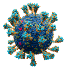
Spike (S) glycoprotein is the largest of the four major structural proteins found in coronaviruses. The spike protein assembles into trimers that form large structures, called spikes or peplomers, that project from the surface of the virion. The distinctive appearance of these spikes when visualized using negative stain transmission electron microscopy, "recalling the solar corona", gives the virus family its main name.

<span class="mw-page-title-main">ORF3a</span> Gene found in coronaviruses of the subgenus Sarbecovirus

ORF3a is a gene found in coronaviruses of the subgenus Sarbecovirus, including SARS-CoV and SARS-CoV-2. It encodes an accessory protein about 275 amino acid residues long, which is thought to function as a viroporin. It is the largest accessory protein and was the first of the SARS-CoV accessory proteins to be described.

ORF7b is a gene found in coronaviruses of the genus Betacoronavirus, which expresses the accessory protein Betacoronavirus NS7b protein. It is a short, highly hydrophobic transmembrane protein of unknown function.

<span class="mw-page-title-main">ORF8</span> Gene that encodes a viral accessory protein

ORF8 is a gene that encodes a viral accessory protein, Betacoronavirus NS8 protein, in coronaviruses of the subgenus Sarbecovirus. It is one of the least well conserved and most variable parts of the genome. In some viruses, a deletion splits the region into two smaller open reading frames, called ORF8a and ORF8b - a feature present in many SARS-CoV viral isolates from later in the SARS epidemic, as well as in some bat coronaviruses. For this reason the full-length gene and its protein are sometimes called ORF8ab. The full-length gene, exemplified in SARS-CoV-2, encodes a protein with an immunoglobulin domain of unknown function, possibly involving interactions with the host immune system. It is similar in structure to the ORF7a protein, suggesting it may have originated through gene duplication.

ORF6 is a gene that encodes a viral accessory protein in coronaviruses of the subgenus Sarbecovirus, including SARS-CoV and SARS-CoV-2. It is not present in MERS-CoV. It is thought to reduce the immune system response to viral infection through interferon antagonism.

<span class="mw-page-title-main">ORF9b</span> Gene

ORF9b is a gene that encodes a viral accessory protein in coronaviruses of the subgenus Sarbecovirus, including SARS-CoV and SARS-CoV-2. It is an overlapping gene whose open reading frame is entirely contained within the N gene, which encodes coronavirus nucleocapsid protein. The encoded protein is 97 amino acid residues long in SARS-CoV and 98 in SARS-CoV-2, in both cases forming a protein dimer.

References

  1. Solodovnikov, Alexey; Arkhipova, Valeria (2021-07-29). "Достоверно красиво: как мы сделали 3D-модель SARS-CoV-2" [Truly beautiful: how we made the SARS-CoV-2 3D model] (in Russian). N+1. Archived from the original on 2021-07-30. Retrieved 30 July 2021.
  2. Hu Y, Wen J, Tang L, Zhang H, Zhang X, Li Y, et al. (May 2003). "The M protein of SARS-CoV: basic structural and immunological properties". Genomics, Proteomics & Bioinformatics. 1 (2): 118–130. doi:10.1016/S1672-0229(03)01016-7. PMC   5172243 . PMID   15626342.
  3. Thomas S. The Structure of the Membrane Protein of SARS-CoV-2 Resembles the Sugar Transporter SemiSWEET. Pathog Immun. 2020 Oct 19;5(1):342-363.
  4. 1 2 3 4 5 6 7 8 9 10 11 12 Wong NA, Saier MH (January 2021). "The SARS-Coronavirus Infection Cycle: A Survey of Viral Membrane Proteins, Their Functional Interactions and Pathogenesis". International Journal of Molecular Sciences. 22 (3): 1308. doi: 10.3390/ijms22031308 . PMC   7865831 . PMID   33525632.
  5. 1 2 3 4 5 6 7 8 Neuman BW, Kiss G, Kunding AH, Bhella D, Baksh MF, Connelly S, et al. (April 2011). "A structural analysis of M protein in coronavirus assembly and morphology". Journal of Structural Biology. 174 (1): 11–22. doi:10.1016/j.jsb.2010.11.021. PMC   4486061 . PMID   21130884.
  6. 1 2 3 4 5 6 7 8 9 Tseng YT, Wang SM, Huang KJ, Lee AI, Chiang CC, Wang CT (April 2010). "Self-assembly of severe acute respiratory syndrome coronavirus membrane protein". The Journal of Biological Chemistry. 285 (17): 12862–12872. doi: 10.1074/jbc.M109.030270 . PMC   2857088 . PMID   20154085.
  7. 1 2 3 4 5 6 7 Schoeman D, Fielding BC (May 2019). "Coronavirus envelope protein: current knowledge". Virology Journal. 16 (1): 69. doi: 10.1186/s12985-019-1182-0 . PMC   6537279 . PMID   31133031.
  8. 1 2 3 4 5 6 7 8 9 Masters PS (2006). "The molecular biology of coronaviruses". Advances in Virus Research. 66: 193–292. doi:10.1016/S0065-3527(06)66005-3. ISBN   9780120398690. PMC   7112330 . PMID   16877062.
  9. 1 2 3 4 5 J Alsaadi EA, Jones IM (April 2019). "Membrane binding proteins of coronaviruses". Future Virology. 14 (4): 275–286. doi:10.2217/fvl-2018-0144. PMC   7079996 . PMID   32201500.
  10. Cao Y, Yang R, Lee I, Zhang W, Sun J, Wang W, Meng X (June 2021). "Characterization of the SARS-CoV-2 E Protein: Sequence, Structure, Viroporin, and Inhibitors". Protein Science. 30 (6): 1114–1130. doi:10.1002/pro.4075. PMC   8138525 . PMID   33813796.
  11. Thomas S. The Structure of the Membrane Protein of SARS-CoV-2 Resembles the Sugar Transporter SemiSWEET. Pathog Immun. 2020 Oct 19;5(1):342-363
  12. 1 2 Voss D, Pfefferle S, Drosten C, Stevermann L, Traggiai E, Lanzavecchia A, Becker S (June 2009). "Studies on membrane topology, N-glycosylation and functionality of SARS-CoV membrane protein". Virology Journal. 6 (1): 79. doi: 10.1186/1743-422X-6-79 . PMC   2705359 . PMID   19534833.
  13. 1 2 3 4 Ujike M, Taguchi F (April 2015). "Incorporation of spike and membrane glycoproteins into coronavirus virions". Viruses. 7 (4): 1700–1725. doi: 10.3390/v7041700 . PMC   4411675 . PMID   25855243.
  14. Perrier A, Bonnin A, Desmarets L, Danneels A, Goffard A, Rouillé Y, et al. (September 2019). "The C-terminal domain of the MERS coronavirus M protein contains a trans-Golgi network localization signal". The Journal of Biological Chemistry. 294 (39): 14406–14421. doi: 10.1074/jbc.RA119.008964 . PMC   6768645 . PMID   31399512.
  15. Goodsell DS, Voigt M, Zardecki C, Burley SK (August 2020). "Integrative illustration for coronavirus outreach". PLOS Biology. 18 (8): e3000815. doi: 10.1371/journal.pbio.3000815 . PMC   7433897 . PMID   32760062.
  16. 1 2 Liu J, Sun Y, Qi J, Chu F, Wu H, Gao F, et al. (October 2010). "The membrane protein of severe acute respiratory syndrome coronavirus acts as a dominant immunogen revealed by a clustering region of novel functionally and structurally defined cytotoxic T-lymphocyte epitopes". The Journal of Infectious Diseases. 202 (8): 1171–1180. doi:10.1086/656315. PMC   7537489 . PMID   20831383.
  17. Zheng Y, Zhuang MW, Han L, Zhang J, Nan ML, Zhan P, et al. (December 2020). "Severe acute respiratory syndrome coronavirus 2 (SARS-CoV-2) membrane (M) protein inhibits type I and III interferon production by targeting RIG-I/MDA-5 signaling". Signal Transduction and Targeted Therapy. 5 (1): 299. doi:10.1038/s41392-020-00438-7. PMC   7768267 . PMID   33372174.
  18. Pang H, Liu Y, Han X, Xu Y, Jiang F, Wu D, et al. (October 2004). "Protective humoral responses to severe acute respiratory syndrome-associated coronavirus: implications for the design of an effective protein-based vaccine". The Journal of General Virology. 85 (Pt 10): 3109–3113. doi: 10.1099/vir.0.80111-0 . PMID   15448374.
  19. Haystead, T., Lee, E., Cho, K., Gullickson, G., Hughes, P., Krafsur, G., ... & Scarneo, S. (2023). Investigation of SARS-CoV-2 individual proteins reveals the in vitro and in vivo immunogenicity of membrane protein. Scientific Reports, 13(1), 22873.
  20. Haystead, T., Lee, E., Cho, K., Gullickson, G., Hughes, P., Krafsur, G., ... & Scarneo, S. (2023). Investigation of SARS-CoV-2 individual proteins reveals the in vitro and in vivo immunogenicity of membrane protein. Scientific Reports, 13(1), 22873.
  21. Naskalska A, Dabrowska A, Szczepanski A, Milewska A, Jasik KP, Pyrc K (October 2019). "Membrane Protein of Human Coronavirus NL63 Is Responsible for Interaction with the Adhesion Receptor". Journal of Virology. 93 (19): e00355-19. doi:10.1128/JVI.00355-19. PMC   6744225 . PMID   31315999.
  22. Shen L, Bard JD, Triche TJ, Judkins AR, Biegel JA, Gai X (December 2021). "Emerging variants of concern in SARS-CoV-2 membrane protein: a highly conserved target with potential pathological and therapeutic implications". Emerging Microbes & Infections. 10 (1): 885–893. doi:10.1080/22221751.2021.1922097. PMC   8118436 . PMID   33896413.
  23. Cagliani R, Forni D, Clerici M, Sironi M (June 2020). "Computational Inference of Selection Underlying the Evolution of the Novel Coronavirus, Severe Acute Respiratory Syndrome Coronavirus 2". Journal of Virology. 94 (12): e00411-20. doi:10.1128/JVI.00411-20. PMC   7307108 . PMID   32238584.