Medical uses
| | This section is empty. You can help by adding to it. (December 2023) |
| Whole lung lavage | |
|---|---|
| Other names | Lung washing |
| ICD-9-CM | 33.99 |
Whole lung lavage (WLL), also called lung washing, is a medical procedure in which the patient's lungs are washed with saline (salt water) by filling and draining repeatedly. It is used to treat pulmonary alveolar proteinosis, in which excess lung surfactant proteins prevent the patient from breathing. [1] [2] Some sources consider it a variation of bronchoalveolar lavage. [3]
WLL has been experimentally used for silicosis, [4] other forms of mineral inhalation, and accidental inhalation of radioactive dust. [5] It appears to effectively remove these foreign particles. [4] [6] WLL treatments may slow down the lung function decline of miners with pneumoconiosis. [7]
| | This section is empty. You can help by adding to it. (December 2023) |
WLL is not a standarized procedure. Patients are usually first put under general anesthesia. A double lumen endotracheal tube is used to keep one lung breathing while the other is being washed. The lung to be washed is filled with fluid by gravity, then drained. Drainage can be done by suction [2] or gravity. [8] Some versions add a shaking step between the filling and draining to help with the washing. [2] The procedure typically uses 10–20 liters of fluid per patient, but severe cases require up to 50. [2]
Variations on the WLL include a "mini-WLL" with reduced infusion volume. [9] Reducing the suction power seems to reduce lung injury. [10]
| | This section is empty. You can help by adding to it. (December 2023) |
| | This section is empty. You can help by adding to it. (December 2023) |
| | This section is empty. You can help by adding to it. (December 2023) |
| | This section is empty. You can help by adding to it. (December 2023) |
| | This section is empty. You can help by adding to it. (December 2023) |

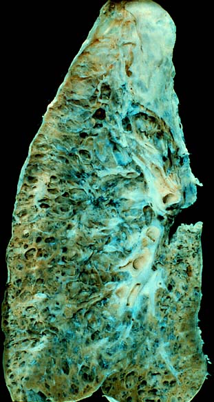
Pneumoconiosis is the general term for a class of interstitial lung disease where inhalation of dust has caused interstitial fibrosis. The three most common types are asbestosis, silicosis, and coal miner's lung. Pneumoconiosis often causes restrictive impairment, although diagnosable pneumoconiosis can occur without measurable impairment of lung function. Depending on extent and severity, it may cause death within months or years, or it may never produce symptoms. It is usually an occupational lung disease, typically from years of dust exposure during work in mining; textile milling; shipbuilding, ship repairing, and/or shipbreaking; sandblasting; industrial tasks; rock drilling ; or agriculture. It is one of the most common occupational diseases in the world.

Silicosis is a form of occupational lung disease caused by inhalation of crystalline silica dust. It is marked by inflammation and scarring in the form of nodular lesions in the upper lobes of the lungs. It is a type of pneumoconiosis. Silicosis, particularly the acute form, is characterized by shortness of breath, cough, fever, and cyanosis. It may often be misdiagnosed as pulmonary edema, pneumonia, or tuberculosis. Using workplace controls, silicosis is almost always a preventable disease.

Pulmonary alveolar proteinosis (PAP) is a rare lung disorder characterized by an abnormal accumulation of surfactant-derived lipoprotein compounds within the alveoli of the lung. The accumulated substances interfere with the normal gas exchange and expansion of the lungs, ultimately leading to difficulty breathing and a predisposition to developing lung infections. The causes of PAP may be grouped into primary, secondary, and congenital causes, although the most common cause is a primary autoimmune condition in an individual.

Interstitial lung disease (ILD), or diffuse parenchymal lung disease (DPLD), is a group of respiratory diseases affecting the interstitium and space around the alveoli of the lungs. It concerns alveolar epithelium, pulmonary capillary endothelium, basement membrane, and perivascular and perilymphatic tissues. It may occur when an injury to the lungs triggers an abnormal healing response. Ordinarily, the body generates just the right amount of tissue to repair damage, but in interstitial lung disease, the repair process is disrupted, and the tissue around the air sacs (alveoli) becomes scarred and thickened. This makes it more difficult for oxygen to pass into the bloodstream. The disease presents itself with the following symptoms: shortness of breath, nonproductive coughing, fatigue, and weight loss, which tend to develop slowly, over several months. The average rate of survival for someone with this disease is between three and five years. The term ILD is used to distinguish these diseases from obstructive airways diseases.

A chest radiograph, called a chest X-ray (CXR), or chest film, is a projection radiograph of the chest used to diagnose conditions affecting the chest, its contents, and nearby structures. Chest radiographs are the most common film taken in medicine.

Pneumonitis describes general inflammation of lung tissue. Possible causative agents include radiation therapy of the chest, exposure to medications used during chemo-therapy, the inhalation of debris, aspiration, herbicides or fluorocarbons and some systemic diseases. If unresolved, continued inflammation can result in irreparable damage such as pulmonary fibrosis.
Siderosis is the deposition of excess iron in body tissue. When used without qualification, it usually refers to an environmental disease of the lung, also known more specifically as pulmonary siderosis or Welder's disease, which is a form of pneumoconiosis.

Cryptogenic organizing pneumonia (COP), formerly known as bronchiolitis obliterans organizing pneumonia (BOOP), is an inflammation of the bronchioles (bronchiolitis) and surrounding tissue in the lungs. It is a form of idiopathic interstitial pneumonia.

Black lung disease (BLD), also known as coal-mine dust lung disease, or simply black lung, is an occupational type of pneumoconiosis caused by long-term inhalation and deposition of coal dust in the lungs and the consequent lung tissue's reaction to its presence. It is common in coal miners and others who work with coal. It is similar to both silicosis from inhaling silica dust and asbestosis from inhaling asbestos dust. Inhaled coal dust progressively builds up in the lungs and leads to inflammation, fibrosis, and in worse cases, necrosis.
Ventilator-associated pneumonia (VAP) is a type of lung infection that occurs in people who are on mechanical ventilation breathing machines in hospitals. As such, VAP typically affects critically ill persons that are in an intensive care unit (ICU) and have been on a mechanical ventilator for at least 48 hours. VAP is a major source of increased illness and death. Persons with VAP have increased lengths of ICU hospitalization and have up to a 20–30% death rate. The diagnosis of VAP varies among hospitals and providers but usually requires a new infiltrate on chest x-ray plus two or more other factors. These factors include temperatures of >38 °C or <36 °C, a white blood cell count of >12 × 109/ml, purulent secretions from the airways in the lung, and/or reduction in gas exchange.
Bronchoalveolar lavage (BAL), also known as bronchoalveolar washing, is a diagnostic method of the lower respiratory system in which a bronchoscope is passed through the mouth or nose into an appropriate airway in the lungs, with a measured amount of fluid introduced and then collected for examination. This method is typically performed to diagnose pathogenic infections of the lower respiratory airways, though it also has been shown to have utility in diagnosing interstitial lung disease. Bronchoalveolar lavage can be a more sensitive method of detection than nasal swabs in respiratory molecular diagnostics, as has been the case with SARS-CoV-2 where bronchoalveolar lavage samples detect copies of viral RNA after negative nasal swab testing.

Alveolar lung diseases, are a group of diseases that mainly affect the alveoli of the lungs.
Occupational lung diseases comprise a broad group of diseases, including occupational asthma, industrial bronchitis, chronic obstructive pulmonary disease (COPD), bronchiolitis obliterans, inhalation injury, interstitial lung diseases, infections, lung cancer and mesothelioma. These can be caused directly or due to immunological response to an exposure to a variety of dusts, chemicals, proteins or organisms. Occupational cases of interstitial lung disease may be misdiagnosed as COPD, idiopathic pulmonary fibrosis, or a myriad of other diseases; leading to a delay in identification of the causative agent.
Chalicosis is a form of pneumoconiosis affecting the lungs or bronchioles, found mainly among stonecutters. The disease is caused by the inhalation of fine particles of stone. The term is from Greek, χάλιξ, gravel.
Stannosis is an occupational, non-fibrotic pneumoconiosis caused by chronic exposure and inhalation of tin. Pneumoconiosis is essentially when inorganic dust is found on the lung tissue; in this case, caused by tin oxide minerals. Dust particles and fumes from tin industries, stannous oxide (SnO) and stannic oxide (SnO2), are specific to stannosis diagnoses. Hazardous occupations such as, tinning, tin-working, and smelting are where most cases of stannosis are documented. When melted tin ions are inhaled as a fume, the tin oxides deposit onto the lung nodules and immune response cells. If a worker is exposed to tin oxides over multiple events for an extended time, they are at risk of developing stannosis.

Emphysema is any air-filled enlargement in the body's tissues. Most commonly emphysema refers to the enlargement of air spaces (alveoli) in the lungs, and is also known as pulmonary emphysema.

William N. Rom is the Sol and Judith Bergstein Professor of Medicine and Environmental Medicine, Emeritus at New York University School of Medicine and former Director of the Division of Pulmonary, Critical Care and Sleep Medicine at New York University and Chief of the Chest Service at Bellevue Hospital Center, 1989–2014. He is Research Scientist at the School of Global Public Health at New York University and Adjunct Professor at the NYU Robert F. Wagner Graduate School of Public Service. He teaches Climate Change and Global Public Health and Environmental Health in a Global World.

Lipid-laden alveolar macrophages, also known as pulmonary foam cells, are cells found in bronchoalveolar lavage (BAL) specimens that consist of macrophages containing deposits of lipids (fats). The lipid content of the macrophages can be demonstrated using a lipid targeting stain like Oil Red O or Nile red. Increased levels of lipid-laden alveolar macrophages are associated with various respiratory conditions, including chronic smoking, gastroesophageal reflux, lipoid pneumonia, fat embolism, pulmonary alveolar proteinosis and pulmonary aspiration. Lipid-laden alveolar macrophages have been reported in cases of vaping-associated pulmonary injury.

Crazy paving refers to a pattern seen on computed tomography of the chest, involving lobular septal thickening with variable alveolar filling. The finding is seen in pulmonary alveolar proteinosis, and other diseases. Its name comes from its resemblance to irregular paving stones, called crazy pavings.
Bat wing appearance is a radiologic sign referring to bilateral perihilar lung shadowing seen in frontal chest X-ray and in chest CT. The most common reason for bat wing appearance is the accumulation of oedema fluid in the lungs. The batwing sign is symmetrical, usually showing ground glass appearance and spares the lung cortices. This sign is seen in individuals with pneumonia, inhalation injuries, pulmonary haemorrhage, sarcoidosis, bronchoalveolar carcinoma and pulmonary alveolar proteinosis.