ANLN

Last updated
ANLN
Available structures
PDB Ortholog search: PDBe RCSB
Identifiers
Aliases ANLN , Scraps, scra, FSGS8, anillin actin binding protein, anillin, actin binding protein
External IDs OMIM: 616027 MGI: 1920174 HomoloGene: 41281 GeneCards: ANLN
Orthologs
SpeciesHumanMouse
Entrez
Ensembl
UniProt
RefSeq (mRNA)

NM_001284301
NM_001284302
NM_018685

NM_028390

RefSeq (protein)

NP_001271230
NP_001271231
NP_061155

NP_082666
NP_001391862

Location (UCSC) Chr 7: 36.39 – 36.45 Mb Chr 9: 22.24 – 22.3 Mb
PubMed search [3] [4]
Wikidata
View/Edit Human View/Edit Mouse

Anillin is a conserved protein implicated in cytoskeletal dynamics during cellularization and cytokinesis. The ANLN gene in humans and the scraps gene in Drosophila encode Anillin. [5] In 1989, anillin was first isolated in embryos of Drosophila melanogaster . It was identified as an F-actin binding protein. [6] Six years later, the anillin gene was cloned from cDNA originating from a Drosophila ovary. Staining with anti-anillin (Antigen 8) antibody showed the anillin localizes to the nucleus during interphase and to the contractile ring during cytokinesis. [7] These observations agree with further research that found anillin in high concentrations near the cleavage furrow coinciding with RhoA, a key regulator of contractile ring formation. [8]

Contents

The name of the protein anillin originates from a Spanish word, anillo. Anillo means ring and shows that the name anillin references the observed enrichment of anillins at the contractile ring during cytokinesis. Anillins are also enriched at other actomyosin rings, most significantly, those at the leading edge of the Drosophila embryo during cellularization. These actomyosin rings invaginate to separate all nuclei for one another in the syncytial blastoderm. [5]

Structure

Anillin has a unique multi-domain structure. At the N-terminus, there is an actin- and myosin-binding domain. At the C-terminus, there is a PH domain. The PH domain is conserved and essential for anillin functionality. [8] The human anillin cDNA, located on Chr7, encodes a 1,125–amino acid protein with a predicted molecular mass of 124 kD and a pI of 8.1. The mouse anillin gene is located on Chromosome 9. [9]

There are also numerous anillin-like protein homologues found outside of metazoans. In Schizosaccharomyces pombe (fission yeast), there are Mid1p and Mid2p. These two anillin-like proteins do not have any overlap in their functions. Mid1p has been characterized as a key regulator in cytokinesis, responsible for arranging contractile ring assembly and positioning. [10] Mid2p acts later in cytokinesis to organize septins during septation, or the invagination of inner membranes, outer membranes, and the cell wall that occurs in order to separate daughter cells completely. [11] Saccharomyces cerevisiae (budding yeast) also have two anillin-like proteins, Boi1p and Boi2p. Boi1p and Boi2p localize to the nucleus and contractile ring at the bud neck, respectively. They are essential for cell growth and bud formation. [12]

The domains found in anillin across species Anillin.jpg
The domains found in anillin across species

Function

Anillins are required for the faithfulness of cytokinesis and its F-actin-, myosin-, and septin-binding domains implicate anillin in actomyosin cytoskeletal organization. In agreement with this belief, anillin-mutant cells have disrupted contractile rings. Additionally, it is hypothesized that anillin couples the actomyosin cytoskeleton to microtubules by binding MgcRacGAP/CYK-4/RacGAP50C. [13]

Anillins have also been shown to organize the actomyosin cytoskeleton into syncytial structures observed in Drosophila embryos or C. elegans gonads. ANI-1 and ANI-2 (proteins homologous to anillin) are essential for embryonic viability in both organisms. ANI-1 is required for cortical ruffling, pseudocleavage, and all contractile events that occur in embryos prior to mitosis. ANI-1 is also crucial for segregation of polar bodies during meiosis. ANI-2 functions in the maintenance of the structure of the central core of the cytoplasm, the rachis, during oogenesis. ANI-2 ensures oocytes do not disconnect prematurely from the rachis, thereby leading to the generation of embryos of varying sizes. [14]

In vitro experiments suggest that anillin drives myosin-independent actin contractility. [15]

Binding Partners

Actin

Anillin specifically binds F-actin, rather than G-actin. Binding of F-actin by anillin only occurs during cell division. Anillin also bundles actin filaments together and drives their relative sliding. [15] This contractile behavior is independent of myosin and ATP and may couple with actin filament disassembly. Amino acids 258-340 are sufficient and necessary for F-actin binding in Drosophila, but amino acids 246-371 are necessary to bundle actin filaments. [7] The ability of anillin to bind to and bundle actin together is conversed through many species. It is hypothesized that by regulating actin bundling, anillin increases the efficiency of actomyosin contractility during cell division. Both anillin and F-actin are found in contractile structures. They are recruited independently to the contractile ring, but F-actin increases the efficiency of anillin targeting. [5] Anillin may also be involved in promoting the polymerization of F-actin by stabilizing formin mDia2 in an active form. [16]

Myosin

Anillin interacts directly with non-muscle myosin II and interacts indirectly with myosin via F-actin. Residues 142-254 (near the N-terminus) are essential for anillin binding myosin in Xenopus. The interaction of anillin and myosin is also dependent on phosphorylation of the myosin light chain. [17] The interaction of myosin and anillin does not seem to serve in recruitment, but rather organization of myosin. In Drosophila, anillin is necessary to organize myosin into rings in the cellularization front. [18] Depletion of anillin in Drosophila and humans leads to changes in the spatial and temporal stability of myosin during cytokinesis. [19] In C. elegans, ANI-1 organizes myosin into foci during cytokinesis and establishment of polarity, whereas, ANI-2 is a requirement for the maintenance of myosin-rich contractile lining of oogenic gonads. [14]

Septins

Septin localization during cytokinesis and cellularization is dependent on its association with anillin. [20] The direct interaction between anillin and septins was first shown by the interaction seen between Xenopus anillin and a minimal reconstituted heterooligomer of human septins 2, 6, and 7. [21] The ability of anillin to bind to septins is dependent on the C-terminal domain, which contains a terminal PH domain and an upstream sequence known as the “Anillin Homology” (AH) domain. [9]

Rho

The AH domain of human anillin is essential for its interaction with RhoA. Depletion of RhoA halts contractile ring assembly and ingression, whereas, anillin depletion leads to a less severe phenotype when the contractile ring forms and ingresses partially. Depletion of anillin in Drosophila spermatocytes greatly reduces the localization of Rho and F-actin to equatorial regions. [19]

Ect2

Anillin interacts with Ect2, further supporting the idea that anillin stabilizes RhoA localization since Ect2 is an activator of RhoA. Independent of RhoA, the interaction between anillin and Ect2 occurs. This interaction is essential of the GEF activity of Ect2 and requires the AH domain of anillin and the PH domain of Ect2. [22]

Cyk-4

Drosophila anillin interacts with Cyk-4, a central spindle protein, indicating that anillin may have a role in determining the division plane during cytokinesis. [23] In anillin-depleted larval cells, the central spindle does not extend to the cortex. [24] Human anillin-depleted cells show improperly positioned and distorted central spindles. [25]

Microtubules

Anillin was first isolated from Drosophila by harnessing its interactions with both F-actin and microtubules. [26] Furthermore, anillin-rich structures that form after Latrunculin A treatment of Drosophila cells localize to the plus-ends of microtubules. [27] The interaction between anillin and microtubules suggest that anillin may serve as a signaling factor to relay the position of the mitotic spindle to the cortex to ensure appropriate contractile ring formation during cytokinesis. [5]

Regulation

Anillins in metazoans are heavily phosphorylated; however, the kinases responsible for the phosphorylation are unknown at the present time. In humans and Drosophila, anillins are recruited to the equatorial cortex in a RhoA-dependent manner. This recruitment is independent of other cytoskeletal Rho targets such as myosin, F-actin, and Rho-kinase. It has been observed that anillin proteolysis is triggered after mitotic exit by the Anaphase Promoting Complex (APC).

Most anillins can be sequestered to the nucleus during interphase, but there are exceptions – Drosophila anilins in the early embryo, C. elegans ANI-1 in early embryos, C. elegans ANI-2 in oogenic gonads, and Mid2p in fission yeast. These anillins that are not sequestered during interphase suggest that anillins may also regulate cytoskeletal dynamics outside the contractile ring during cytokinesis. [6]

Role in Diseases

Anillin is critical for cell division and therefore development and homeostasis in metazoans. In recent years, the expression levels of anillin have been shown to correlate to the metastatic potential of human tumours. In colorectal cancer, expression levels of anillin are higher in tumours and when anillin was over-expressed in HT29 cells, a classical colorectal cancer cell line, the cells showed faster replication kinetics due to the lengthening of G2/M phase. Increasing the expression of anillin also led to further invasiveness and migration of numerous colorectal cancer cell lines. The hypothesis from such observations is that anillin promotes EMT and cell migration through cytoskeletal remodeling, leading to enhanced proliferation, invasion, and mobility of tumour cells. [28] [29]

Related Research Articles

<span class="mw-page-title-main">Cytoskeleton</span> Network of filamentous proteins that forms the internal framework of cells

The cytoskeleton is a complex, dynamic network of interlinking protein filaments present in the cytoplasm of all cells, including those of bacteria and archaea. In eukaryotes, it extends from the cell nucleus to the cell membrane and is composed of similar proteins in the various organisms. It is composed of three main components: microfilaments, intermediate filaments, and microtubules, and these are all capable of rapid growth or disassembly depending on the cell's requirements.

<span class="mw-page-title-main">Cytokinesis</span> Part of the cell division process

Cytokinesis is the part of the cell division process and part of mitosis during which the cytoplasm of a single eukaryotic cell divides into two daughter cells. Cytoplasmic division begins during or after the late stages of nuclear division in mitosis and meiosis. During cytokinesis the spindle apparatus partitions and transports duplicated chromatids into the cytoplasm of the separating daughter cells. It thereby ensures that chromosome number and complement are maintained from one generation to the next and that, except in special cases, the daughter cells will be functional copies of the parent cell. After the completion of the telophase and cytokinesis, each daughter cell enters the interphase of the cell cycle.

<span class="mw-page-title-main">Cleavage furrow</span> Plasma membrane invagination at the cell division site

In cell biology, the cleavage furrow is the indentation of the cell's surface that begins the progression of cleavage, by which animal and some algal cells undergo cytokinesis, the final splitting of the membrane, in the process of cell division. The same proteins responsible for muscle contraction, actin and myosin, begin the process of forming the cleavage furrow, creating an actomyosin ring. Other cytoskeletal proteins and actin binding proteins are involved in the procedure.

<span class="mw-page-title-main">Motor protein</span> Class of molecular proteins

Motor proteins are a class of molecular motors that can move along the cytoplasm of cells. They convert chemical energy into mechanical work by the hydrolysis of ATP. Flagellar rotation, however, is powered by a proton pump.

<span class="mw-page-title-main">Cell cortex</span> Layer on the inner face of a cell membrane

The cell cortex, also known as the actin cortex, cortical cytoskeleton or actomyosin cortex, is a specialized layer of cytoplasmic proteins on the inner face of the cell membrane. It functions as a modulator of membrane behavior and cell surface properties. In most eukaryotic cells lacking a cell wall, the cortex is an actin-rich network consisting of F-actin filaments, myosin motors, and actin-binding proteins. The actomyosin cortex is attached to the cell membrane via membrane-anchoring proteins called ERM proteins that plays a central role in cell shape control. The protein constituents of the cortex undergo rapid turnover, making the cortex both mechanically rigid and highly plastic, two properties essential to its function. In most cases, the cortex is in the range of 100 to 1000 nanometers thick.

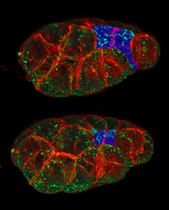
An asymmetric cell division produces two daughter cells with different cellular fates. This is in contrast to symmetric cell divisions which give rise to daughter cells of equivalent fates. Notably, stem cells divide asymmetrically to give rise to two distinct daughter cells: one copy of the original stem cell as well as a second daughter programmed to differentiate into a non-stem cell fate.

The LIM kinases are a family of actin-binding kinases that phosphorylate members of the ADF/cofilin family of actin binding and filament severing proteins. The LIM kinase family is made up of two proteins: LIM kinase-1 (LIMK1) and LIM kinase-2 (LIMK2)

Septins are a group of GTP-binding proteins expressed in all eukaryotic cells except plants. Different septins form protein complexes with each other. These complexes can further assemble into filaments, rings and gauzes. Assembled as such, septins function in cells by localizing other proteins, either by providing a scaffold to which proteins can attach, or by forming a barrier preventing the diffusion of molecules from one compartment of the cell to another, or in the cell cortex as a barrier to the diffusion of membrane-bound proteins.

<span class="mw-page-title-main">Aurora kinase B</span> Protein

Aurora kinase B is a protein that functions in the attachment of the mitotic spindle to the centromere.

Polo-like kinases (Plks) are regulatory serine/threonine kinases of the cell cycle involved in mitotic entry, mitotic exit, spindle formation, cytokinesis, and meiosis. Only one Plk is found in the genomes of the fly Drosophila melanogaster (Polo), budding yeast (Cdc5) and fission yeast (Plo1). Vertebrates and other animals, however, have many Plk family members including Plk1, Plk2/Snk, Plk3/Prk/FnK, Plk4/Sak and Plk5. Of the vertebrate Plk family members, the mammalian Plk1 has been most extensively studied. During mitosis and cytokinesis, Plks associate with several structures including the centrosome, kinetochores, and the central spindle.

<span class="mw-page-title-main">Transforming protein RhoA</span> Protein and coding gene in humans

Transforming protein RhoA, also known as Ras homolog family member A (RhoA), is a small GTPase protein in the Rho family of GTPases that in humans is encoded by the RHOA gene. While the effects of RhoA activity are not all well known, it is primarily associated with cytoskeleton regulation, mostly actin stress fibers formation and actomyosin contractility. It acts upon several effectors. Among them, ROCK1 and DIAPH1 are the best described. RhoA, and the other Rho GTPases, are part of a larger family of related proteins known as the Ras superfamily, a family of proteins involved in the regulation and timing of cell division. RhoA is one of the oldest Rho GTPases, with homologues present in the genomes since 1.5 billion years. As a consequence, RhoA is somehow involved in many cellular processes which emerged throughout evolution. RhoA specifically is regarded as a prominent regulatory factor in other functions such as the regulation of cytoskeletal dynamics, transcription, cell cycle progression and cell transformation.

<span class="mw-page-title-main">MYH10</span> Protein-coding gene in the species Homo sapiens

Myosin-10 also known as myosin heavy chain 10 or non-muscle myosin IIB (NM-IIB) is a protein that in humans is encoded by the MYH10 gene. Non-muscle myosins are expressed in a wide variety of tissues, but NM-IIB is the only non-muscle myosin II isoform expressed in cardiac muscle, where it localizes to adherens junctions within intercalated discs. NM-IIB is essential for normal development of cardiac muscle and for integrity of intercalated discs. Mutations in MYH10 have been identified in patients with left atrial enlargement.

<span class="mw-page-title-main">Citron kinase</span> Enzyme found in humans

Citron Rho-interacting kinase is an enzyme that in humans is encoded by the CIT gene.

<span class="mw-page-title-main">SEPT7</span> Protein-coding gene in the species Homo sapiens

Septin-7 is a protein that in humans is encoded by the SEPT7 gene.

<span class="mw-page-title-main">Apical constriction</span>

Apical constriction is the process in which contraction of the apical side of a cell causes the cell to take on a wedged shape. Generally, this shape change is coordinated across many cells of an epithelial layer, generating forces that can bend or fold the cell sheet.

<span class="mw-page-title-main">Rho-associated protein kinase</span>

Rho-associated protein kinase (ROCK) is a kinase belonging to the AGC family of serine-threonine specific protein kinases. It is involved mainly in regulating the shape and movement of cells by acting on the cytoskeleton.

<span class="mw-page-title-main">MDia1</span> Protein

mDia1 is a member of the protein family called the formins and is a Rho effector. It is the mouse version of the diaphanous homolog 1 of Drosophila. mDia1 localizes to cells' mitotic spindle and midbody, plays a role in stress fiber and filopodia formation, phagocytosis, activation of serum response factor, formation of adherens junctions, and it can act as a transcription factor. mDia1 accelerates actin nucleation and elongation by interacting with barbed ends of actin filaments. The gene encoding mDia1 is located on Chromosome 18 of Mus musculus and named Diap1.

Centralspindlin is a motor complex implicated in cell division. It contributes to virtually every step in cytokinesis, It is highly conserved in animal cells as a component of the spindle midzone and midbody. Centralspindlin is required for the assembly of the mitotic spindle as well as for microtubule bundling and anchoring of midbody microtubules to the plasma membrane. This complex is also implicated in tethering the spindle apparatus to the plasma membrane during cytokinesis This interaction permits cleavage furrow ingression. In addition, centralspindlin's interaction with the ESCRT III allows for abscission to occur.

<span class="mw-page-title-main">Actomyosin ring</span> Cellular formation during cytokinesis

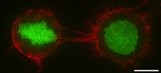
In molecular biology, an actomyosin ring or contractile ring, is a prominent structure during cytokinesis. It forms perpendicular to the axis of the spindle apparatus towards the end of telophase, in which sister chromatids are identically separated at the opposite sides of the spindle forming nuclei. The actomyosin ring follows an orderly sequence of events: identification of the active division site, formation of the ring, constriction of the ring, and disassembly of the ring. It is composed of actin and myosin II bundles, thus the term actomyosin. The actomyosin ring operates in contractile motion, although the mechanism on how or what triggers the constriction is still an evolving topic. Other cytoskeletal proteins are also involved in maintaining the stability of the ring and driving its constriction. Apart from cytokinesis, in which the ring constricts as the cells divide, actomyosin ring constriction has also been found to activate during wound closure. During this process, actin filaments are degraded, preserving the thickness of the ring. After cytokinesis is complete, one of the two daughter cells inherits a remnant known as the midbody ring.

Edwin W. Taylor is an adjunct professor of cell and developmental biology at Northwestern University. He was elected to the National Academy of Sciences in 2001. Taylor received a BA in physics and chemistry from the University of Toronto in 1952; an MSc in physical chemistry from McMaster University in 1955, and a PhD in biophysics from the University of Chicago in 1957. In 2001 Taylor was elected to the National Academy of Scineces in Cellular and Developmental Biology and Biochemistry.

References

  1. 1 2 3 GRCh38: Ensembl release 89: ENSG00000011426 - Ensembl, May 2017
  2. 1 2 3 GRCm38: Ensembl release 89: ENSMUSG00000036777 - Ensembl, May 2017
  3. "Human PubMed Reference:". National Center for Biotechnology Information, U.S. National Library of Medicine.
  4. "Mouse PubMed Reference:". National Center for Biotechnology Information, U.S. National Library of Medicine.
  5. 1 2 3 4 Piekny AJ, Maddox AS (December 2010). "The myriad roles of Anillin during cytokinesis" (PDF). Seminars in Cell & Developmental Biology. 21 (9): 881–91. doi:10.1016/j.semcdb.2010.08.002. PMID   20732437.
  6. 1 2 Zhang L, Maddox AS (February 2010). "Anillin". Current Biology. 20 (4): R135-6. Bibcode:2010CBio...20.R135Z. doi: 10.1016/j.cub.2009.12.017 . PMID   20178751.
  7. 1 2 Field CM, Alberts BM (October 1995). "Anillin, a contractile ring protein that cycles from the nucleus to the cell cortex". The Journal of Cell Biology. 131 (1): 165–78. doi: 10.1083/jcb.131.1.165 . PMC   2120607 . PMID   7559773.
  8. 1 2 Piekny AJ, Glotzer M (January 2008). "Anillin is a scaffold protein that links RhoA, actin, and myosin during cytokinesis". Current Biology. 18 (1): 30–6. Bibcode:2008CBio...18...30P. doi: 10.1016/j.cub.2007.11.068 . PMID   18158243. S2CID   6310134.
  9. 1 2 Oegema K, Savoian MS, Mitchison TJ, Field CM (August 2000). "Functional analysis of a human homologue of the Drosophila actin binding protein anillin suggests a role in cytokinesis". The Journal of Cell Biology. 150 (3): 539–52. doi: 10.1083/jcb.150.3.539 . PMC   2175195 . PMID   10931866.
  10. Saha S, Pollard TD (October 2012). "Characterization of structural and functional domains of the anillin-related protein Mid1p that contribute to cytokinesis in fission yeast". Molecular Biology of the Cell. 23 (20): 3993–4007. doi: 10.1091/mbc.E12-07-0536 . PMC   3469515 . PMID   22918954.
  11. Tasto JJ, Morrell JL, Gould KL (March 2003). "An anillin homologue, Mid2p, acts during fission yeast cytokinesis to organize the septin ring and promote cell separation". The Journal of Cell Biology. 160 (7): 1093–103. doi: 10.1083/jcb.200211126 . PMC   2172762 . PMID   12668659.
  12. Toya M, Iino Y, Yamamoto M (August 1999). "Fission yeast Pob1p, which is homologous to budding yeast Boi proteins and exhibits subcellular localization close to actin patches, is essential for cell elongation and separation". Molecular Biology of the Cell. 10 (8): 2745–57. doi:10.1091/mbc.10.8.2745. PMC   25510 . PMID   10436025.
  13. D'Avino PP, Takeda T, Capalbo L, Zhang W, Lilley KS, Laue ED, et al. (April 2008). "Interaction between Anillin and RacGAP50C connects the actomyosin contractile ring with spindle microtubules at the cell division site". Journal of Cell Science. 121 (Pt 8): 1151–8. doi: 10.1242/jcs.026716 . PMID   18349071.
  14. 1 2 Maddox AS, Habermann B, Desai A, Oegema K (June 2005). "Distinct roles for two C. elegans anillins in the gonad and early embryo". Development. 132 (12): 2837–48. doi: 10.1242/dev.01828 . PMID   15930113.
  15. 1 2 Kučera O, Siahaan V, Janda D, Dijkstra SH, Pilátová E, Zatecka E, et al. (2021). "Anillin propels myosin-independent constriction of actin rings". Nature Communications. 12 (1): 4595. Bibcode:2021NatCo..12.4595K. doi:10.1038/s41467-021-24474-1. PMC   8319318 . PMID   34321459.
  16. Watanabe S, Okawa K, Miki T, Sakamoto S, Morinaga T, Segawa K, et al. (September 2010). "Rho and anillin-dependent control of mDia2 localization and function in cytokinesis". Molecular Biology of the Cell. 21 (18): 3193–204. doi:10.1091/mbc.E10-04-0324. PMC   2938385 . PMID   20660154.
  17. Straight AF, Field CM, Mitchison TJ (January 2005). "Anillin binds nonmuscle myosin II and regulates the contractile ring". Molecular Biology of the Cell. 16 (1): 193–201. doi:10.1091/mbc.E04-08-0758. PMC   539163 . PMID   15496454.
  18. Field CM, Coughlin M, Doberstein S, Marty T, Sullivan W (June 2005). "Characterization of anillin mutants reveals essential roles in septin localization and plasma membrane integrity". Development. 132 (12): 2849–60. doi: 10.1242/dev.01843 . PMID   15930114.
  19. 1 2 Goldbach P, Wong R, Beise N, Sarpal R, Trimble WS, Brill JA (May 2010). "Stabilization of the actomyosin ring enables spermatocyte cytokinesis in Drosophila". Molecular Biology of the Cell. 21 (9): 1482–93. doi:10.1091/mbc.E09-08-0714. PMC   2861608 . PMID   20237160.
  20. Versele M, Thorner J (August 2005). "Some assembly required: yeast septins provide the instruction manual". Trends in Cell Biology. 15 (8): 414–24. doi:10.1016/j.tcb.2005.06.007. PMC   1761124 . PMID   16009555.
  21. Kinoshita M, Field CM, Coughlin ML, Straight AF, Mitchison TJ (December 2002). "Self- and actin-templated assembly of Mammalian septins". Developmental Cell. 3 (6): 791–802. doi: 10.1016/S1534-5807(02)00366-0 . PMID   12479805.
  22. Solski PA, Wilder RS, Rossman KL, Sondek J, Cox AD, Campbell SL, et al. (June 2004). "Requirement for C-terminal sequences in regulation of Ect2 guanine nucleotide exchange specificity and transformation". The Journal of Biological Chemistry. 279 (24): 25226–33. doi: 10.1074/jbc.M313792200 . PMID   15073184.
  23. Glotzer M (January 2009). "The 3Ms of central spindle assembly: microtubules, motors and MAPs". Nature Reviews. Molecular Cell Biology. 10 (1): 9–20. doi:10.1038/nrm2609. PMC   2789570 . PMID   19197328.
  24. Gregory SL, Ebrahimi S, Milverton J, Jones WM, Bejsovec A, Saint R (January 2008). "Cell division requires a direct link between microtubule-bound RacGAP and Anillin in the contractile ring". Current Biology. 18 (1): 25–9. Bibcode:2008CBio...18...25G. doi: 10.1016/j.cub.2007.11.050 . PMID   18158242. S2CID   17517089.
  25. Zhao WM, Fang G (September 2005). "Anillin is a substrate of anaphase-promoting complex/cyclosome (APC/C) that controls spatial contractility of myosin during late cytokinesis". The Journal of Biological Chemistry. 280 (39): 33516–24. doi: 10.1074/jbc.M504657200 . PMID   16040610.
  26. Sisson JC, Field C, Ventura R, Royou A, Sullivan W (November 2000). "Lava lamp, a novel peripheral golgi protein, is required for Drosophila melanogaster cellularization". The Journal of Cell Biology. 151 (4): 905–18. doi: 10.1083/jcb.151.4.905 . PMC   2169433 . PMID   11076973.
  27. Hickson GR, O'Farrell PH (January 2008). "Rho-dependent control of anillin behavior during cytokinesis". The Journal of Cell Biology. 180 (2): 285–94. doi: 10.1083/jcb.200709005 . PMC   2213597 . PMID   18209105.
  28. Chuang HY, Ou YH (2014). Overexpression of anillin in colorectal cancer promoter the cell proliferation, cell mobility and cell invasion. Proceedings of the 105th Annual Meeting of the American Association for Cancer Research. San Diego, CA.
  29. Wang G, Shen W, Cui L, Chen W, Hu X, Fu J (2016). "Overexpression of Anillin (ANLN) is correlated with colorectal cancer progression and poor prognosis". Cancer Biomarkers. 16 (3): 459–65. doi:10.3233/CBM-160585. PMID   27062703.

Further reading