Airway basal cell

Last updated

Airway basal cells are found deep in the respiratory epithelium, attached to, and lining the basement membrane. [1]

Contents

Basal cells are the stem cells or progenitors of the airway epithelium and can differentiate to replenish all of the epithelial cells including the ciliated cells, and secretory goblet cells. [2] [3] This repairs the protective functions of the epithelial barrier. [3]

Basal cells are cuboidal with a large nucleus, few organelles, and scattered microvilli. [1] Basal cells are the first cells to be affected by exposure to cigarette smoke. Their disorganisation is seen to be responsible for the major airway changes that are characteristic of COPD. [4]

Structure

Cells of the respiratory epithelium. Basal cells shown in purple, ciliated cells shown in brown, goblet cells shown in green, and submucosal gland shown in blue. Respiratory Tract Cells.png
Cells of the respiratory epithelium. Basal cells shown in purple, ciliated cells shown in brown, goblet cells shown in green, and submucosal gland shown in blue.

Basal cells are cuboidal, with a large nucleus, few organelles, and scattered microvilli. Basal cells are attached to, and line the basement membrane. [1]

The numbers of basal cells are highest in the large airways and become increasingly decreased in the smaller airways. Their percentage in the trachea is 34%, in the large bronchi 27%, and 10% in the larger of the bronchioles. [1] Basal cells can express a number of different receptors, notably EGFR. [1]

Basal cell derived precursors are found as intermediate cells (also known as parabasal, or indetermined cells) between the basal cells and the differentiated cells. [1]

Function

Basal cells are the stem cells, or progenitors of all the cells in the respiratory epithelium. The ciliated cells and secretory cells that form the epithelial barrier, and function in mucociliary clearance, are terminally differentiated meaning that they cannot self-renew. These cells are vulnerable to damage and the basal cells can replace damaged cells by differentiation. [3]

Typically the airway epithelium turns over fairly slowly with the basal cells in a quiescent state, and just a few intermediate cells being seen. [3] In response to damage to the differentiated cells, or to stress caused by smoking, the basal cells become active, proliferate, and form clonal patches, and the numbers of intermediate cells increase. [3] [1] When activated the basal cells acquire damage-associated phenotypes to enable differentiation to the particular cell type that has been damaged. [3] The intermediate cells differentiate under the control of FOXJ1 into ciliated cells; and into secretory cells under the control of Notch signalling, and the transcription factors SPDEF and FOXA3. [1] The regeneration of cells can be normal or in the case of smokers the regeneration may be of altered histologic phenotypes. [1]

Basal cells can also provide a defence function upregulating innate immune mediators such as RNase7, an antimicrobial protein. [3] RNase7, mediates tissue repair, and the production of inflammatory cytokines induced either by tobacco smoke, or by microbial pattern recognition. This is seen as a possible defence response of basal cells to injury that would be associated with microbial attack. [3] The response requires signalling via EGFR, which is highly expressed in basal cells. [3] Other innate immune mediators include beta-defensin 2, and lipocalin-2; pro-inflammatory cytokines, interleukins IL6, and IL8; and chemokine CCL20. [3]

Dysfunction

SEM image of syncytial virions taken from A549 cells in the lung epithelium. Human Respiratory Syncytial Virus (RSV) (33114415716).jpg
SEM image of syncytial virions taken from A549 cells in the lung epithelium.

Following injury to airway epithelium, the basal cells can become infected by the respiratory syncytial virus. When this happens the basal cell can be skewed to favour the differentiation of mucous-producing (secretory goblet cells) over that of ciliated cells. The normal ratio of ciliated cells to secretory cells is 10 to 1, and this is highly controlled throughout most of the respiratory tree. [3] When this ratio is upset in this way, the effect is of generating the mucus hyperplasia associated with a number of respiratory diseases, due to the insufficient ciliary action needed for mucociliary clearance. The release of antimicrobials may occur as a second-line defence with cell differentiation. [3] [5]

Disruption of the tight-junction barrier is a common feature of asthma, and of smoking-related respiratory diseases. The persistent activation of the innate immune response in basal cells, may contribute to the characteristic chronic inflammation seen in these disorders. [3]

Clinical significance

The hyperplasia of airway basal cells is the earliest indication of smoking-related abnormality in the lung. [4] [1] This is followed by shortened cilia, loss of ciliated cells, mucous cell hyperplasia, and loss of cell junctions giving a leaky epithelial barrier. With persistent stress from smoking, the basal cells become disarranged and lose their regenerative ability needed to repair the barrier. The disorganised basal cells are seen to be responsible for the major airway changes that are characteristic of COPD, and with continued stress can undergo a malignant transformation. Studies have shown that the initial development of emphysema is centred on the early changes in the airway epithelium of the small airways. [1] Basal cells become further deranged in a smoker’s transition to clinically defined COPD. [1]

Image showing the accumulation of mucus in the airways. Respiratory Tract.jpg
Image showing the accumulation of mucus in the airways.

Basal cell dysfunction causes the overproduction and secretion of mucus, and decreased mucus clearance that leads to the characteristic features of mucus hypersecretion, and productive cough of chronic bronchitis and chronic obstructive pulmonary disease (COPD). [6] Mucus accumulation in COPD influences lung function, quality of life, exacerbations, hospital stays, and mortality, for the worse. [6]

In response to a viral infection the basal cells may be activated to produce an increased production of Interleukin 33 and mucins, and these activated cells may become a long-lived population that promote chronic inflammation; [7] as can influence the occurrence of an exacerbation of COPD. [6]

Related Research Articles

<span class="mw-page-title-main">Lung</span> Primary organ of the respiratory system

The lungs are the primary organs of the respiratory system in humans and most other animals, including some snails and a small number of fish. In mammals and most other vertebrates, two lungs are located near the backbone on either side of the heart. Their function in the respiratory system is to extract oxygen from the air and transfer it into the bloodstream, and to release carbon dioxide from the bloodstream into the atmosphere, in a process of gas exchange. The pleurae, which are thin, smooth, and moist, serve to reduce friction between the lungs and chest wall during breathing, allowing for easy and effortless movements of the lungs.

<span class="mw-page-title-main">Respiratory tract</span> Organs involved in transmission of air to and from the point where gases diffuse into tissue

The respiratory tract is the subdivision of the respiratory system involved with the process of respiration in mammals. The respiratory tract is lined with respiratory epithelium as respiratory mucosa.

<span class="mw-page-title-main">Bronchus</span> Airway in the respiratory tract

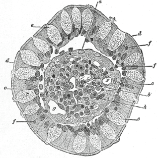
A bronchus is a passage or airway in the lower respiratory tract that conducts air into the lungs. The first or primary bronchi to branch from the trachea at the carina are the right main bronchus and the left main bronchus. These are the widest bronchi, and enter the right lung, and the left lung at each hilum. The main bronchi branch into narrower secondary bronchi or lobar bronchi, and these branch into narrower tertiary bronchi or segmental bronchi. Further divisions of the segmental bronchi are known as 4th order, 5th order, and 6th order segmental bronchi, or grouped together as subsegmental bronchi. The bronchi, when too narrow to be supported by cartilage, are known as bronchioles. No gas exchange takes place in the bronchi.

<span class="mw-page-title-main">Bronchiole</span> Passageways by which air passes through the nose or mouth to the alveoli of the lungs

The bronchioles or bronchioli are the smaller branches of the bronchial airways in the lower respiratory tract. They include the terminal bronchioles, and finally the respiratory bronchioles that mark the start of the respiratory zone delivering air to the gas exchanging units of the alveoli. The bronchioles no longer contain the cartilage that is found in the bronchi, or glands in their submucosa.

<span class="mw-page-title-main">Mucus</span> Secretion produced by mucous membranes

Mucus is a slippery aqueous secretion produced by, and covering, mucous membranes. It is typically produced from cells found in mucous glands, although it may also originate from mixed glands, which contain both serous and mucous cells. It is a viscous colloid containing inorganic salts, antimicrobial enzymes, immunoglobulins, and glycoproteins such as lactoferrin and mucins, which are produced by goblet cells in the mucous membranes and submucosal glands. Mucus serves to protect epithelial cells in the linings of the respiratory, digestive, and urogenital systems, and structures in the visual and auditory systems from pathogenic fungi, bacteria and viruses. Most of the mucus in the body is produced in the gastrointestinal tract.

Club cells, also known as bronchiolar exocrine cells, and formerly known as Clara cells, are low columnar/cuboidal cells with short microvilli, found in the small airways (bronchioles) of the lungs.

<span class="mw-page-title-main">Goblet cell</span> Epithelial cells that secrete mucins

Goblet cells are simple columnar epithelial cells that secrete gel-forming mucins, like mucin 5AC. The goblet cells mainly use the merocrine method of secretion, secreting vesicles into a duct, but may use apocrine methods, budding off their secretions, when under stress. The term goblet refers to the cell's goblet-like shape. The apical portion is shaped like a cup, as it is distended by abundant mucus laden granules; its basal portion lacks these granules and is shaped like a stem.

<span class="mw-page-title-main">Respiratory disease</span> Disease of the respiratory system

Respiratory diseases, or lung diseases, are pathological conditions affecting the organs and tissues that make gas exchange difficult in air-breathing animals. They include conditions of the respiratory tract including the trachea, bronchi, bronchioles, alveoli, pleurae, pleural cavity, the nerves and muscles of respiration. Respiratory diseases range from mild and self-limiting, such as the common cold, influenza, and pharyngitis to life-threatening diseases such as bacterial pneumonia, pulmonary embolism, tuberculosis, acute asthma, lung cancer, and severe acute respiratory syndromes, such as COVID-19. Respiratory diseases can be classified in many different ways, including by the organ or tissue involved, by the type and pattern of associated signs and symptoms, or by the cause of the disease.

<span class="mw-page-title-main">Respiratory epithelium</span> Mucosa that serves to moisten and protect the airways

Respiratory epithelium, or airway epithelium, is a type of ciliated columnar epithelium found lining most of the respiratory tract as respiratory mucosa, where it serves to moisten and protect the airways. It is not present in the vocal cords of the larynx, or the oropharynx and laryngopharynx, where instead the epithelium is stratified squamous. It also functions as a barrier to potential pathogens and foreign particles, preventing infection and tissue injury by the secretion of mucus and the action of mucociliary clearance.

<span class="mw-page-title-main">Erdosteine</span> Chemical to treat chronic bronchitis

Erdosteine is a molecule with mucolytic activity. Structurally it is a thiol derivative characterized by the presence of two thiol groups. These two functional sulfhydryl groups contained in the molecule are released following first-pass metabolism with the conversion of erdosteine into its pharmacologically active metabolite Met-I.

<span class="mw-page-title-main">Bronchitis</span> Inflammation of the large airways in the lungs

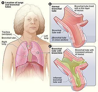
Bronchitis is inflammation of the bronchi in the lungs that causes coughing. Bronchitis usually begins as an infection in the nose, ears, throat, or sinuses. The infection then makes its way down to the bronchi. Symptoms include coughing up sputum, wheezing, shortness of breath, and chest pain. Bronchitis can be acute or chronic.

<span class="mw-page-title-main">Obstructive lung disease</span> Category of respiratory disease characterized by airway obstruction

Obstructive lung disease is a category of respiratory disease characterized by airway obstruction. Many obstructive diseases of the lung result from narrowing (obstruction) of the smaller bronchi and larger bronchioles, often because of excessive contraction of the smooth muscle itself. It is generally characterized by inflamed and easily collapsible airways, obstruction to airflow, problems exhaling, and frequent medical clinic visits and hospitalizations. Types of obstructive lung disease include; asthma, bronchiectasis, bronchitis and chronic obstructive pulmonary disease (COPD). Although COPD shares similar characteristics with all other obstructive lung diseases, such as the signs of coughing and wheezing, they are distinct conditions in terms of disease onset, frequency of symptoms, and reversibility of airway obstruction. Cystic fibrosis is also sometimes included in obstructive pulmonary disease.

<span class="mw-page-title-main">Intestinal epithelium</span> Single-cell layer lining the intestines

The intestinal epithelium is the single cell layer that form the luminal surface (lining) of both the small and large intestine (colon) of the gastrointestinal tract. Composed of simple columnar epithelial cells, it serves two main functions: absorbing useful substances into the body and restricting the entry of harmful substances. As part of its protective role, the intestinal epithelium forms an important component of the intestinal mucosal barrier. Certain diseases and conditions are caused by functional defects in the intestinal epithelium. On the other hand, various diseases and conditions can lead to its dysfunction which, in turn, can lead to further complications.

<span class="mw-page-title-main">Mucociliary clearance</span>

Mucociliary clearance (MCC), mucociliary transport, or the mucociliary escalator, describes the self-clearing mechanism of the airways in the respiratory system. It is one of the two protective processes for the lungs in removing inhaled particles including pathogens before they can reach the delicate tissue of the lungs. The other clearance mechanism is provided by the cough reflex. Mucociliary clearance has a major role in pulmonary hygiene.

<span class="mw-page-title-main">Acute exacerbation of chronic obstructive pulmonary disease</span> Medical condition

An acute exacerbation of chronic obstructive pulmonary disease, or acute exacerbations of chronic bronchitis (AECB), is a sudden worsening of chronic obstructive pulmonary disease (COPD) symptoms including shortness of breath, quantity and color of phlegm that typically lasts for several days.

<span class="mw-page-title-main">Chronic obstructive pulmonary disease</span> Lung disease involving long-term poor airflow

Chronic obstructive pulmonary disease (COPD) is a type of progressive lung disease characterized by long-term respiratory symptoms and airflow limitation. The main symptoms of COPD include shortness of breath and a cough, which may or may not produce mucus. COPD progressively worsens, with everyday activities such as walking or dressing becoming difficult. While COPD is incurable, it is preventable and treatable. The two most common types of COPD are emphysema and chronic bronchitis and have been the two classic COPD phenotypes. Emphysema is defined as enlarged airspaces (alveoli) whose walls have broken down resulting in permanent damage to the lung tissue. Chronic bronchitis is defined as a productive cough that is present for at least three months each year for two years. Both of these conditions can exist without airflow limitation when they are not classed as COPD. Emphysema is just one of the structural abnormalities that can limit airflow and can exist without airflow limitation in a significant number of people. Chronic bronchitis does not always result in airflow limitation but in young adults who smoke the risk of developing COPD is high. Many definitions of COPD in the past included emphysema and chronic bronchitis, but these have never been included in GOLD report definitions. Emphysema and chronic bronchitis remain the predominant phenotypes of COPD but there is often overlap between them and a number of other phenotypes have also been described. COPD and asthma may coexist and converge in some individuals. COPD is associated with low-grade systemic inflammation.

The lung microbiota is the pulmonary microbial community consisting of a complex variety of microorganisms found in the lower respiratory tract particularly on the mucous layer and the epithelial surfaces. These microorganisms include bacteria, fungi, viruses and bacteriophages. The bacterial part of the microbiota has been more closely studied. It consists of a core of nine genera: Prevotella, Sphingomonas, Pseudomonas, Acinetobacter, Fusobacterium, Megasphaera, Veillonella, Staphylococcus, and Streptococcus. They are aerobes as well as anaerobes and aerotolerant bacteria. The microbial communities are highly variable in particular individuals and compose of about 140 distinct families. The bronchial tree for instance contains a mean of 2000 bacterial genomes per cm2 surface. The harmful or potentially harmful bacteria are also detected routinely in respiratory specimens. The most significant are Moraxella catarrhalis, Haemophilus influenzae, and Streptococcus pneumoniae. They are known to cause respiratory disorders under particular conditions namely if the human immune system is impaired. The mechanism by which they persist in the lower airways in healthy individuals is unknown.

<span class="mw-page-title-main">Intestinal mucosal barrier</span>

The intestinal mucosal barrier, also referred to as intestinal barrier, refers to the property of the intestinal mucosa that ensures adequate containment of undesirable luminal contents within the intestine while preserving the ability to absorb nutrients. The separation it provides between the body and the gut prevents the uncontrolled translocation of luminal contents into the body proper. Its role in protecting the mucosal tissues and circulatory system from exposure to pro-inflammatory molecules, such as microorganisms, toxins, and antigens is vital for the maintenance of health and well-being. Intestinal mucosal barrier dysfunction has been implicated in numerous health conditions such as: food allergies, microbial infections, irritable bowel syndrome, inflammatory bowel disease, celiac disease, metabolic syndrome, non-alcoholic fatty liver disease, diabetes, and septic shock.

<span class="mw-page-title-main">Emphysema</span> Medical condition

Emphysema is any air-filled enlargement in the body's tissues. Most commonly emphysema refers to the enlargement of air spaces (alveoli) in the lungs, and is also known as pulmonary emphysema.

Donna Elizabeth Davies is a British biochemist and professor of respiratory cell and molecular biology at the University of Southampton. In 2003, Davies was the co-founder of Synairgen, an interferon-beta drug designed to treat patients with asthma and chronic obstructive pulmonary disease.

References

  1. 1 2 3 4 5 6 7 8 9 10 11 12 Crystal, RG (15 December 2014). "Airway basal cells. The "smoking gun" of chronic obstructive pulmonary disease". American Journal of Respiratory and Critical Care Medicine. 190 (12): 1355–62. doi:10.1164/rccm.201408-1492PP. PMC   4299651 . PMID   25354273.
  2. Weinberger, S (2019). Principles of pulmonary medicine (Seventh ed.). Elsevier. p. 67. ISBN   9780323523714.
  3. 1 2 3 4 5 6 7 8 9 10 11 12 13 Shaykhiev, R (October 2015). "Multitasking basal cells: combining stem cell and innate immune duties". The European Respiratory Journal. 46 (4): 894–7. doi:10.1183/13993003.00521-2015. PMC   4732698 . PMID   26424520.
  4. 1 2 Shaykhiev, R; Crystal, RG (December 2014). "Early events in the pathogenesis of chronic obstructive pulmonary disease. Smoking-induced reprogramming of airway epithelial basal progenitor cells". Annals of the American Thoracic Society. 11 (Suppl 5): S252-8. doi:10.1513/AnnalsATS.201402-049AW. PMC   4298974 . PMID   25525728.
  5. Amatngalim, GD; van Wijck, Y; de Mooij-Eijk, Y; Verhoosel, RM; Harder, J; Lekkerkerker, AN; Janssen, RA; Hiemstra, PS (1 April 2015). "Basal cells contribute to innate immunity of the airway epithelium through production of the antimicrobial protein RNase 7". Journal of Immunology. 194 (7): 3340–50. doi: 10.4049/jimmunol.1402169 . PMID   25712218.
  6. 1 2 3 Ramos, FL; Krahnke, JS; Kim, V (2014). "Clinical issues of mucus accumulation in COPD". International Journal of Chronic Obstructive Pulmonary Disease. 9: 139–50. doi: 10.2147/COPD.S38938 . PMC   3908831 . PMID   24493923.
  7. Hiemstra, PS; McCray PB, Jr; Bals, R (April 2015). "The innate immune function of airway epithelial cells in inflammatory lung disease". The European Respiratory Journal. 45 (4): 1150–62. doi:10.1183/09031936.00141514. PMC   4719567 . PMID   25700381.