Aminoallyl nucleotide

Last updated
Aminoallyl uridine (aa-UTP) Aminoallyl-UTP.svg
Aminoallyl uridine (aa-UTP)

Aminoallyl nucleotide is a nucleotide with a modified base containing an allylamine. They are used in post-labeling of nucleic acids by fluorescence detection in microarray. They are reactive with N-Hydroxysuccinimide ester group which helps attach a fluorescent dye to the primary amino group on the nucleotide. These nucleotides are known as 5-(3-aminoallyl)-nucleotides since the aminoallyl group is usually attached to carbon 5 of the pyrimidine ring of uracil or cytosine. The primary amine group in the aminoallyl moiety is aliphatic and thus more reactive compared to the amine groups that are directly attached to the rings (aromatic) of the bases. Common names of aminoallyl nucleosides are initially abbreviated with aa- or AA- to indicate aminoallyl. The 5-carbon sugar is indicated with or without the lowercase "d" indicating deoxyribose if included or ribose if not. Finally the nitrogenous base and number of phosphates are indicated (i.e. aa-UTP = aminoallyl uridine triphosphate).

Contents

History

Hierarchically clustered heat map using aa-dUTPs Heatmap using aminoallyl nucleotide.png
Hierarchically clustered heat map using aa-dUTPs

The goal of combining fluorescence and nucleic acids has been to provide a non-isotopic tag that is detectable to study DNA or RNA. This type of labeling allows scientists to study DNA or RNA in their structure, function, or formation with other nucleic acids. [2] The first base modification for fluorescent labeling occurred in 1971 with a 4-thiouridine and 4-thiouracil. [3] This research along with others, which included various types of direct and non-direct labeling via: analogs, addition via enzymes, or other methods made labeling of nucleotides much safer for scientist to study DNA. [2]

As instrumentation and technologies become more advanced in the field of DNA microarray, better reagents and techniques will be needed to further scientific studies. Fluorescent labeling with Cy3 was shown to be more insufficient and skew results; the method of aminoallyl nucleotide incorporation was opted instead. Using aminoallyl nucleotides as indirect fluorescent labeling seemed to nullify the sensitivity issues seen in cyanine-labeling. [4]

Synthesis

Aminoallyl nucleosides can be synthesized via Heck coupling as shown in the image below. [5]

Heck coupling aminoallyl nucleotide reaction.jpg

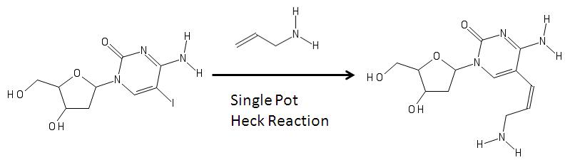
In the image above, on the left is a modified nucleoside with an iodine (the iodine is added via electrophilic halogenation) in the fifth carbon in the pyrimidine ring. Its formation can be associated with a reaction with an allylamine and various reagents via heck coupling are able to remove the halogen group from the base and add the allylamine to become the aminoallyl nucleoside shown on the right. [5] The product on the right is then used to in molecular biology in RNA synthesis. [4] [6] [7]

Other reactions include using a single pot synthesis with other halogens. [8]

Reaction

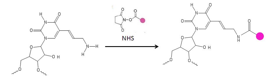
The primary amine on the aminoallyl nucleotide reacts with amino-reactive dyes [9] such as a cyanine and patented dyes [10] [11] which contain a reactive leaving group, such as a succinimidyl ester (NHS).The amine groups directly attached to the ring of the base are not affected. These nucleotides are used for labeling DNA. [4] [6] [10] [11] [12]

Tagging modified uridine.jpg

Uses

Aminoallyl NTPs are used for indirect DNA labeling in PCR, nick translation, primer extensions and cDNA synthesis. [13] These labeled NTPs are helpful because of their application in molecular biology labs where they do not have the capacity to handle radioactive material. For example, 5-(3-Aminoallyl)-Uridine(AA-UTPs) are more effective for high density labeling of DNA than pre-labeling the DNA. After the enzymatic addition of the NTPs, amine reactant fluorescent dyes can be added for detection of the DNA molecule. [7] When incorporated into DNA or RNA molecules by DNA/RNA polymerase, 5-(3-aminoallyl)-UTP provide a reactive group for the addition of other chemical groups. Thus aminoallyl modified DNA or RNA can be labeled with any compound which has an amine-reactive group. aa-NTPs incorporated into DNA/RNA in combination with a secondary dye coupling reagents can probe for an array analysis. [6]

cDNA relies on aminoallyl labeling for detection purposes. Although direct labeling of dNTP is the quickest and cheapest method of fluorescent labeling, it is disadvantageous as the sequence allows for only one modified nucleotide for use. Another disadvantage of direct labeling is the bulky nucleotides, however this can be overcome by indirect labeling using aminoallyl modified nucleotides. [14] An easy way to check for labeling success is the color;Good labeling will result in visible blue (Cy5) or red (Cy3) color in the final material. [15]

Process of preparing amino-allyl labeled cDNA Process of preparing amino allyl-labelled cDNA.jpg
Process of preparing amino-allyl labeled cDNA

Another process which uses aminoallyl labeling is NASBA ( Nucleic Acid Sequence Based Amplification), a highly sensitive technique for amplifying RNA. In this specific case, the aaUTP modified RNAs were tagged with fluorescent market Cy3. NASBA combined with aminoallyl-UTP labeling is very useful for many different areas of microbial diagnostics including environmental monitoring, bio threat detection, industrial process monitoring and clinical microbiology. [16] DNA microarray is another method which utilizes specifically AA-NTP's making DNA microarray testing quicker and cheaply. [12]

Post-synthesis labeling avoids the problems found in direct enzymatic incorporation of Cy-labeled dNTPs by generating probes with equal labeling effectiveness. With indirect labeling, amine-modified NTPs are incorporated during reverse transcription, RNA amplification, or PCR. Amino allyl-NTPs are incorporated with similar efficiency as unmodified NTPs during polymerization. [17] [18]

Concerns with labeling: The amine group, in aminoallyl-modified nucleotide, is reactive with dyes such as the cyanine series, or other patented dyes. A problem arises when the dyes react with buffering agents which are necessary for the proper storage of the nucleotides. However, a carbonate buffer can be used to overcome this problem. [19]

See also

Related Research Articles

<span class="mw-page-title-main">Nucleotide</span> Biological molecules that form the building blocks of nucleic acids

Nucleotides are organic molecules composed of a nitrogenous base, a pentose sugar and a phosphate. They serve as monomeric units of the nucleic acid polymers – deoxyribonucleic acid (DNA) and ribonucleic acid (RNA), both of which are essential biomolecules within all life-forms on Earth. Nucleotides are obtained in the diet and are also synthesized from common nutrients by the liver.

<span class="mw-page-title-main">Nucleobase</span> Nitrogen-containing biological compounds that form nucleosides

Nucleobases are nitrogen-containing biological compounds that form nucleosides, which, in turn, are components of nucleotides, with all of these monomers constituting the basic building blocks of nucleic acids. The ability of nucleobases to form base pairs and to stack one upon another leads directly to long-chain helical structures such as ribonucleic acid (RNA) and deoxyribonucleic acid (DNA). Five nucleobases—adenine (A), cytosine (C), guanine (G), thymine (T), and uracil (U)—are called primary or canonical. They function as the fundamental units of the genetic code, with the bases A, G, C, and T being found in DNA while A, G, C, and U are found in RNA. Thymine and uracil are distinguished by merely the presence or absence of a methyl group on the fifth carbon (C5) of these heterocyclic six-membered rings. In addition, some viruses have aminoadenine (Z) instead of adenine. It differs in having an extra amine group, creating a more stable bond to thymine.

<span class="mw-page-title-main">Nucleoside</span> Any of several glycosylamines comprising a nucleobase and a sugar molecule

Nucleosides are glycosylamines that can be thought of as nucleotides without a phosphate group. A nucleoside consists simply of a nucleobase and a five-carbon sugar whereas a nucleotide is composed of a nucleobase, a five-carbon sugar, and one or more phosphate groups. In a nucleoside, the anomeric carbon is linked through a glycosidic bond to the N9 of a purine or the N1 of a pyrimidine. Nucleotides are the molecular building blocks of DNA and RNA.

Oligonucleotides are short DNA or RNA molecules, oligomers, that have a wide range of applications in genetic testing, research, and forensics. Commonly made in the laboratory by solid-phase chemical synthesis, these small fragments of nucleic acids can be manufactured as single-stranded molecules with any user-specified sequence, and so are vital for artificial gene synthesis, polymerase chain reaction (PCR), DNA sequencing, molecular cloning and as molecular probes. In nature, oligonucleotides are usually found as small RNA molecules that function in the regulation of gene expression, or are degradation intermediates derived from the breakdown of larger nucleic acid molecules.

<span class="mw-page-title-main">DNA microarray</span> Collection of microscopic DNA spots attached to a solid surface

A DNA microarray is a collection of microscopic DNA spots attached to a solid surface. Scientists use DNA microarrays to measure the expression levels of large numbers of genes simultaneously or to genotype multiple regions of a genome. Each DNA spot contains picomoles of a specific DNA sequence, known as probes. These can be a short section of a gene or other DNA element that are used to hybridize a cDNA or cRNA sample under high-stringency conditions. Probe-target hybridization is usually detected and quantified by detection of fluorophore-, silver-, or chemiluminescence-labeled targets to determine relative abundance of nucleic acid sequences in the target. The original nucleic acid arrays were macro arrays approximately 9 cm × 12 cm and the first computerized image based analysis was published in 1981. It was invented by Patrick O. Brown. An example of its application is in SNPs arrays for polymorphisms in cardiovascular diseases, cancer, pathogens and GWAS analysis. It is also used for the identification of structural variations and the measurement of gene expression.

<span class="mw-page-title-main">Fluorescent tag</span>

In molecular biology and biotechnology, a fluorescent tag, also known as a fluorescent label or fluorescent probe, is a molecule that is attached chemically to aid in the detection of a biomolecule such as a protein, antibody, or amino acid. Generally, fluorescent tagging, or labeling, uses a reactive derivative of a fluorescent molecule known as a fluorophore. The fluorophore selectively binds to a specific region or functional group on the target molecule and can be attached chemically or biologically. Various labeling techniques such as enzymatic labeling, protein labeling, and genetic labeling are widely utilized. Ethidium bromide, fluorescein and green fluorescent protein are common tags. The most commonly labelled molecules are antibodies, proteins, amino acids and peptides which are then used as specific probes for detection of a particular target.

<span class="mw-page-title-main">Ribonucleotide</span> Nucleotide containing ribose as its pentose component

In biochemistry, a ribonucleotide is a nucleotide containing ribose as its pentose component. It is considered a molecular precursor of nucleic acids. Nucleotides are the basic building blocks of DNA and RNA. Ribonucleotides themselves are basic monomeric building blocks for RNA. Deoxyribonucleotides, formed by reducing ribonucleotides with the enzyme ribonucleotide reductase (RNR), are essential building blocks for DNA. There are several differences between DNA deoxyribonucleotides and RNA ribonucleotides. Successive nucleotides are linked together via phosphodiester bonds.

A nucleoside triphosphate is a nucleoside containing a nitrogenous base bound to a 5-carbon sugar, with three phosphate groups bound to the sugar. They are the molecular precursors of both DNA and RNA, which are chains of nucleotides made through the processes of DNA replication and transcription. Nucleoside triphosphates also serve as a source of energy for cellular reactions and are involved in signalling pathways.

Cyanines, also referred to as tetramethylindo(di)-carbocyanines are a synthetic dye family belonging to the polymethine group. Although the name derives etymologically from terms for shades of blue, the cyanine family covers the electromagnetic spectrum from near IR to UV.

<span class="mw-page-title-main">Real-time polymerase chain reaction</span> Laboratory technique of molecular biology

A real-time polymerase chain reaction is a laboratory technique of molecular biology based on the polymerase chain reaction (PCR). It monitors the amplification of a targeted DNA molecule during the PCR, not at its end, as in conventional PCR. Real-time PCR can be used quantitatively and semi-quantitatively.

<span class="mw-page-title-main">Molecular beacon</span>

Molecular beacons, or molecular beacon probes, are oligonucleotide hybridization probes that can report the presence of specific nucleic acids in homogenous solutions. Molecular beacons are hairpin-shaped molecules with an internally quenched fluorophore whose fluorescence is restored when they bind to a target nucleic acid sequence. This is a novel non-radioactive method for detecting specific sequences of nucleic acids. They are useful in situations where it is either not possible or desirable to isolate the probe-target hybrids from an excess of the hybridization probes.

<span class="mw-page-title-main">Nucleic acid metabolism</span> Process

Nucleic acid metabolism is a collective term that refers to the variety of chemical reactions by which nucleic acids are either synthesized or degraded. Nucleic acids are polymers made up of a variety of monomers called nucleotides. Nucleotide synthesis is an anabolic mechanism generally involving the chemical reaction of phosphate, pentose sugar, and a nitrogenous base. Degradation of nucleic acids is a catabolic reaction and the resulting parts of the nucleotides or nucleobases can be salvaged to recreate new nucleotides. Both synthesis and degradation reactions require multiple enzymes to facilitate the event. Defects or deficiencies in these enzymes can lead to a variety of diseases.

<span class="mw-page-title-main">Nucleoside phosphoramidite</span>

Nucleoside phosphoramidites are derivatives of natural or synthetic nucleosides. They are used to synthesize oligonucleotides, relatively short fragments of nucleic acid and their analogs. Nucleoside phosphoramidites were first introduced in 1981 by Beaucage and Caruthers. To avoid undesired side reactions, reactive hydroxy and exocyclic amino groups present in natural or synthetic nucleosides are appropriately protected. As long as a nucleoside analog contains at least one hydroxy group, the use of the appropriate protecting strategy allows one to convert that to the respective phosphoramidite and to incorporate the latter into synthetic nucleic acids. To be incorporated in the middle of an oligonucleotide chain using phosphoramidite strategy, the nucleoside analog must possess two hydroxy groups or, less often, a hydroxy group and another nucleophilic group (amino or mercapto). Examples include, but are not limited to, alternative nucleotides, LNA, morpholino, nucleosides modified at the 2'-position (OMe, protected NH2, F), nucleosides containing non-canonical bases (hypoxanthine and xanthine contained in natural nucleosides inosine and xanthosine, respectively, tricyclic bases such as G-clamp, etc.) or bases derivatized with a fluorescent group or a linker arm.

Oligonucleotide synthesis is the chemical synthesis of relatively short fragments of nucleic acids with defined chemical structure (sequence). The technique is extremely useful in current laboratory practice because it provides a rapid and inexpensive access to custom-made oligonucleotides of the desired sequence. Whereas enzymes synthesize DNA and RNA only in a 5' to 3' direction, chemical oligonucleotide synthesis does not have this limitation, although it is most often carried out in the opposite, 3' to 5' direction. Currently, the process is implemented as solid-phase synthesis using phosphoramidite method and phosphoramidite building blocks derived from protected 2'-deoxynucleosides, ribonucleosides, or chemically modified nucleosides, e.g. LNA or BNA.

<span class="mw-page-title-main">Nucleic acid analogue</span> Compound analogous to naturally occurring RNA and DNA

Nucleic acid analogues are compounds which are analogous to naturally occurring RNA and DNA, used in medicine and in molecular biology research. Nucleic acids are chains of nucleotides, which are composed of three parts: a phosphate backbone, a pentose sugar, either ribose or deoxyribose, and one of four nucleobases. An analogue may have any of these altered. Typically the analogue nucleobases confer, among other things, different base pairing and base stacking properties. Examples include universal bases, which can pair with all four canonical bases, and phosphate-sugar backbone analogues such as PNA, which affect the properties of the chain . Nucleic acid analogues are also called Xeno Nucleic Acid and represent one of the main pillars of xenobiology, the design of new-to-nature forms of life based on alternative biochemistries.

<span class="mw-page-title-main">MAGIChip</span>

MAGIChips, also known as "microarrays of gel-immobilized compounds on a chip" or "three-dimensional DNA microarrays", are devices for molecular hybridization produced by immobilizing oligonucleotides, DNA, enzymes, antibodies, and other compounds on a photopolymerized micromatrix of polyacrylamide gel pads of 100x100x20µm or smaller size. This technology is used for analysis of nucleic acid hybridization, specific binding of DNA, and low-molecular weight compounds with proteins, and protein-protein interactions.

The FluoProbes series of fluorescent dyes were developed by Interchim to improve performances of standard fluorophores. They are designed for labeling biomolecules, cells, tissues or beads in advanced fluorescent detection techniques.

<span class="mw-page-title-main">Illumina dye sequencing</span> DNA sequencing method

Illumina dye sequencing is a technique used to determine the series of base pairs in DNA, also known as DNA sequencing. The reversible terminated chemistry concept was invented by Bruno Canard and Simon Sarfati at the Pasteur Institute in Paris. It was developed by Shankar Balasubramanian and David Klenerman of Cambridge University, who subsequently founded Solexa, a company later acquired by Illumina. This sequencing method is based on reversible dye-terminators that enable the identification of single nucleotides as they are washed over DNA strands. It can also be used for whole-genome and region sequencing, transcriptome analysis, metagenomics, small RNA discovery, methylation profiling, and genome-wide protein-nucleic acid interaction analysis.

A bridged nucleic acid (BNA) is a modified RNA nucleotide. They are sometimes also referred to as constrained or inaccessible RNA molecules. BNA monomers can contain a five-membered, six-membered or even a seven-membered bridged structure with a "fixed" C3'-endo sugar puckering. The bridge is synthetically incorporated at the 2', 4'-position of the ribose to afford a 2', 4'-BNA monomer. The monomers can be incorporated into oligonucleotide polymeric structures using standard phosphoramidite chemistry. BNAs are structurally rigid oligo-nucleotides with increased binding affinities and stability.

<span class="mw-page-title-main">Michal Hocek</span> Czech chemist

Michal Hocek is a Czech chemist. He is a group leader at the Institute of Organic Chemistry and Biochemistry of the Czech Academy of Sciences and a professor of organic chemistry at Charles University in Prague. He specializes in the chemistry and chemical biology of nucleosides, nucleotides, and nucleic acids.

References

  1. Hogan, Daniel J.; Riordan, Daniel P.; Gerber, André P.; Herschlag, Daniel; Brown, Patrick O. (2008). "Diverse RNA-Binding Proteins Interact with Functionally Related Sets of RNAs, Suggesting an Extensive Regulatory System". PLOS Biology. 6 (10): e255. doi: 10.1371/journal.pbio.0060255 . PMC   2573929 . PMID   18959479.
  2. 1 2 Kricka, LJ; Fortina, P (Apr 2009). "Analytical ancestry: "firsts" in fluorescent labeling of nucleosides, nucleotides, and nucleic acids". Clinical Chemistry. 55 (4): 670–83. doi: 10.1373/clinchem.2008.116152 . PMID   19233914.
  3. Secrist III, John A.; Jorge R. Barrio; Nelson J. Leonard (3 December 1971). "Attachment of a fluorescent label to 4-thiouracil and 4-thiouridine". Biochemical and Biophysical Research Communications. 45 (5): 1262–1270. doi:10.1016/0006-291x(71)90154-9. PMID   4332594.
  4. 1 2 3 Farrell, Robert (2010-07-22). RNA Methodologies: A Laboratory Guide for Isolation and Characterization. Elsevier. p. 597. ISBN   9780080454764.
  5. 1 2 Reddington, Mark; Daniel Cunninghan-Bryant (12 January 2011). "Convenient synthesis of (E)-5-aminoallyl-2′-deoxycytidine and some related derivatives". Tetrahedron Letters. 52 (2): 181–183. doi:10.1016/j.tetlet.2010.10.137.
  6. 1 2 3 Biosystems, Applied. "Modified Nucleotide 5-(3-aminoallyl)-UTP" (PDF).
  7. 1 2 Biotechnology, Trilink. "Modified Nucleotides" (PDF). Archived from the original (PDF) on 2017-02-23. Retrieved 2014-04-22.
  8. Kore, Anilkumar R.; Bo Yang; Balasubramanian Srinivasan (13 November 2013). "Fluorous-assisted synthesis of (E)-5-[3-Aminoallyl]-uridine-5′-triphosphate". Tetrahedron Letters. 54 (46): 6264–6266. doi:10.1016/j.tetlet.2013.09.026.
  9. DeRisi, Joseph. "Amino-allyl Dye Coupling Protocol" (PDF). Retrieved 9 April 2014.
  10. 1 2 AnaSpec, Inc. "Hiyte Fluor Brochure" (PDF). Archived from the original (PDF) on 13 April 2014. Retrieved 9 April 2014.
  11. 1 2 life technologies. "Aminoallyl dUTP" . Retrieved 24 March 2014.
  12. 1 2 Xiang, CC; Kozhich, OA; Chen, M; Inman, JM; Phan, QN; Chen, Y; Brownstein, MJ (Jul 2002). "Amine-modified random primers to label probes for DNA microarrays". Nature Biotechnology. 20 (7): 738–42. doi:10.1038/nb0702-738. PMID   12089562.
  13. Gibriel, Abdullah (17 April 2012). "Options available for labeling nucleic acid samples in DNA micro-array-based detection methods". Briefings in Functional Genomics. II (4): 311–318. doi: 10.1093/bfgp/els015 . PMID   22510454.
  14. Green, Michael. "Molecular Cloning-A Laboratory Manual". Cold Spring Harbor Laboratory Press.
  15. Ott, Scheler; Barry Glynn (2009). "Fluorescent labeling of NASBA amplified tmRNA molecules for microarray applications". BMC Biotechnology.
  16. 't Hoen, PA; de Kort, F; van Ommen, GJ; den Dunnen, JT (Mar 1, 2003). "Fluorescent labelling of cRNA for microarray applications". Nucleic Acids Research. 31 (5): e20. doi:10.1093/nar/gng020. PMC   149842 . PMID   12595569.
  17. Kaposi-Novak, P; Lee, JS; Mikaelyan, A; Patel, V; Thorgeirsson, SS (Oct 2004). "Oligonucleotide microarray analysis of aminoallyl-labeled cDNA targets from linear RNA amplification". BioTechniques. 37 (4): 580, 582–6, 588. doi: 10.2144/04374ST02 . PMID   15517970.
  18. Soundy, P; Wheeler, C.; Latham, H. (2001). "Preparing highly fluorescent, evenly labelled probes for microarray hybridization using the amino allyl method with CyScribe Post-Labelling Kit". Life Science News. 9: 17–19.
  1. Seidel, Chris. "Fluorescent Probe Preparation" . Retrieved 24 March 2014.