
Metastasis is a pathogenic agent's spread from an initial or primary site to a different or secondary site within the host's body; the term is typically used when referring to metastasis by a cancerous tumor. The newly pathological sites, then, are metastases (mets). It is generally distinguished from cancer invasion, which is the direct extension and penetration by cancer cells into neighboring tissues.
Hypercalcemia, also spelled hypercalcaemia, is a high calcium (Ca2+) level in the blood serum. The normal range is 2.1–2.6 mmol/L (8.8–10.7 mg/dL, 4.3–5.2 mEq/L), with levels greater than 2.6 mmol/L defined as hypercalcemia. Those with a mild increase that has developed slowly typically have no symptoms. In those with greater levels or rapid onset, symptoms may include abdominal pain, bone pain, confusion, depression, weakness, kidney stones or an abnormal heart rhythm including cardiac arrest.
Disorders of calcium metabolism occur when the body has too little or too much calcium. The serum level of calcium is closely regulated within a fairly limited range in the human body. In a healthy physiology, extracellular calcium levels are maintained within a tight range through the actions of parathyroid hormone, vitamin D and the calcium sensing receptor. Disorders in calcium metabolism can lead to hypocalcemia, decreased plasma levels of calcium or hypercalcemia, elevated plasma calcium levels.

Anal cancer is a cancer which arises from the anus, the distal opening of the gastrointestinal tract. Symptoms may include bleeding from the anus or a lump near the anus. Other symptoms may include pain, itchiness, or discharge from the anus. A change in bowel movements may also occur.

Hyperparathyroidism is an increase in parathyroid hormone (PTH) levels in the blood. This occurs from a disorder either within the parathyroid glands or as response to external stimuli. Symptoms of hyperparathyroidism are caused by inappropriately normal or elevated blood calcium leaving the bones and flowing into the blood stream in response to increased production of parathyroid hormone. In healthy people, when blood calcium levels are high, parathyroid hormone levels should be low. With long-standing hyperparathyroidism, the most common symptom is kidney stones. Other symptoms may include bone pain, weakness, depression, confusion, and increased urination. Both primary and secondary may result in osteoporosis.
A mastocytoma or mast cell tumor is a type of round-cell tumor consisting of mast cells. It is found in humans and many animal species; it also can refer to an accumulation or nodule of mast cells that resembles a tumor.
The anal glands or anal sacs are small glands near the anus in many mammals, including dogs and cats. They are paired sacs on either side of the anus between the external and internal sphincter muscles. Sebaceous glands within the lining secrete a liquid that is used for identification of members within a species. These sacs are found in many species of carnivorans, including wolves, bears, sea otters and kinkajous.

The health of dogs is a well studied area in veterinary medicine.

Lymphoma (lymphosarcoma) in animals is a type of cancer defined by a proliferation of malignant lymphocytes within solid organs such as the lymph nodes, bone marrow, liver and spleen. The disease also may occur in the eye, skin, and gastrointestinal tract.

A mammary tumor is a neoplasm originating in the mammary gland. It is a common finding in older female dogs and cats that are not spayed, but they are found in other animals as well. The mammary glands in dogs and cats are associated with their nipples and extend from the underside of the chest to the groin on both sides of the midline. There are many differences between mammary tumors in animals and breast cancer in humans, including tumor type, malignancy, and treatment options. The prevalence in dogs is about three times that of women. In dogs, mammary tumors are the second most common tumor over all and the most common tumor in female dogs with a reported incidence of 3.4%. Multiple studies have documented that spaying female dogs when young greatly decreases their risk of developing mammary neoplasia when aged. Compared with female dogs left intact, those spayed before puberty have 0.5% of the risk, those spayed after one estrous cycle have 8.0% of the risk, and dogs spayed after two estrous cycles have 26.0% of the risk of developing mammary neoplasia later in life. Overall, unspayed female dogs have a seven times greater risk of developing mammary neoplasia than do those that are spayed. While the benefit of spaying decreases with each estrous cycle, some benefit has been demonstrated in female dogs even up to 9 years of age. There is a much lower risk in male dogs and a risk in cats about half that of dogs.

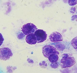
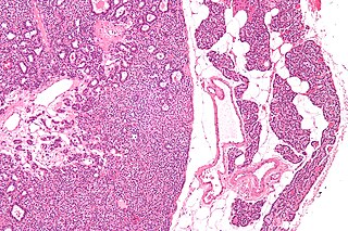
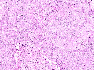
A perianal gland tumor is a type of tumor found near the anus in dogs that arises from specialized glandular tissue found in the perineum. It is also known as a hepatoid tumor because of the similarity in cell shape to hepatocytes. It is most commonly seen in intact dogs and is the third most common tumor type in intact male dogs. There are two types of perianal gland tumors, perianal gland adenomas, which are benign, and perianal gland adenocarcinomas, which are malignant. Both have receptors for testosterone. Perianal gland adenomas are three times more likely to be found in intact male dogs than females, and perianal gland adenocarcinomas are ten times more common in male dogs than females. The most commonly affected breeds for adenomas are the Siberian Husky, Cocker Spaniel, Pekingese, and Samoyed; for adenocarcinomas the most commonly affected breeds are the Siberian Husky, Bulldog, and Alaskan Malamute.
The uterine sarcomas form a group of malignant tumors that arises from the smooth muscle or connective tissue of the uterus.

Bladder stones or uroliths are a common occurrence in animals, especially in domestic animals such as dogs and cats. Occurrence in other species, including tortoises, has been reported as well. The stones form in the urinary bladder in varying size and numbers secondary to infection, dietary influences, and genetics. Stones can form in any part of the urinary tract in dogs and cats, but unlike in humans, stones of the kidney are less common and do not often cause significant disease, although they can contribute to pyelonephritis and chronic kidney disease. Types of stones include struvite, calcium oxalate, urate, cystine, calcium phosphate, and silicate. Struvite and calcium oxalate stones are by far the most common. Bladder stones are not the same as bladder crystals but if the crystals coalesce unchecked in the bladder they can become stones.
Dogs, as with all mammals, have natural odors. Natural dog odor can be unpleasant to dog owners especially when dogs are kept inside the home, as some people are not used to being exposed to the natural odor of a non-human species living in proximity to them. Dogs may also develop unnatural odors as a result of skin disease or other disorders or may become contaminated with odors from other sources in their environment.

Supraclavicular lymph nodes are lymph nodes found above the clavicle, that can be felt in the supraclavicular fossa. The supraclavicular lymph nodes on the left side are called Virchow's nodes. It leads to an appreciable mass that can be recognized clinically, called Troisier sign.

Parathyroid carcinoma is a rare cancer resulting in parathyroid adenoma to carcinoma progression. It forms in tissues of one or more of the parathyroid glands.
Many conditions are associated with disorders of the function of the parathyroid gland. Some disorders may be purely anatomical resulting in an enlarged gland which will raise concern. Such benign disorders, such as parathyroid cyst, are not discussed here. Parathyroid diseases can be divided into those causing hyperparathyroidism, and those causing hypoparathyroidism.

Spiradenomas (SA) are rare, benign cutaneous adnexal tumors that may progress to become their malignant counterparts, i.e. spiradenocarcinomas (SAC). Cutaneous adnexal tumors are a group of skin tumors consisting of tissues that have differentiated towards one of the four primary adnexal structures found in normal skin: hair follicles, sebaceous sweat glands, apocrine sweat glands, and eccrine sweat glands. SA and SAC tumors were regarded as eccrine gland tumors and termed eccrine spiradenomas and eccrine spiradenocarcinomas, respectively. However, more recent studies have found them to be hair follicle tumors and commonly term them spiradenomas and spiradenocarcinomas, respectively. Further confusing the situation, SA-like and SAC-like tumors are also 1) manifestations of the inherited disorder, CYLD cutaneous syndrome (CCS), and 2) have repeatedly been confused with an entirely different tumor, adenoid cystic carcinomas of the salivary gland. Here, SA and SAC are strictly defined as sporadic hair follicle tumors that do not include the hereditary CCS spiradenomas and heridtary spiradenocarcinoms of CCS or the adenoid cystic carcinomas.
Pure apocrine carcinoma of the breast (PACB) is a rare carcinoma derived from the epithelial cells in the lactiferous ducts of the mammary gland. The mammary gland is an apocrine gland. Its lactiferous ducts have two layers of epithelial cells, a luminal layer which faces the duct's lumen and a basal layer which lies beneath the luminal layer. There are at least 4 subtypes of epithelial cells in these ducts: luminal progenitor cells and luminal mature cells which reside in the luminal layer and mammary stem cells and basal cells which reside in the basal layer. Examination of the genes expressed in PACB cancer cells indicate that most of these tumors consist of cells derived from luminal cells but a minority of these tumors consist of cells derived from basal cells.
