
The macula or macula lutea is an oval-shaped pigmented area near the center of the retina of the human eye and some other animalian eyes. The macula in humans has a diameter of around 5.5 mm (0.22 in) and is subdivided into the umbo, foveola, foveal avascular zone, fovea, parafovea, and perifovea areas.
Retinoschisis is an eye disease characterized by the abnormal splitting of the retina's neurosensory layers, usually in the outer plexiform layer. Most common forms are asymptomatic, some rarer forms result in a loss of vision in the corresponding visual field.

The juxtaglomerular apparatus is a structure in the kidney that regulates the function of each nephron, the functional units of the kidney. The juxtaglomerular apparatus is named because it is next to (juxta-) the glomerulus.

The utricle and saccule are the two otolith organs in the vertebrate inner ear. They are part of the balancing system in the vestibule of the bony labyrinth. They use small stones and a viscous fluid to stimulate hair cells to detect motion and orientation. The utricle detects linear accelerations and head-tilts in the horizontal plane. The word utricle comes from Latin uter, meaning 'leather bag'.
In the kidney, the macula densa is an area of closely packed specialized cells lining the wall of the distal tubule, at the point where the thick ascending limb meets the distal convoluted tubule. The macula densa is the thickening where the distal tubule touches the glomerulus.

The juxtaglomerular cells are cells in the kidney that synthesize, store, and secrete the enzyme renin. They are specialized smooth muscle cells mainly in the walls of the afferent arterioles, and some in the efferent arterioles, that deliver blood to the glomerulus. In synthesizing renin, they play a critical role in the renin–angiotensin system and thus in autoregulation of the kidney.

The afferent arterioles are a group of blood vessels that supply the nephrons in many excretory systems. They play an important role in the regulation of blood pressure as a part of the tubuloglomerular feedback mechanism.

Vitelliform macular dystrophy, is an irregular autosomal dominant eye disorder which can cause progressive vision loss. This disorder affects the retina, specifically cells in a small area near the center of the retina called the macula. The macula is responsible for sharp central vision, which is needed for detailed tasks such as reading, driving, and recognizing faces. The condition is characterized by yellow, slightly elevated, round structures similar to the yolk of an egg.
In the physiology of the kidney, tubuloglomerular feedback (TGF) is a feedback system inside the kidneys. Within each nephron, information from the renal tubules is signaled to the glomerulus. Tubuloglomerular feedback is one of several mechanisms the kidney uses to regulate glomerular filtration rate (GFR). It involves the concept of purinergic signaling, in which an increased distal tubular sodium chloride concentration causes a basolateral release of adenosine from the macula densa cells. This initiates a cascade of events that ultimately brings GFR to an appropriate level.
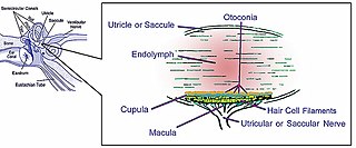
The macula of the utricle, or utricular macula is the region of the utricle that receives the utricular filaments of the vestibulocochlear nerve. The portion of the utricle that forms the macula forms a sort of pouch or cul-de-sac, with a thickened floor and anterior wall. The macula of utricle allows a person to perceive changes in longitudinal acceleration.
The saccule is the smaller sized vestibular sac ; it is globular in form, and lies in the recessus sphæricus near the opening of the scala vestibuli of the cochlea. Its anterior part exhibits an oval thickening, the macula of saccule, to which are distributed the saccular filaments of the acoustic nerve.

The foveola is located within a region called the macula, a yellowish, cone photo receptor filled portion of the human retina. The foveola is approximately 0.35 mm in diameter and lies in the center of the fovea and contains only cone cells, and a cone-shaped zone of Müller cells. In this region the cone receptors are found to be longer, slimmer and more densely packed than anywhere else in the retina, thus allowing that region to have the potential to have the highest visual acuity in the eye.
Astylopsis is a genus of longhorn beetles of the subfamily Lamiinae. It was described by Casey in 1913.
Astylopsis arcuata is a species of longhorn beetle of the subfamily Lamiinae. It was described by John Lawrence LeConte in 1878.
Astylopsis collaris is a species of longhorn beetles of the subfamily Lamiinae. It was described by Haldeman in 1847.
Astylopsis perplexa is a species of longhorn beetles of the subfamily Lamiinae. It was described by Haldeman in 1847.
Astylopsis sexguttata is a species of longhorn beetles of the subfamily Lamiinae. It was described by Say in 1826.
Parafovea or the parafoveal belt is a region in the retina that circumscribes the fovea and is part of the macula lutea. It is circumscribed by the perifovea.
Perifovea is a region in the retina that circumscribes the parafovea and fovea and is a part of the macula lutea. The perifovea is a belt that covers a 10° radius around the fovea and is 1.5 mm wide. The perifovea ends when the Henle's fiber layer disappears and the ganglion cells are one-layered.

Cthulhu Macula is a prominent surface feature of the dwarf planet Pluto, that is reminiscent of a whale in shape. It is an elongated dark region along Pluto's equator, 2,990 km (1,860 mi) long and one of the darkest features on Pluto. It is west of the Sputnik Planitia region of Tombaugh Regio, also known as Pluto's "heart", and to the east of Meng-P'o, the easternmost of Pluto's "Brass Knuckles".


