Auger therapy

Last updated
Auger therapy
Other namesAT
Specialty Radioligand Therapy

Auger therapy is a form of radiation therapy for the treatment of cancer which relies on low-energy electrons (emitted by the Auger effect) to damage cancer cells, rather than the high-energy radiation used in traditional radiation therapy. [1] [2] Similar to other forms of radiation therapy, Auger therapy relies on radiation-induced damage to cancer cells (particularly DNA damage) to arrest cell division, stop tumor growth and metastasis and kill cancerous cells. It differs from other types of radiation therapy in that electrons emitted via the Auger effect (Auger electrons) are released with low kinetic energy. In contrast to traditional α- and β-particle emitters, Auger electron emitters exhibit low cellular toxicity during transit in blood or bone marrow. [3]

Contents

Due to their low kinetic energy, emitted Auger electrons travel over a very short range: way less than the size of a single cell, on the order of less than a few-hundred nanometers. [4] This very short-range delivery of energy permits highly targeted therapies, since the radiation-emitting nuclide will be in close proximity to the delivery site (e.g., a DNA strand) to cause cytotoxicity. [5] However, this is a technical challenge; Auger therapeutics must enter their cell-nuclear targets to be most effective. [4] [6] Auger therapeutics are radiolabelled biomolecules, capable of entering cells of interest and binding to specific sub-cellular components. These typically carry a radioactive atom capable of emitting Auger electrons. The Auger electron emission from the atom is stimulated by radioactive decay, or by external pst (primary system therapy, such as X-ray) excitation. [6]

Auger dose

Simulated radiation dose of an electron in water, where the ionization energy of water at ~10 eV shows a resonant dose enhancement. The upper and lower curves are the short and long limiting ranges, respectively. In a vacuum, the kinetic energy
.mw-parser-output .frac{white-space:nowrap}.mw-parser-output .frac .num,.mw-parser-output .frac .den{font-size:80%;line-height:0;vertical-align:super}.mw-parser-output .frac .den{vertical-align:sub}.mw-parser-output .sr-only{border:0;clip:rect(0,0,0,0);clip-path:polygon(0px 0px,0px 0px,0px 0px);height:1px;margin:-1px;overflow:hidden;padding:0;position:absolute;width:1px}
1/2mev = 1 eV implies an electron velocity of 6x10 cm/s, or 0.2 per cent of the speed of light. Auger Therapy1.jpg
Simulated radiation dose of an electron in water, where the ionization energy of water at ~10 eV shows a resonant dose enhancement. The upper and lower curves are the short and long limiting ranges, respectively. In a vacuum, the kinetic energy 12mev = 1 eV implies an electron velocity of 6×10 cm/s, or 0.2 per cent of the speed of light.

The electron energy in a vacuum may be accurately measured with an electron detector in a Faraday cage, where the bias placed on the cage will accurately define the particle energy reaching the detector. The range of low-energy electrons in tissue or water, particularly electrons at the nanometer scale, cannot be easily measured; it must be inferred, since low-energy electrons scatter at large angles and travel in a zigzag path whose termination distance must be considered statistically and from differential measurements of higher-energy electrons at a much higher range. A 20  eV electron in water, for example, could have a range of 20 nm for 103  Gy or 5 nm for 104.7 Gy. For a group of 9–12 Auger electrons with energies at 12–18 eV in water (including the effect of water ionization at approximately 10 eV), an estimate of 106 Gy is probably sufficiently accurate. The illustration shows the simulated dose calculation in water for an electron using a Monte Carlo random walk [7] which gives up to 0.1 MGy. For a moderately-heavy atom to yield a dozen or more Auger electrons from its inner-shell ionization, the Auger dose becomes 106 Gy per event.

Candidates for molecular modification with in situ dose

With a large, localized dose in situ for molecular modification, the most obvious target molecule is the DNA duplex (where the complementary strands are separated by several nanometers). However, DNA duplex atoms are light elements (with only a few electrons each). Even if they could be induced by a photon beam to deliver Auger electrons, at under 1 keV they would be too soft to penetrate tissue sufficiently for therapy. Mid-range or heavy atoms (from bromine to platinum, for example) which could be induced by sufficiently hard X-ray photons to generate enough electrons to provide low-energy charges in an Auger cascade, will be considered for therapy.

Bromine electrons disrupting herpes-specific gene expression

When a normal cell transforms, replicating uncontrollably, many unusual genes (including viral material such as herpes genes which are not normally expressed) are expressed with viral-specific functions. The molecule proposed to disrupt the herpes gene is BrdC, where Br replaces a methyl (CH3) with nearly the same ionic radius and location (at the 5th position for BrdU, which has an oxygen molecule at the top). Therefore, BrdC could be oxidized and used as BrdU. Before oxidation, BrdC was unusable as dC or dU in mammalian cells (except for the herpes gene, which could incorporate the BrdC). The bromine atom is made from arsenic, with the addition of an alpha particle in a particle accelerator to form 77
Br
. It has a half-life of 57 hours and undergoes electron capture: the K-electron is captured by a proton in an unstable nucleus, creating a K hole in Br, and leading to its Auger cascade and disrupting the herpes gene without killing the cell.

This experiment was performed during the 1970s at Memorial Sloan Kettering Cancer Center by Lawrance Helson and C. G. Wang, using 10 neuroblastoma cell cultures, Two cultures were successful in terminating the cell replication with 77
Br
in vitro, and the experiments were followed by a group of nude mice with implanted tumors.

The in vivo mouse experiments were complicated when the mouse livers cleaved off the sugar component of BrdC rendering the mammalian and herpes genes to incorporate the 77
Br
-containing base, making no distinction between them. However, the Auger dose with 77BrdC disrupted the herpes-specific gene in several transformed cell cultures.[ citation needed ]

DNA-targeted dose using cisplatin

The group of metal-based anticancer drugs originated with cisplatin, one of the leading agents in clinical use. Cisplatin acts by binding to DNA, forming one or two intrastrand cross-links of the G-G adduct at 70% and the A-G adduct at ~20% of the major grooves of the double helix. The planar cis compound (on the same side) is composed of a square molecule with two chloride atoms on one side and two ammonia groups on the other side, centered around the heavy platinum (Pt) which could initiate the Auger dose in situ. Entering a cell with a low NaCl concentration, the aqua-chloride group would detach from the compound (allowing the missing chloride to link the G-G or A-G bases and bend the DNA helixes 45 degrees, damaging them). Although platinum-based antineoplastics are used in as much as 70 percent of all chemotherapy, they are not particularly effective against certain cancers (such as breast and prostate tumors).

The aqua-Cl rationale, detaching the chloride atom from the cisplatin when it enters a cell and binding them to G-G or A-G adducts in the major grooves of the DNA helixes, could be applied to other metals—such as ruthenium (Ru)-chemically similar to platinum. Ruthenium is used to coat the anode target of a mammography X-ray tube, enabling operation at any voltage (22–28  kVp) depending on the compressed thickness of the breast and delivering a high-contrast image. Although ruthenium is lighter than platinum, it can be induced to provide an Auger dose in situ to the DNA adducts and deliver localized chemotherapy. [8] [9]

Monochromatic X-rays to induce inner-shell ionization

X-ray tube with transmission target for line emissions

Monochromatic X-rays may be channeled from synchrotron radiation, obtained from filtered Coolidge X-ray tubes or from the preferred transmission X-ray tubes. To induce inner-shell ionization with resonant scattering from a moderately-heavy atom with dozens of electrons, the X-ray photon energy must be 30 keV or higher to penetrate tissue in therapeutic applications. Although synchrotron radiation is extremely bright and monochromatic without thermal scattering, its brightness falls off at the fourth power of photon energy. At 15–20 kV or higher an X-ray tube with a molybdenum target, for example, could deliver as much X-ray fluence as a typical synchrotron. A Coolidge X-ray tube brightens by 1.7 kVp and synchrotron brightness decreases by 4 kV, implying that it is not useful for Auger therapy.[ citation needed ]

Related Research Articles

<span class="mw-page-title-main">Auger effect</span> Physical phenomenon

The Auger effect or Auger−Meitner effect is a physical phenomenon in which the filling of an inner-shell vacancy of an atom is accompanied by the emission of an electron from the same atom. When a core electron is removed, leaving a vacancy, an electron from a higher energy level may fall into the vacancy, resulting in a release of energy. For light atoms (Z<12), this energy is most often transferred to a valence electron which is subsequently ejected from the atom. This second ejected electron is called an Auger electron. For heavier atomic nuclei, the release of the energy in the form of an emitted photon becomes gradually more probable.

<span class="mw-page-title-main">Radiation</span> Waves or particles moving through space

In physics, radiation is the emission or transmission of energy in the form of waves or particles through space or a material medium. This includes:

<span class="mw-page-title-main">Radiation therapy</span> Therapy using ionizing radiation, usually to treat cancer

Radiation therapy or radiotherapy is a treatment using ionizing radiation, generally provided as part of cancer therapy to either kill or control the growth of malignant cells. It is normally delivered by a linear particle accelerator. Radiation therapy may be curative in a number of types of cancer if they are localized to one area of the body, and have not spread to other parts. It may also be used as part of adjuvant therapy, to prevent tumor recurrence after surgery to remove a primary malignant tumor. Radiation therapy is synergistic with chemotherapy, and has been used before, during, and after chemotherapy in susceptible cancers. The subspecialty of oncology concerned with radiotherapy is called radiation oncology. A physician who practices in this subspecialty is a radiation oncologist.

<span class="mw-page-title-main">X-ray</span> Form of short-wavelength electromagnetic radiation

X-ray is a high-energy electromagnetic radiation. In many languages, it is referred to as Röntgen radiation, after the German scientist Wilhelm Conrad Röntgen, who discovered it in 1895 and named it X-radiation to signify an unknown type of radiation.

<span class="mw-page-title-main">Beta particle</span> Ionizing radiation

A beta particle, also called beta ray or beta radiation, is a high-energy, high-speed electron or positron emitted by the radioactive decay of an atomic nucleus during the process of beta decay. There are two forms of beta decay, β decay and β+ decay, which produce electrons and positrons respectively.

<span class="mw-page-title-main">X-ray fluorescence</span> Emission of secondary X-rays from a material excited by high-energy X-rays

X-ray fluorescence (XRF) is the emission of characteristic "secondary" X-rays from a material that has been excited by being bombarded with high-energy X-rays or gamma rays. The phenomenon is widely used for elemental analysis and chemical analysis, particularly in the investigation of metals, glass, ceramics and building materials, and for research in geochemistry, forensic science, archaeology and art objects such as paintings.

Ionizing radiation, including nuclear radiation, consists of subatomic particles or electromagnetic waves that have sufficient energy to ionize atoms or molecules by detaching electrons from them. Some particles can travel up to 99% of the speed of light, and the electromagnetic waves are on the high-energy portion of the electromagnetic spectrum.

<span class="mw-page-title-main">External beam radiotherapy</span> Treatment of cancer with ionized radiation

External beam radiation therapy (EBRT) is a form of radiotherapy that utilizes a high-energy collimated beam of ionizing radiation, from a source outside the body, to target and kill cancer cells. A radiotherapy beam is composed of particles which travel in a consistent direction; each radiotherapy beam consists of one type of particle intended for use in treatment, though most beams contain some contamination by other particle types.

<span class="mw-page-title-main">Synchrotron light source</span> Particle accelerator designed to produce intense x-ray beams

A synchrotron light source is a source of electromagnetic radiation (EM) usually produced by a storage ring, for scientific and technical purposes. First observed in synchrotrons, synchrotron light is now produced by storage rings and other specialized particle accelerators, typically accelerating electrons. Once the high-energy electron beam has been generated, it is directed into auxiliary components such as bending magnets and insertion devices in storage rings and free electron lasers. These supply the strong magnetic fields perpendicular to the beam that are needed to stimulate the high energy electrons to emit photons.

In radiation physics, kerma is an acronym for "kinetic energy released per unit mass", defined as the sum of the initial kinetic energies of all the charged particles liberated by uncharged ionizing radiation in a sample of matter, divided by the mass of the sample. It is defined by the quotient .

<span class="mw-page-title-main">Cisplatin</span> Chemical compound and pharmaceutical drug

Cisplatin is a chemical compound with formula cis-[Pt(NH3)2Cl2]. It is a coordination complex of platinum that is used as a chemotherapy medication used to treat a number of cancers. These include testicular cancer, ovarian cancer, cervical cancer, bladder cancer, head and neck cancer, esophageal cancer, lung cancer, mesothelioma, brain tumors and neuroblastoma. It is given by injection into a vein.

Iodine-125 (125I) is a radioisotope of iodine which has uses in biological assays, nuclear medicine imaging and in radiation therapy as brachytherapy to treat a number of conditions, including prostate cancer, uveal melanomas, and brain tumors. It is the second longest-lived radioisotope of iodine, after iodine-129.

<span class="mw-page-title-main">Extreme ultraviolet</span> Ultraviolet light with a wavelength of 10–121nm

Extreme ultraviolet radiation or high-energy ultraviolet radiation is electromagnetic radiation in the part of the electromagnetic spectrum spanning wavelengths shorter that the hydrogen Lyman-alpha line from 121 nm down to the X-ray band of 10 nm, and therefore having photons with energies from 10.26 eV up to 124.24 eV. EUV is naturally generated by the solar corona and artificially by plasma, high harmonic generation sources and synchrotron light sources. Since UVC extends to 100 nm, there is some overlap in the terms.

Electron spectroscopy refers to a group formed by techniques based on the analysis of the energies of emitted electrons such as photoelectrons and Auger electrons. This group includes X-ray photoelectron spectroscopy (XPS), which also known as Electron Spectroscopy for Chemical Analysis (ESCA), Electron energy loss spectroscopy (EELS), Ultraviolet photoelectron spectroscopy (UPS), and Auger electron spectroscopy (AES). These analytical techniques are used to identify and determine the elements and their electronic structures from the surface of a test sample. Samples can be solids, gases or liquids.

X-ray absorption near edge structure (XANES), also known as near edge X-ray absorption fine structure (NEXAFS), is a type of absorption spectroscopy that indicates the features in the X-ray absorption spectra (XAS) of condensed matter due to the photoabsorption cross section for electronic transitions from an atomic core level to final states in the energy region of 50–100 eV above the selected atomic core level ionization energy, where the wavelength of the photoelectron is larger than the interatomic distance between the absorbing atom and its first neighbour atoms.

Particle therapy is a form of external beam radiotherapy using beams of energetic neutrons, protons, or other heavier positive ions for cancer treatment. The most common type of particle therapy as of August 2021 is proton therapy.

In radiobiology, the relative biological effectiveness is the ratio of biological effectiveness of one type of ionizing radiation relative to another, given the same amount of absorbed energy. The RBE is an empirical value that varies depending on the type of ionizing radiation, the energies involved, the biological effects being considered such as cell death, and the oxygen tension of the tissues or so-called oxygen effect.

<span class="mw-page-title-main">Gamma ray</span> Energetic electromagnetic radiation arising from radioactive decay of atomic nuclei

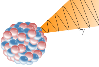
A gamma ray, also known as gamma radiation (symbol γ or ), is a penetrating form of electromagnetic radiation arising from the radioactive decay of atomic nuclei. It consists of the shortest wavelength electromagnetic waves, typically shorter than those of X-rays. With frequencies above 30 exahertz (3×10¹⁹ Hz), it imparts the highest photon energy. Paul Villard, a French chemist and physicist, discovered gamma radiation in 1900 while studying radiation emitted by radium. In 1903, Ernest Rutherford named this radiation gamma rays based on their relatively strong penetration of matter; in 1900 he had already named two less penetrating types of decay radiation (discovered by Henri Becquerel) alpha rays and beta rays in ascending order of penetrating power.

Ionizing radiation can cause biological effects which are passed on to offspring through the epigenome. The effects of radiation on cells has been found to be dependent on the dosage of the radiation, the location of the cell in regards to tissue, and whether the cell is a somatic or germ line cell. Generally, ionizing radiation appears to reduce methylation of DNA in cells.

<span class="mw-page-title-main">X-ray emission spectroscopy</span>

X-ray emission spectroscopy (XES) is a form of X-ray spectroscopy in which a core electron is excited by an incident x-ray photon and then this excited state decays by emitting an x-ray photon to fill the core hole. The energy of the emitted photon is the energy difference between the involved electronic levels. The analysis of the energy dependence of the emitted photons is the aim of the X-ray emission spectroscopy.

References

  1. Unak, P. (2002) Targeted tumor radiotherapy. Brazilian Archives of Biology and Technology, 45(spe) 97-110.
  2. Persson, L. The Auger Electron Effect in Radiation Dosimetry–A Review. Swedish Radiation Protection Institute, S-17116 Stockholm, Sweden.
  3. Knapp, Jr., F. F. (Russ) (2016). "2.5 Low-Energy Electron Emitters". Radiopharmaceuticals for Therapy. Springer, New Delhi. doi:10.1007/978-81-322-2607-9. ISBN   978-81-322-2607-9.
  4. 1 2 Kassis, A. (2003) Cancer therapy with Auger electrons: are we almost there? Journal of Nuclear Medicine, 44(9) 1479-1481.
  5. Giacomo Pirovano, Thomas C. Wilson, Thomas Reiner, Auger: The future of precision medicine, Nuclear Medicine and Biology, Volumes 96–97, 2021, Pages 50-53, ISSN 0969-8051.
  6. 1 2 Sastry, KSR. Biological effects of Auger emitter iodine-125. A review. Report No. 1 of AAPM Nuclear medicine Task Group No. 6. Med. Phys. 19(6), 1361-1383, 1992.
  7. J.Messungnoenab et al, Radiation Research 158, 657-660; 2002
  8. Wang, CG; US Patent 8,278,315; "Radiotherapy Method using X-rays"; 2012.
  9. Hannon, Michael J. "Metal-based Anticancer Drugs: From a past anchored in platinum chemistry to a post-genomic future of diverse chemistry and biology", Pure Appl. Chem. Vol.79, No. 12 pp 2243-2261, 2007.