Related Research Articles

Microscopy is the technical field of using microscopes to view objects and areas of objects that cannot be seen with the naked eye. There are three well-known branches of microscopy: optical, electron, and scanning probe microscopy, along with the emerging field of X-ray microscopy.

A microscope is a laboratory instrument used to examine objects that are too small to be seen by the naked eye. Microscopy is the science of investigating small objects and structures using a microscope. Microscopic means being invisible to the eye unless aided by a microscope.
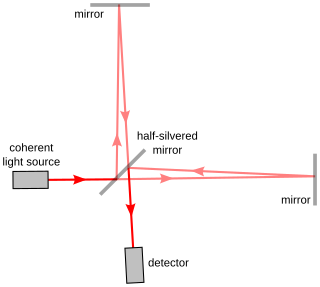
Interferometry is a technique in which waves are superimposed to cause the phenomenon of interference, which is used to extract information. Interferometry typically uses electromagnetic waves and is an important investigative technique in the fields of astronomy, fiber optics, engineering metrology, optical metrology, oceanography, seismology, spectroscopy, quantum mechanics, nuclear and particle physics, plasma physics, remote sensing, biomolecular interactions, surface profiling, microfluidics, mechanical stress/strain measurement, velocimetry, optometry, and making holograms.

Adaptive optics (AO) is a technology used to improve the performance of optical systems by reducing the effect of incoming wavefront distortions by deforming a mirror in order to compensate for the distortion. It is used in astronomical telescopes and laser communication systems to remove the effects of atmospheric distortion, in microscopy, optical fabrication and in retinal imaging systems to reduce optical aberrations. Adaptive optics works by measuring the distortions in a wavefront and compensating for them with a device that corrects those errors such as a deformable mirror or a liquid crystal array.

Optical coherence tomography (OCT) is an imaging technique that uses low-coherence light to capture micrometer-resolution, two- and three-dimensional images from within optical scattering media. It is used for medical imaging and industrial nondestructive testing (NDT). Optical coherence tomography is based on low-coherence interferometry, typically employing near-infrared light. The use of relatively long wavelength light allows it to penetrate into the scattering medium. Confocal microscopy, another optical technique, typically penetrates less deeply into the sample but with higher resolution.

Scanning laser ophthalmoscopy (SLO) is a method of examination of the eye. It uses the technique of confocal laser scanning microscopy for diagnostic imaging of the retina or cornea of the human eye.

The point spread function (PSF) describes the response of an imaging system to a point source or point object. A more general term for the PSF is a system's impulse response, the PSF being the impulse response of a focused optical system. The PSF in many contexts can be thought of as the extended blob in an image that represents a single point object. In functional terms, it is the spatial domain version of the optical transfer function of the imaging system. It is a useful concept in Fourier optics, astronomical imaging, medical imaging, electron microscopy and other imaging techniques such as 3D microscopy and fluorescence microscopy.

Confocal microscopy, most frequently confocal laser scanning microscopy (CLSM) or laser confocal scanning microscopy (LCSM), is an optical imaging technique for increasing optical resolution and contrast of a micrograph by means of using a spatial pinhole to block out-of-focus light in image formation. Capturing multiple two-dimensional images at different depths in a sample enables the reconstruction of three-dimensional structures within an object. This technique is used extensively in the scientific and industrial communities and typical applications are in life sciences, semiconductor inspection and materials science.
Medical optical imaging is the use of light as an investigational imaging technique for medical applications. Examples include optical microscopy, spectroscopy, endoscopy, scanning laser ophthalmoscopy, laser Doppler imaging, and optical coherence tomography. Because light is an electromagnetic wave, similar phenomena occur in X-rays, microwaves, and radio waves.

Two-photon excitation microscopy is a fluorescence imaging technique that allows imaging of living tissue up to about one millimeter in thickness. Unlike traditional fluorescence microscopy, in which the excitation wavelength is shorter than the emission wavelength, two-photon excitation requires simultaneous excitation by two photons with longer wavelength than the emitted light. Two-photon excitation microscopy typically uses near-infrared (NIR) excitation light which can also excite fluorescent dyes. However, for each excitation, two photons of NIR light are absorbed. Using infrared light minimizes scattering in the tissue. Due to the multiphoton absorption, the background signal is strongly suppressed. Both effects lead to an increased penetration depth for this technique. Two-photon excitation can be a superior alternative to confocal microscopy due to its deeper tissue penetration, efficient light detection, and reduced photobleaching.
Surface metrology is the measurement of small-scale features on surfaces, and is a branch of metrology. Surface primary form, surface fractality and surface roughness are the parameters most commonly associated with the field. It is important to many disciplines and is mostly known for the machining of precision parts and assemblies which contain mating surfaces or which must operate with high internal pressures.
Laser scanning is the controlled deflection of laser beams, visible or invisible. Scanned laser beams are used in some 3-D printers, in rapid prototyping, in machines for material processing, in laser engraving machines, in ophthalmological laser systems for the treatment of presbyopia, in confocal microscopy, in laser printers, in laser shows, in Laser TV, and in barcode scanners.

Deformable mirrors (DM) are mirrors whose surface can be deformed, in order to achieve wavefront control and correction of optical aberrations. Deformable mirrors are used in combination with wavefront sensors and real-time control systems in adaptive optics. In 2006 they found a new use in femtosecond pulse shaping.
The Gemini Planet Imager (GPI) is a high contrast imaging instrument that was built for the Gemini South Telescope in Chile. The instrument achieves high contrast at small angular separations, allowing for the direct imaging and integral field spectroscopy of extrasolar planets around nearby stars. The collaboration involved in planning and building the Gemini Planet imager includes the American Museum of Natural History (AMNH), Dunlap Institute, Gemini Observatory, Herzberg Institute of Astrophysics (HIA), Jet Propulsion Laboratory, Lawrence Livermore National Lab (LLNL), Lowell Observatory, SETI Institute, The Space Telescope Science Institute (STSCI), the University of Montreal, University of California, Berkeley, University of California, Los Angeles (UCLA), University of California, Santa Cruz (UCSC), University of Georgia.
Nikon Instruments is a division of the Nikon Corporation, which is headquartered in Tokyo. Its US operations are based in Melville, New York and its European operations in Amstelveen, Netherlands. Nikon Instruments is a specialist in optical instrumentation and the only microscope company to manufacture its own glass.
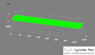
Digital holographic microscopy (DHM) is digital holography applied to microscopy. Digital holographic microscopy distinguishes itself from other microscopy methods by not recording the projected image of the object. Instead, the light wave front information originating from the object is digitally recorded as a hologram, from which a computer calculates the object image by using a numerical reconstruction algorithm. The image forming lens in traditional microscopy is thus replaced by a computer algorithm. Other closely related microscopy methods to digital holographic microscopy are interferometric microscopy, optical coherence tomography and diffraction phase microscopy. Common to all methods is the use of a reference wave front to obtain amplitude (intensity) and phase information. The information is recorded on a digital image sensor or by a photodetector from which an image of the object is created (reconstructed) by a computer. In traditional microscopy, which do not use a reference wave front, only intensity information is recorded and essential information about the object is lost.

A microscanner, or micro scanning mirror, is a microoptoelectromechanical system (MOEMS) in the category of micromirror actuators for dynamic light modulation. Depending upon the type of microscanner the modulatory movement of a single mirror can be either translatory or rotational, on one or two axes. In the first case, a phase shifting effect takes place. In the second case, the incident light wave is deflected.
Endomicroscopy is a technique for obtaining histology-like images from inside the human body in real-time, a process known as ‘optical biopsy’. It generally refers to fluorescence confocal microscopy, although multi-photon microscopy and optical coherence tomography have also been adapted for endoscopic use. Commercially available clinical and pre-clinical endomicroscopes can achieve a resolution on the order of a micrometre, have a field-of-view of several hundred µm, and are compatible with fluorophores which are excitable using 488 nm laser light. The main clinical applications are currently in imaging of the tumour margins of the brain and gastro-intestinal tract, particularly for the diagnosis and characterisation of Barrett’s Esophagus, pancreatic cysts and colorectal lesions. A number of pre-clinical and transnational applications have been developed for endomicroscopy as it enables researchers to perform live animal imaging. Major pre-clinical applications are in gastro-intestinal tract, toumour margin detection, uterine complications, ischaemia, live imaging of cartilage and tendon and organoid imaging.
ALPAO is a company which manufactures a range of adaptive optics products for use in research and industry, including deformable mirrors with large strokes, wavefront sensors, and adaptive optics loops. These products are designed for astronomy, vision science, microscopy, wireless optical communications, and laser applications.
Iris AO, Inc. manufactures small-scale, microelectromechanical systems (MEMS)-based deformable mirrors (DM) and adaptive optics systems that offer radical advantages in cost, size, durability, flexibility, and power consumption. Iris AO systems make adaptive optics (AO) practical for a host of new applications, including astronomy, retinal and biomedical imaging, beam shaping, portable laser communications, and horizontal-path imaging.
References
- ↑ CLEO Conference: Boston Micromachines Awarded Grant from NASA for Space Imaging Research. " Archived 2016-10-12 at the Wayback Machine "
- ↑ Boston Micromachines Introduces High Performance Reflective Optical Chopper for Laser Science Applications. " Archived 2016-10-12 at the Wayback Machine "
- ↑ Boston Micromachines, Applications and Customer Profiles. ""
- ↑ "Retinal Imaging Systems". Boston Mircomachines Corporation. Retrieved 5 October 2016.CS1 maint: discouraged parameter (link)
- ↑ Andrew Norton, Donald Gavel, Daren Dillon and Steven Cornelissen, 2010, High-power visible-laser effect on a Boston Micromachines MEMS deformable mirror, ""
- ↑ GPI , GPI Adaptive Optics Subsystem , ""
- ↑ "high actuator count" . Retrieved 5 October 2016.
- ↑ Delphine Débarre, Edward J. Botcherby, Martin J. Booth, and Tony Wilson, Adaptive optics for structured illumination microscopy, 2008, ""
- ↑ Weiyao Zou and Stephen A. Burns ,High-accuracy wavefront control for retinal imaging with Adaptive-Influence-Matrix Adaptive Optics, 2009, ""
- ↑ Steven Menn, Steven A. Cornelissen, Paul A. Bierden , 2007, Advances in MEMS deformable mirror technology for laser beam shaping, ""