Cell mechanics

Last updated

Cell mechanics is a sub-field of biophysics that focuses on the mechanical properties and behavior of living cells and how it relates to cell function. [1] It encompasses aspects of cell biophysics, biomechanics, soft matter physics and rheology, mechanobiology and cell biology.

Contents

Eukaryotic

Eukaryotic cells [2] are cells that consist of membrane-bound organelles, a membrane-bound nucleus, and more than one linear chromosome. Being much more complex than prokaryotic cells, cells without a true nucleus, eukaryotes must protect its organelles from outside forces.

Plant cell structure Plant cell structure-en.svg
Plant cell structure

Plant

Plant cell mechanics combines principles of biomechanics and mechanobiology to investigate the growth and shaping of the plant cells. Plant cells, similar to animal cells, respond to externally applied forces, such as by reorganization of their cytoskeletal network. The presence of a considerably rigid extracellular matrix, the cell wall, however, bestows the plant cells with a set of particular properties. Mainly, the growth of plant cells is controlled by the mechanics and chemical composition of the cell wall. [3] A major part of research in plant cell mechanics is put toward the measurement and modeling of the cell wall mechanics to understand how modification of its composition and mechanical properties affects the cell function, growth and morphogenesis. [4] [5]

Animal

Animal cell structure Animal cell structure en.svg
Animal cell structure

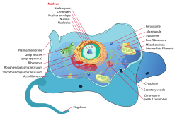
Because animal cells [6] do not have cell walls to protect them like plant cells, they require other specialized structures to sustain external mechanical forces. All animal cells are encased within a cell membrane made of a thin lipid bilayer that protects the cell from exposure to the outside environment. Using receptors composed of protein structures, the cell membrane is able to let selected molecules within the cell. Inside the cell membrane includes the cytoplasm, which contains the cytoskeleton. [7] A network of filamentous proteins including microtubules, intermediate filaments, and actin filaments makes up the cytoskeleton and helps maintain the cell's shape. By working together, the three types of polymers can organize themselves to counter the applied external forces and resist deformation. However, there are differences between the three polymers.

The primary structural component of the cytoskeleton is actin filaments. Being the narrowest with a diameter of 7 nm and most flexible out of the three types of polymers, actin filaments are typically found at the very edge of the cytoplasm in animal cells. [1] Formed by the linking of polymers of a protein called actin, they help give cells shape and structure and are able to transport protein packages and organelles. Furthermore, actin filaments have the ability to be assembled and disassembled quickly, allowing them to take part in cell mobility. [8]

On the other hand, intermediate filaments are more permanent structures with a diameter of 8 to 10 nm. [9] Composed of numerous fibrous protein strands wound together, intermediate proteins’ main role is bearing tension and retaining the shape and structure of the cell by securing the nucleus and other organelles in their designated areas.

The largest cytoskeletal structure of the three types of polymers is the microtubules with a diameter of 25 nm. [8] Unlike actin filaments, microtubules are stiff, hollow structures that radiate outwards from the microtubule organizing center (MTOC). Composed of tubulin proteins, microtubules are dynamic structures that allows them to shrink or grow with the addition or removal of tubulin proteins. In terms of cell mechanics, microtubules’ main purpose is to resist compressive cellular forces and act as a transportation system for motor proteins. [8]

It was shown that melanin also can have an impact on mechanic properties of cells. The research done by Sarna's team proved that heavily pigmented melanoma cells have Young's modulus about 4.93, when in non-pigmented ones it was only 0.98. [10] In another experiment they found that elasticity of melanoma cells is important for its metastasis and growth: non-pigmented tumors were bigger than pigmented and it was much easier for them to spread. They shown that there are both pigmented and non-pigmented cells in melanoma tumors, so that they can both be drug-resistant and metastatic. [10]

Measuring

Because cells are tiny, soft objects that must be measured differently than materials like metal, plastic, and glass, new techniques have been developed for the accurate measurement of cell mechanics. The variety of techniques can be divided into two categories: force application techniques and force sensing techniques. [1] In case of walled cells, such as plant or fungal cells, due to existence of a stiff, anisotropic and curved cell wall encapsulating the cells, special considerations and tailored approaches may be required compared to the methods used to measure the mechanics of animal cells. [11]

Force application

Force application techniques uses the cell's response of deformation to force applied onto the cell as a way to measure cell mechanical properties. [12] There are several different types of force application techniques including:

  1. Micropipette aspiration uses applied suction pressure with a small diameter glass pipet. The measurement of the length of aspiration caused by the suction pressure can reveal several cell mechanical properties. [13]
  2. Cantilever manipulation operates through an magnetic, electrical, or mechanical interaction between a probe and the surface of the cell that gives off a signal that can be used to measure mechanical properties. [14]
  3. Optical techniques involves the usage of trapped photons to manipulate cells. The photons will change in direction based on the cell's refractive index, which will cause a change in momentum, leading to a force applied upon the cell. [12]
  4. Mechanical techniques utilizes the incorporation of ferromagnetic beads into the cell or attached to specific receptors on the cell. When a magnetic force is applied, the stretch of the membrane can be measured to calculate mechanical properties. [12]
  5. Substrate strain measures elasticity through stretching the cell. The elasticity of the cell provides information that can determine motility and adhesion. [12] [15]
  6. Compression requires the usage of applied pressure onto the entire cell. By calculating the changes of the cell's shape, compression is a way to measure mechanical responses to force. [12]
  7. Flow technique uses Reynold's number, a dimensionless number in fluid mechanics, to distinguish whether the cell is subject to laminar, transitional, or turbulent flow. [12]
  8. Acoustic force spectroscopy can be used to extract mechanical properties of single cells. [16]

Force sensing

  1. Wrinkling membranes requires putting the cell into a flexible silicon envelope. As the cell contracts, the magnitude of the forces can be estimated by utilizing the length and number of wrinkles. [12]
  2. Traction force microscopy [17] detects deformations through comparison of images the movement of fluorescent beads that have been adhered to the cell. [18]
  3. Cantilever sensing can detect surface stresses with the attachment of micromechanical beams on one end of the cell. [19]
  4. Bioreactors allow the measurement of multicellular forces in a three-dimensional system, while external forces are applied at the same time. This enables better results and more accurate data from complex experiments. [12]
  5. When adherent cells are excited by acoustic waves, they start to generate acoustic microstreaming flow. The velocity magnitude of this flow near the cell membrane is directly proportional to the stiffness (i.e., modulus of elasticity) of the cell. [20]

Research

Researchers who study cell mechanics are interested in the mechanics and dynamics of the assemblies and structures that make up the cell including membranes, cytoskeleton, organelles, and cytoplasm, and how they interact to give rise to the emergent properties of the cell as a whole. [21]

A particular focus of many cell mechanical studies has been the cytoskeleton, which (in animal cells) can be thought to consist of:

  1. actomyosin assemblies (F-actin, myosin motors, and associated binding, nucleating, capping, stabilizing, and crosslinking proteins),
  2. microtubules and their associated motor proteins (kinesins and dyneins),
  3. intermediate filaments,
  4. other assemblies such as spectrins and septins.

The active non-equilibrium and non-linear rheological properties of cellular assemblies have been keen point of research in recent times. [22] [23] Another point of interest has been how cell cycle-related changes in cytoskeletal activity affect global cell properties, such as intracellular pressure increase during mitotic cell rounding. [24]

Related Research Articles

<span class="mw-page-title-main">Cytoplasm</span> All of the contents of a eukaryotic cell except the nucleus.

In cell biology, the cytoplasm describes all material within a eukaryotic cell, enclosed by the cell membrane, except for the cell nucleus. The material inside the nucleus and contained within the nuclear membrane is termed the nucleoplasm. The main components of the cytoplasm are the cytosol, the organelles, and various cytoplasmic inclusions. The cytoplasm is about 80% water and is usually colorless.

<span class="mw-page-title-main">Microtubule</span> Polymer of tubulin that forms part of the cytoskeleton

Microtubules are polymers of tubulin that form part of the cytoskeleton and provide structure and shape to eukaryotic cells. Microtubules can be as long as 50 micrometres, as wide as 23 to 27 nm and have an inner diameter between 11 and 15 nm. They are formed by the polymerization of a dimer of two globular proteins, alpha and beta tubulin into protofilaments that can then associate laterally to form a hollow tube, the microtubule. The most common form of a microtubule consists of 13 protofilaments in the tubular arrangement.

<span class="mw-page-title-main">Cytoskeleton</span> Network of filamentous proteins that forms the internal framework of cells

The cytoskeleton is a complex, dynamic network of interlinking protein filaments present in the cytoplasm of all cells, including those of bacteria and archaea. In eukaryotes, it extends from the cell nucleus to the cell membrane and is composed of similar proteins in the various organisms. It is composed of three main components: microfilaments, intermediate filaments, and microtubules, and these are all capable of rapid growth or disassembly depending on the cell's requirements.

<span class="mw-page-title-main">Cytokinesis</span> Part of the cell division process

Cytokinesis is the part of the cell division process and part of mitosis during which the cytoplasm of a single eukaryotic cell divides into two daughter cells. Cytoplasmic division begins during or after the late stages of nuclear division in mitosis and meiosis. During cytokinesis the spindle apparatus partitions and transports duplicated chromatids into the cytoplasm of the separating daughter cells. It thereby ensures that chromosome number and complement are maintained from one generation to the next and that, except in special cases, the daughter cells will be functional copies of the parent cell. After the completion of the telophase and cytokinesis, each daughter cell enters the interphase of the cell cycle.

<span class="mw-page-title-main">Microfilament</span> Filament in the cytoplasm of eukaryotic cells

Microfilaments, also called actin filaments, are protein filaments in the cytoplasm of eukaryotic cells that form part of the cytoskeleton. They are primarily composed of polymers of actin, but are modified by and interact with numerous other proteins in the cell. Microfilaments are usually about 7 nm in diameter and made up of two strands of actin. Microfilament functions include cytokinesis, amoeboid movement, cell motility, changes in cell shape, endocytosis and exocytosis, cell contractility, and mechanical stability. Microfilaments are flexible and relatively strong, resisting buckling by multi-piconewton compressive forces and filament fracture by nanonewton tensile forces. In inducing cell motility, one end of the actin filament elongates while the other end contracts, presumably by myosin II molecular motors. Additionally, they function as part of actomyosin-driven contractile molecular motors, wherein the thin filaments serve as tensile platforms for myosin's ATP-dependent pulling action in muscle contraction and pseudopod advancement. Microfilaments have a tough, flexible framework which helps the cell in movement.

<span class="mw-page-title-main">Cleavage furrow</span> Plasma membrane invagination at the cell division site

In cell biology, the cleavage furrow is the indentation of the cell's surface that begins the progression of cleavage, by which animal and some algal cells undergo cytokinesis, the final splitting of the membrane, in the process of cell division. The same proteins responsible for muscle contraction, actin and myosin, begin the process of forming the cleavage furrow, creating an actomyosin ring. Other cytoskeletal proteins and actin binding proteins are involved in the procedure.

<span class="mw-page-title-main">Actin</span> Family of proteins

Actin is a family of globular multi-functional proteins that form microfilaments in the cytoskeleton, and the thin filaments in muscle fibrils. It is found in essentially all eukaryotic cells, where it may be present at a concentration of over 100 μM; its mass is roughly 42 kDa, with a diameter of 4 to 7 nm.

<span class="mw-page-title-main">Cytoplasmic streaming</span> Flow of the cytoplasm inside the cell

Cytoplasmic streaming, also called protoplasmic streaming and cyclosis, is the flow of the cytoplasm inside the cell, driven by forces from the cytoskeleton. It is likely that its function is, at least in part, to speed up the transport of molecules and organelles around the cell. It is usually observed in large plant and animal cells, greater than approximately 0.1 mm. In smaller cells, the diffusion of molecules is more rapid, but diffusion slows as the size of the cell increases, so larger cells may need cytoplasmic streaming for efficient function.

Cell migration is a central process in the development and maintenance of multicellular organisms. Tissue formation during embryonic development, wound healing and immune responses all require the orchestrated movement of cells in particular directions to specific locations. Cells often migrate in response to specific external signals, including chemical signals and mechanical signals. Errors during this process have serious consequences, including intellectual disability, vascular disease, tumor formation and metastasis. An understanding of the mechanism by which cells migrate may lead to the development of novel therapeutic strategies for controlling, for example, invasive tumour cells.

<span class="mw-page-title-main">Spectrin</span>

Spectrin is a cytoskeletal protein that lines the intracellular side of the plasma membrane in eukaryotic cells. Spectrin forms pentagonal or hexagonal arrangements, forming a scaffold and playing an important role in maintenance of plasma membrane integrity and cytoskeletal structure. The hexagonal arrangements are formed by tetramers of spectrin subunits associating with short actin filaments at either end of the tetramer. These short actin filaments act as junctional complexes allowing the formation of the hexagonal mesh. The protein is named spectrin since it was first isolated as a major protein component of human red blood cells which had been treated with mild detergents; the detergents lysed the cells and the hemoglobin and other cytoplasmic components were washed out. In the light microscope the basic shape of the red blood cell could still be seen as the spectrin-containing submembranous cytoskeleton preserved the shape of the cell in outline. This became known as a red blood cell "ghost" (spectre), and so the major protein of the ghost was named spectrin.

<span class="mw-page-title-main">Motor protein</span> Class of molecular proteins

Motor proteins are a class of molecular motors that can move along the cytoplasm of cells. They convert chemical energy into mechanical work by the hydrolysis of ATP. Flagellar rotation, however, is powered by a proton pump.

<span class="mw-page-title-main">Growth cone</span> Large actin extension of a developing neurite seeking its synaptic target

A growth cone is a large actin-supported extension of a developing or regenerating neurite seeking its synaptic target. It is the growth cone that drives axon growth. Their existence was originally proposed by Spanish histologist Santiago Ramón y Cajal based upon stationary images he observed under the microscope. He first described the growth cone based on fixed cells as "a concentration of protoplasm of conical form, endowed with amoeboid movements". Growth cones are situated on the tips of neurites, either dendrites or axons, of the nerve cell. The sensory, motor, integrative, and adaptive functions of growing axons and dendrites are all contained within this specialized structure.

<span class="mw-page-title-main">Cell cortex</span> Layer on the inner face of a cell membrane

The cell cortex, also known as the actin cortex, cortical cytoskeleton or actomyosin cortex, is a specialized layer of cytoplasmic proteins on the inner face of the cell membrane. It functions as a modulator of membrane behavior and cell surface properties. In most eukaryotic cells lacking a cell wall, the cortex is an actin-rich network consisting of F-actin filaments, myosin motors, and actin-binding proteins. The actomyosin cortex is attached to the cell membrane via membrane-anchoring proteins called ERM proteins that plays a central role in cell shape control. The protein constituents of the cortex undergo rapid turnover, making the cortex both mechanically rigid and highly plastic, two properties essential to its function. In most cases, the cortex is in the range of 100 to 1000 nanometers thick.

<span class="mw-page-title-main">Protein filament</span> Long chain of protein monomers

In biology, a protein filament is a long chain of protein monomers, such as those found in hair, muscle, or in flagella. Protein filaments form together to make the cytoskeleton of the cell. They are often bundled together to provide support, strength, and rigidity to the cell. When the filaments are packed up together, they are able to form three different cellular parts. The three major classes of protein filaments that make up the cytoskeleton include: actin filaments, microtubules and intermediate filaments.

The mechanome consists of the body, or ome, of data including cell and molecular processes relating to force and mechanical systems at molecular, cellular and tissue length scales - the fundamental "machine code" structures of the cell. The mechanome encompasses biological motors, like kinesin, myosin, RNAP, and Ribosome mechanical structures, like actin or the cytoskeleton and also proteomic and genomic components that are mechanosensitive and are involved in the response of cells to externally applied force.

<span class="mw-page-title-main">Stress fiber</span> Contractile actin bundles found in non-muscle cells

Stress fibers are contractile actin bundles found in non-muscle cells. They are composed of actin (microfilaments) and non-muscle myosin II (NMMII), and also contain various crosslinking proteins, such as α-actinin, to form a highly regulated actomyosin structure within non-muscle cells. Stress fibers have been shown to play an important role in cellular contractility, providing force for a number of functions such as cell adhesion, migration and morphogenesis.

The LINC complex is a protein complex associated with both inner and outer membranes of the nucleus. It is composed of SUN-domain proteins and KASH-domain proteins. The SUN-domain proteins are associated with both nuclear lamins and chromatin and cross the inner nuclear membrane. They interact with the KASH domain proteins in the perinuclear (lumen) space between the two membranes. The KASH domain proteins cross the outer nuclear membrane and interact with actin filaments, microtubule filaments, intermediate filaments, centrosomes and cytoplasmic organelles. The number of SUN-domain and KASH-domain proteins increased in evolution.

Force Spectrum Microscopy (FSM) is an application of active microrheology developed to measure aggregate random forces in the cytoplasm. Large, inert flow tracers are injected into live cells and become lodged inside the cytoskeletal mesh, wherein it is oscillated by repercussions from active motor proteins. The magnitude of these random forces can be inferred from the frequency of oscillation of tracer particles. Tracking the fluctuations of tracer particles using optical microscopy can isolate the contribution of active random forces to intracellular molecular transport from that of Brownian motion.

<span class="mw-page-title-main">Intracellular transport</span> Directed movement of vesicles and substances within a cell

Intracellular transport is the movement of vesicles and substances within a cell. Intracellular transport is required for maintaining homeostasis within the cell by responding to physiological signals. Proteins synthesized in the cytosol are distributed to their respective organelles, according to their specific amino acid’s sorting sequence. Eukaryotic cells transport packets of components to particular intracellular locations by attaching them to molecular motors that haul them along microtubules and actin filaments. Since intracellular transport heavily relies on microtubules for movement, the components of the cytoskeleton play a vital role in trafficking vesicles between organelles and the plasma membrane by providing mechanical support. Through this pathway, it is possible to facilitate the movement of essential molecules such as membrane‐bounded vesicles and organelles, mRNA, and chromosomes.

A microtubular membrane is a type of membrane made up of small tubular structures. Microtubular associated membranes are found in various cell types and are essential for maintaining cell structure and function. Synthetic membranes are used in chemical separation processes and in flow batteries.

References

  1. 1 2 3 Moeendarbary E, Harris AR (2014). "Cell mechanics: principles, practices, and prospects". Wiley Interdisciplinary Reviews. Systems Biology and Medicine. 6 (5): 371–88. doi:10.1002/wsbm.1275. PMC   4309479 . PMID   25269160.
  2. "Intro to eukaryotic cells". Khan Academy.
  3. Bidhendi AJ, Altartouri B, Gosselin FP, Geitmann A (July 2019). "Mechanical Stress Initiates and Sustains the Morphogenesis of Wavy Leaf Epidermal Cells". Cell Reports. 28 (5): 1237–1250.e6. doi: 10.1016/j.celrep.2019.07.006 . PMID   31365867.
  4. Bidhendi AJ, Geitmann A (January 2016). "Relating the mechanics of the primary plant cell wall to morphogenesis". Journal of Experimental Botany. 67 (2): 449–61. doi: 10.1093/jxb/erv535 . PMID   26689854.
  5. Bidhendi AJ, Geitmann A (January 2018). "Finite Element Modeling of Shape Changes in Plant Cells". Plant Physiology. 176 (1): 41–56. doi:10.1104/pp.17.01684. PMC   5761827 . PMID   29229695.
  6. McGregor J (6 August 2018). "The Parts Of An Animal Cell". Science Trends. doi: 10.31988/SciTrends.24128 .
  7. Clark AG, Wartlick O, Salbreux G, Paluch EK (May 2014). "Stresses at the cell surface during animal cell morphogenesis". Current Biology. 24 (10): R484-94. doi: 10.1016/j.cub.2014.03.059 . PMID   24845681.
  8. 1 2 3 "Microtubules and Filaments". Scitable by Nature Education.
  9. "What are intermediate filaments? | MBInfo". www.mechanobio.info.
  10. 1 2 Sarna M, Krzykawska-Serda M, Jakubowska M, Zadlo A, Urbanska K (June 2019). "Melanin presence inhibits melanoma cell spread in mice in a unique mechanical fashion". Scientific Reports. 9 (1): 9280. Bibcode:2019NatSR...9.9280S. doi:10.1038/s41598-019-45643-9. PMC   6594928 . PMID   31243305.
  11. Bidhendi AJ, Geitmann A (July 2019). "Methods to quantify primary plant cell wall mechanics". Journal of Experimental Botany. 70 (14): 3615–3648. doi: 10.1093/jxb/erz281 . PMID   31301141.
  12. 1 2 3 4 5 6 7 8 Rodriguez ML, McGarry PJ, Sniadecki NJ (15 October 2013). "Review on Cell Mechanics: Experimental and Modeling Approaches". Applied Mechanics Reviews. 65 (6): 060801–060801–41. Bibcode:2013ApMRv..65f0801R. doi:10.1115/1.4025355.
  13. "Lecture 17: Cell Mechanics" (PDF).
  14. Jalili N (10 November 2012). "Nanomechanical Cantilever-Based Manipulation for Sensing and Imaging". Nanorobotics. Springer New York. pp. 29–40. doi:10.1007/978-1-4614-2119-1_2. ISBN   978-1-4614-2118-4.
  15. Ghassemi S, Meacci G, Liu S, Gondarenko AA, Mathur A, Roca-Cusachs P, et al. (April 2012). "Cells test substrate rigidity by local contractions on submicrometer pillars". Proceedings of the National Academy of Sciences of the United States of America. 109 (14): 5328–33. Bibcode:2012PNAS..109.5328G. doi: 10.1073/pnas.1119886109 . PMC   3325713 . PMID   22431603.
  16. Evers, Tom M.J.; Sheikhhassani, Vahid; Haks, Mariëlle C.; Storm, Cornelis; Ottenhoff, Tom H.M.; Mashaghi, Alireza (2022). "Single-cell analysis reveals chemokine-mediated differential regulation of monocyte mechanics". iScience. 25 (1): 103555. Bibcode:2022iSci...25j3555E. doi:10.1016/j.isci.2021.103555. PMC   8693412 . PMID   34988399.
  17. Lekka, Małgorzata; Gnanachandran, Kajangi; Kubiak, Andrzej; Zieliński, Tomasz; Zemła, Joanna (November 2021). "Traction force microscopy – Measuring the forces exerted by cells" (PDF). Micron. 150: 103138. doi: 10.1016/j.micron.2021.103138 .
  18. Plotnikov SV, Sabass B, Schwarz US, Waterman CM (2014). "High-resolution traction force microscopy". Quantitative Imaging in Cell Biology. Methods in Cell Biology. Vol. 123. pp. 367–94. doi:10.1016/B978-0-12-420138-5.00020-3. ISBN   9780124201385. PMC   4699589 . PMID   24974038.
  19. Datar R, Kim S, Jeon S, Hesketh P, Manalis S, Boisen A, Thundat T. "Cantilever Sensors: Nanomechanical Tools for Diagnostics" (PDF).
  20. Salari A, Appak-Baskoy S, Ezzo M, Hinz B, Kolios MC, Tsai SS (March 2020). "Dancing with the Cells: Acoustic Microflows Generated by Oscillating Cells". Small. 16 (9): e1903788. doi:10.1002/smll.201903788. PMID   31829522. S2CID   209330149.
  21. Fletcher DA, Mullins RD (January 2010). "Cell mechanics and the cytoskeleton". Nature. 463 (7280): 485–92. Bibcode:2010Natur.463..485F. doi:10.1038/nature08908. PMC   2851742 . PMID   20110992.
  22. Mizuno D, Tardin C, Schmidt CF, Mackintosh FC (January 2007). "Nonequilibrium mechanics of active cytoskeletal networks". Science. 315 (5810): 370–3. Bibcode:2007Sci...315..370M. doi:10.1126/science.1134404. PMID   17234946. S2CID   20272334.
  23. Guo M, Ehrlicher AJ, Jensen MH, Renz M, Moore JR, Goldman RD, et al. (August 2014). "Probing the stochastic, motor-driven properties of the cytoplasm using force spectrum microscopy". Cell. 158 (4): 822–832. doi:10.1016/j.cell.2014.06.051. PMC   4183065 . PMID   25126787.
  24. Stewart MP, Helenius J, Toyoda Y, Ramanathan SP, Muller DJ, Hyman AA (January 2011). "Hydrostatic pressure and the actomyosin cortex drive mitotic cell rounding". Nature. 469 (7329): 226–30. Bibcode:2011Natur.469..226S. doi:10.1038/nature09642. PMID   21196934. S2CID   4425308.

See also