Central nervous system primitive neuroectodermal tumor

Last updated
Central nervous system primitive neuroectodermal tumor
PNET.jpg
Primitive neuroectodermal tumor of the central nervous system in a 5-year-old

A central nervous system primitive neuroectodermal tumor, often abbreviated as PNET, supratentorial PNET, or CNS-PNET, [1] is one of the 3 types of embryonal central nervous system tumors (medulloblastoma, atypical teratoid rhabdoid tumor, and PNET). [2] It is considered an embryonal tumor because it arises from cells partially differentiated or still undifferentiated from birth. [1] Those cells are usually neuroepithelial cells, [1] [2] [3] stem cells destined to turn into glia or neurons. [4] It can occur anywhere within the spinal cord and cerebrum and can have multiple sites of origins, with a high probability of metastasis through cerebrospinal fluid (CSF). [1] [2]

Contents

PNET has five subtypes of tumors: neuroblastoma, ganglioneuroblastoma, medulloepithelioma, ependymoblastoma, and not otherwise specified PNET. [1] It is similar to medulloblastoma regarding histology but different regarding genetic factors and tumor site. It is a rare disease occurring mostly among children, [1] [2] accounting for 1.9 to 7% of childhood brain tumors. [2] Symptoms involve emotional, visual, motor, and speech defects. [2] Magnetic resonance imaging (MRI) and computed tomography (CT) are used to diagnose PNETs. [2] Even though a universal treatment plan hasn't been stablished yet, common strategies involve chemotherapy and radiotherapy for individuals older than 3 years of age. [1] [2] Their efficacy, however, is still controversial. [2] Surgery can be used to remove mass affected by tumorous cells. [2] The prognosis of the disease is more positive for adults than for children, who have a higher probability of having sequelae from the tumor. [1] [2]

It is important to note that this classification term has been removed from the latest WHO classification of CNS tumors as of 2016. Instead PNETs are now included into the category of "Embryonal Tumors with Multilayered Rosettes" along with ependymoblastoma and embryonal tumor with abundant neuropil and true rosettes (ETANTR). [5]

Classification

Histology of Medulloepithelioma Medulloepithelioma Histology.jpg
Histology of Medulloepithelioma

The World Health Organization has classified the central nervous system primitive neuroectodermal tumors into five subtypes: neuroblastoma, ganglioneuroblastoma, medulloepithelioma, ependymoblastoma, and not otherwise specified PNET. [1] The last one encompasses the PNETs with varying characteristics that hasn't been well defined yet. [1] Neuroblastomas are PNETS that involve the process of cell differentiation into neurons, [1] [2] while ganglioneuroblastomas are PNETs that involve ganglion cells. [1]

Medulloepithelioma, on the other hand, are tumors involving the constant cell division on the epithelium tissue where bundle of neuron endings are located. [1] Such tissue will differentiate into a similar form as the embryonic neural tube, also known as the starting structure of the central nervous system. [1] [2] [3] Medulloepitheliomas also present a pattern known as rosettes, characterized by the arrangement of a bundle of cells into circular shapes and around a center or a neuropil. [1] Ependymoblastoma also present rosettes as well as a higher density of cells. [1] [3] It involves the process of differentiation into ependymal cells. [2] [3]

Rosettes in Ependymoblastoma histology Ependymoblastomatous Rosette.jpg
Rosettes in Ependymoblastoma histology

Further classification types have come up but not yet approved by the World Health Organization. [1] The term "embryonal tumor with abundant neuropil and true rosettes", or ETANTR, has been proposed as a sixth subtype of PNET. [1] However, the still unofficial term "embryonal tumor with multilayered rosettes" (ETMR) has been more frequently used and encompasses ETANTRs, medulloepitheliomas, ependymoblastomas, and variants of PNETs with presence of rosettes and with no well defined classification. [3]

Risk factors

The rate of PNETs in not correlated with sex, but it shows a correlation with age. [1] Most cases occur in children around 5 years of age, having a very low frequency in adults. [1] Regarding genetic mutations, a specific type of gene alteration that directly leads to this tumor hasn't been defined yet. [1] However, a positive correlation between individuals with Li-Fraumeni syndrome with a mutation in the gene p53 and PNET has been reported. [2] A significant number of individuals with mutations on the rb tumor suppressor gene have also developed the tumor. [2] Such gene encodes for the protein Rb responsible for stopping the cell cycle at the G1 phase. [6] Another possible contributing factor are mutations in the CREB-binding protein, whose function includes activating transcription, [6] but this interaction still need to be studied further. [2] It has also been presumed that the tumor can arise from cranial irradiation. [2]

Diagnosis

Magnetic resonance image of PNET MRI of PNET.jpg
Magnetic resonance image of PNET

Most children that develop primitive neuroectodermal tumors are diagnosed early in life, usually at around 3–6.8 years of age. [2] Symptoms patients present at time of diagnosis include irritable mood, visual difficulties, lethargy, and ataxia. [2] The circumference of the patient's head might also become enlarged and they might be subject to seizures, especially if they have less than one year of life. [2]

Several analysis can be used to determine the presence of the disease. Physical examinations showing papilledema, visual field defects, cranial nerves palsy, dysphasia, and focal neurological deficits are evidences for possible tumor. [2] PNETs can also be spotted through computed tomography (CT) and magnetic resonance imaging (MRI). [2] In images produced by MRIs, an irregular augmentation among a solid mass will indicated the presence of tumor. [3] However, the results of MRIs are usually ambiguous in defining the presence for this specific tumor. [2] In CT scans, the presence of PNETs will be indicated by an elevated density and an increase in volume of the brain. [2] The CT scan can also show calcification, [3] which is present in 41-44% of PNET cases. [2] Since the tumor can be replicated in other parts of the nervous system through the cerebrospinal fluid (CSF), a CSF analysis can also be conducted. [2] A spinal MRI is a fourth type of analysis that is useful in investigating the level of tumor propagation to the spinal cord. [2]

Treatment

There is not a standardized procedure to treat primitive neuroectodermal tumors. [2] Common strategies involve risk-adapted radiotherapy combined with chemotherapy and stem cell rescue. [1] For patients younger than 2–3 years of age, treatment with radiation is not used, once they are in a more vulnerable phase and, thus, more prone to risks in development. [1] Examinations such as CSF analysis and spinal MRIs are used to investigate the effectiveness of treatment in preventing metastasis. [2]

A method for eliminating tumorous mass is surgery, where the best outcome would be total resection, meaning the complete removal of the tumor. [2] Along with the surgery, several measures that contribute to a safe procedure can be taken: urine exams, transfusion, and the constant supervision of arterial pressure. [2] Possible problems that arise from the surgery include hemorrhage, brain edema, and hemiparesis. [2] MRIs are typically done after 1 or 2 days of postoperative in order to inspect the amount of tumor remaining. [2]

Prognosis

The probability of primitive neuroectodermal tumors to have recurrence and metastasize through cerebrospinal fluid is relatively high. [3] The outcome of PNET is more positive when the individual is an adult, independent of age subgroups, or an older child. [2] Less than 50% of children survive more than 5 years, [1] while the majority of adults live to 7 years. [2] The reason the prognosis for such tumor is worst in children is due to the higher probability of the tumor spreading to the rest of the nervous system through the cerebrospinal fluid and growing again. [2] Moreover, children have the probability of developing deficiencies in cognitive processes, problems in the endocrine system, and psychological obstacles after the disease. [2] Adults, on the other hand, don't show such propensity. [2] As a consequence, 37.7% of children affected by the tumor live to 4 years. [2]

The effect of treatment strategies such as chemotherapy and radiation therapy on the prognosis of the disease is still controversial, with studies claiming either their benefits or their ineffectiveness. [2] The same holds true for the relationship between volume of tumor removed by surgery and survival. [2] Furthermore, factors such as tumor size, location of origin, race, and sex of individual don't show any influence on the outcome of the disease. [2] However, interactions of some factors such as tumor site, age, and treatment strategy can affect one's prognosis. [2] For instance, when younger children below the age of three develop tumors originating in places other than the pineal gland are treated with chemotherapy, they present better outcomes than those with pineal tumors and treated with chemotherapy. [2]

Related Research Articles

<span class="mw-page-title-main">Cerebrospinal fluid</span> Clear, colorless bodily fluid found in the brain and spinal cord

Cerebrospinal fluid (CSF) is a clear, colorless body fluid found within the tissue that surrounds the brain and spinal cord of all vertebrates.

<span class="mw-page-title-main">Brain tumor</span> Neoplasm in the brain

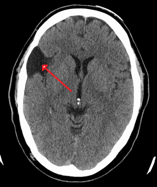
A brain tumor occurs when abnormal cells form within the brain. There are two main types of tumors: malignant tumors and benign (non-cancerous) tumors. These can be further classified as primary tumors, which start within the brain, and secondary tumors, which most commonly have spread from tumors located outside the brain, known as brain metastasis tumors. All types of brain tumors may produce symptoms that vary depending on the size of the tumor and the part of the brain that is involved. Where symptoms exist, they may include headaches, seizures, problems with vision, vomiting and mental changes. Other symptoms may include difficulty walking, speaking, with sensations, or unconsciousness.

<span class="mw-page-title-main">Retinoblastoma</span> Medical condition

Retinoblastoma (Rb) is a rare form of cancer that rapidly develops from the immature cells of a retina, the light-detecting tissue of the eye. It is the most common primary malignant intraocular cancer in children, and it is almost exclusively found in young children.

<span class="mw-page-title-main">Ependymoma</span> Medical condition

An ependymoma is a tumor that arises from the ependyma, a tissue of the central nervous system. Usually, in pediatric cases the location is intracranial, while in adults it is spinal. The common location of intracranial ependymomas is the fourth ventricle. Rarely, ependymomas can occur in the pelvic cavity.

<span class="mw-page-title-main">Neuroblastoma</span> Medical condition

Neuroblastoma (NB) is a type of cancer that forms in certain types of nerve tissue. It most frequently starts from one of the adrenal glands but can also develop in the head, neck, chest, abdomen, or spine. Symptoms may include bone pain, a lump in the abdomen, neck, or chest, or a painless bluish lump under the skin.

A blastoma is a type of cancer, more common in children, that is caused by malignancies in precursor cells, often called blasts. Examples are nephroblastoma, medulloblastoma, and retinoblastoma. The suffix -blastoma is used to imply a tumor of primitive, incompletely differentiated cells, e.g., chondroblastoma is composed of cells resembling the precursor of chondrocytes.

<span class="mw-page-title-main">Medulloblastoma</span> Most common type of primary brain cancer in children

Medulloblastoma is a common type of primary brain cancer in children. It originates in the part of the brain that is towards the back and the bottom, on the floor of the skull, in the cerebellum, or posterior fossa.

<span class="mw-page-title-main">Primitive neuroectodermal tumor</span> Medical condition

Primitive neuroectodermal tumor is a malignant (cancerous) neural crest tumor. It is a rare tumor, usually occurring in children and young adults under 25 years of age. The overall 5 year survival rate is about 53%.

<span class="mw-page-title-main">Atypical teratoid rhabdoid tumor</span> Medical condition

An atypical teratoid rhabdoid tumor (AT/RT) is a rare tumor usually diagnosed in childhood. Although usually a brain tumor, AT/RT can occur anywhere in the central nervous system (CNS), including the spinal cord. About 60% will be in the posterior cranial fossa. One review estimated 52% in the posterior fossa, 39% are supratentorial primitive neuroectodermal tumors (sPNET), 5% are in the pineal, 2% are spinal, and 2% are multifocal.

The Ewing family of tumors (EFTs) is a group of small cell sarcomas including Ewing sarcoma of the bone, extra osseous Ewing tumors, and primitive neuroectodermal tumors. They are rare cancers, usually diagnosed in peoples' twenties. The sarcoma of bone is the most common of the variants. All forms are predisposed to metastasis and have had historically high rates of mortality. The family of tumors shares a common translocation mutation of the EWS gene on chromosome 22 to an ETS-type gene, most commonly the FLI1 gene. EFTs are highly malignant, with 5-year survival for patients with metastatic disease at 20%. The current standard of care includes resection, radiation, and chemotherapy.

<span class="mw-page-title-main">Leptomeningeal cancer</span> Medical condition

Leptomeningeal cancer is a rare complication of cancer in which the disease spreads from the original tumor site to the meninges surrounding the brain and spinal cord. This leads to an inflammatory response, hence the alternative names neoplastic meningitis (NM), malignant meningitis, or carcinomatous meningitis. The term leptomeningeal describes the thin meninges, the arachnoid and the pia mater, between which the cerebrospinal fluid is located. The disorder was originally reported by Eberth in 1870.

<span class="mw-page-title-main">Choroid plexus carcinoma</span> Medical condition

A choroid plexus carcinoma is a type of choroid plexus tumor that affects the choroid plexus of the brain. It is considered the worst of the three grades of chord plexus tumors, having a much poorer prognosis than choroid atypical plexus papilloma and choroid plexus papilloma. The disease creates lesions in the brain and increases cerebrospinal fluid volume, resulting in hydrocephalus.

<span class="mw-page-title-main">Central nervous system cyst</span> Medical condition

A central nervous system cyst is a type of cyst that presents and affects part of the central nervous system (CNS). They are usually benign and filled with either cerebrospinal fluid, blood, or tumor cells. CNS cysts are classified into two categories: cysts that originate from non-central nervous system tissue, migrate to, and form on a portion of the CNS, and cysts that originate within central nervous system tissue itself. Within these two categories, there are many types of CNS cysts that have been identified from previous studies.

<span class="mw-page-title-main">Pineoblastoma</span> Medical condition

Pineoblastoma is a malignant tumor of the pineal gland. A pineoblastoma is a supratentorial midline primitive neuroectodermal tumor. Pineoblastoma can present at any age, but is most common in young children. They account for 0.001% of all primary CNS neoplasms.

<span class="mw-page-title-main">Medulloepithelioma</span> Medical condition

Medulloepithelioma is a rare, primitive, fast-growing brain tumour thought to stem from cells of the embryonic medullary cavity. Tumours originating in the ciliary body of the eye are referred to as embryonal medulloepitheliomas, or diktyomas.

Neuro-oncology is the study of brain and spinal cord neoplasms, many of which are very dangerous and life-threatening. Among the malignant brain cancers, gliomas of the brainstem and pons, glioblastoma multiforme, and high-grade astrocytoma/oligodendroglioma are among the worst. In these cases, untreated survival usually amounts to only a few months, and survival with current radiation and chemotherapy treatments may extend that time from around a year to a year and a half, possibly two or more, depending on the patient's condition, immune function, treatments used, and the specific type of malignant brain neoplasm. Surgery may in some cases be curative, but, as a general rule, malignant brain cancers tend to regenerate and emerge from remission easily, especially highly malignant cases. In such cases, the goal is to excise as much of the mass and as much of the tumor margin as possible without endangering vital functions or other important cognitive abilities. The Journal of Neuro-Oncology is the longest continuously published journal in the field and serves as a leading reference to those practicing in the area of neuro-oncology.

Embryonal rhabdomyosarcoma (EMRS) is a rare histological form of cancer in the connective tissue wherein the mesenchymally-derived malignant cells resemble the primitive developing skeletal muscle of the embryo. It is the most common soft tissue sarcoma occurring in children. Embryonal rhabdomyosarcoma is also known as PAX-fusion negative or fusion-negative rhabdomyosarcoma, as tumors of this subtype are unified by their lack of a PAX3-FOXO1 fusion oncogene. Fusion status refers to the presence or absence of a fusion gene, which is a gene formed from joining two different genes together through DNA rearrangements. These types of tumors are classified as embryonal rhabdomyosarcoma "because of their remarkable resemblance to developing embryonic and fetal skeletal muscle."

Bing–Neel syndrome (BNS) is an extremely rare neurologic complication of Waldenström macroglobulinemia (WM), which is a chronic lymphoproliferative disorder. There's no clear definition of BNS but what is known so far is that unlike WM, It involves the central nervous system (CNS), infiltrated by differentiated malignant B cells and by having hyperglobulinemia. This infiltration increases blood viscosity, which impairs blood circulation through small blood vessels of the brain and the eye. Some scientists proposed that a person diagnosed with BNS is typically classified into Group A and Group B depending on whether or not plasma cells are present within the brain parenchyma, leptomeninges, dura, and/or the cerebral spinal fluid (CSF). Symptoms are diverse and nonspecific, and they can vary depending on which aspect of the CNS is being affected. Symptoms can include a range of severity of nausea and seizures. Since the symptoms vary, there are multiple treatment options to treat the symptoms for this non-curable disease. Although there is no specific set of diagnosis for BNS, different combinations of diagnostic tools are used to narrow down and conclude the presence of BNS.

<span class="mw-page-title-main">Palisade (pathology)</span> Microscopic view of single layer of cells

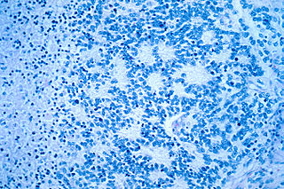
In histopathology, a palisade is a single layer of relatively long cells, arranged loosely perpendicular to a surface and parallel to each other. A rosette is a palisade in a halo or spoke-and-wheel arrangement, surrounding a central core or hub. A pseudorosette is a perivascular radial arrangement of neoplastic cells around a small blood vessel.

Embryonal tumor with multilayered rosettes (ETMR) is an embryonal central nervous system tumor. It is considered an embryonal tumor because it arises from cells partially differentiated or still undifferentiated from birth, usually neuroepithelial cells, stem cells destined to turn into glia or neurons. It can occur anywhere within the brain and can have multiple sites of origins, with a high probability of metastasis through cerebrospinal fluid (CSF). Metastases outside the central nervous system have been reported, but remain rare.

References

  1. 1 2 3 4 5 6 7 8 9 10 11 12 13 14 15 16 17 18 19 20 21 22 23 24 Karajannis, Matthias A.; Zagzag, David, eds. (2015). Molecular Pathology of Nervous System Tumors. Molecular Pathology Library. Vol. 8. doi:10.1007/978-1-4939-1830-0. ISBN   978-1-4939-1829-4. ISSN   1935-987X.
  2. 1 2 3 4 5 6 7 8 9 10 11 12 13 14 15 16 17 18 19 20 21 22 23 24 25 26 27 28 29 30 31 32 33 34 35 36 37 38 39 40 41 42 43 44 45 Hayat, M.A., ed. (2014). Tumors of the Central Nervous System, Volume 13. Vol. 13. doi:10.1007/978-94-007-7602-9. ISBN   978-94-007-7601-2. ISSN   2215-096X.
  3. 1 2 3 4 5 6 7 8 Fuller, Christine E. (2009-10-23), "Oligodendroglial Tumors", Atlas of Pediatric Brain Tumors, Springer New York, pp. 39–46, doi:10.1007/978-1-4419-1062-2_4, ISBN   9781441910615
  4. Nelesen, Richard A (March 2000). "Biological Psychology: An Introduction to Behavioral, Cognitive, and Clinical Neuroscience, 2nd edition. Mark R. Rosenweig, Arnold L. Leiman, and S. Marc Breedlove, Sinauer Associates, Inc., Sunderland MA, 1999. 561+92 pp. ISBN 0-87893-791-9". Biological Psychology. 52 (2): 185–186. doi:10.1016/s0301-0511(99)00025-3. ISSN   0301-0511. S2CID   54349873.
  5. Louis DN, Ohgaki H, Wiestler OD, Cavenee WK "WHO Classification of Tumours of the Central Nervous System. 4th Edition Revised"
  6. 1 2 Baker, Henry V (June 2003). "Essential Genetics: A Genomics Perspective . Third Edition. By Daniel L Hartl and , Elizabeth W Jones. Sudbury (Massachusetts): Jones and Bartlett Publishers . $78.95 (Paper). Xxvi + 613 p; ill.; index. ISBN: 0–7637–1852–1. 2002" . The Quarterly Review of Biology. 78 (2): 225–226. doi:10.1086/377959. ISSN   0033-5770.