Neuroepithelial cell

Last updated

Neuroepithelial cells, or neuroectodermal cells, form the wall of the closed neural tube in early embryonic development. The neuroepithelial cells span the thickness of the tube's wall, connecting with the pial surface and with the ventricular or lumenal surface. They are joined at the lumen of the tube by junctional complexes, where they form a pseudostratified layer of epithelium called neuroepithelium. [1]

Contents

Neuroepithelial cells are the stem cells of the central nervous system, known as neural stem cells, and generate the intermediate progenitor cells known as radial glial cells, that differentiate into neurons and glia in the process of neurogenesis. [1]

Embryonic neural development

Brain development

Development of the neural tube Development of the neural tube.png
Development of the neural tube

During the third week of embryonic growth, the brain begins to develop in the early fetus in a process called morphogenesis. [2] Neuroepithelial cells of the ectoderm begin multiplying rapidly and fold in forming the neural plate, which invaginates during the fourth week of embryonic growth and forms the neural tube. [2] The formation of the neural tube polarizes the neuroepithelial cells by orienting the apical side of the cell to face inward, which later becomes the ventricular zone, and the basal side is oriented outward, which contacts the pia, or outer surface of the developing brain. [3] As part of this polarity, neuroepithelial cells express prominin-1 in the apical plasma membrane as well as tight junctions to maintain the cell polarity. [4] Integrin alpha 6 anchors the neuroepithelial cells to the basal lamina. [4] The neural tube begins as a single layer of pseudostratified epithelial cells, but rapid proliferation of neuroepithelial cells creates additional layers and eventually three distinct regions of growth. [2] [4] As these additional layers form the apical-basal polarity must be downregulated. [3] Further proliferation of the cells in these regions gives rise to three distinct areas of the brain: the forebrain, midbrain, and hindbrain. The neural tube also gives rise to the spinal cord. [2]

Neuroepithelial cell proliferation

Neuroepithelial cells symmetrically divide or differentiate into progenitor cells called radial glial cells in asymmetric cell division. These can further differentiate into neurons or glial cells. Stem cell division and differentiation.svg
Neuroepithelial cells symmetrically divide or differentiate into progenitor cells called radial glial cells in asymmetric cell division. These can further differentiate into neurons or glial cells.

Neuroepithelial cells are a class of stem cell and have the ability to self-renew. During the formation of the neural tube, neuroepithelial cells undergo symmetric proliferative divisions that give rise to two new neuroepithelial cells. At a later stage of brain development, neuroepithelial cells begin to self renew and give rise to non-stem cell progenitors, such as radial glial cells simultaneously by undergoing asymmetric division. Expression of Tis21, an antiproliferative gene, causes the neuroepithelial cell to make the switch from proliferative division to neuronic division. Many of the neuroepithelial cells also divide into radial glial cells, a similar, but more fate restricted cell. Being a more fate restricted cell the radial glial cell will either generate postmitotic neurons, intermediate progenitor cells, or astrocytes in gliogenesis. During neuroepithelial cell division, interkinetic nuclear migration allows the cells to divide unrestricted while maintaining a dense packing. During G1 the cell nucleus migrates to the basal side of the cell and remains there for S phase and migrates to the apical side for G2 phase. This migration requires the help of microtubules and actin filaments. [4]

Radial glial cell transition

Neuroepithelial cells give rise to radial glial progenitor cells in early embryonic development. To make this change, neuroepithelial cells begin to downregulate their epithelial features, by stopping the expression of occludin, a tight junction protein. [3] Loss of occludin causes a loss of the previous tight junction seals which is required for the generation of neuroblasts. Another tight junction protein, PARD3, remains at the apical side of the cell co-localizing with N-cadherin and keeps the apical face of the neuroepithelial cell intact. [4] In the absence of occludin some polarity is still lost and the neuroepithelial cell gives rise to the radial glial cell. [4]

Adult neurogenesis

Genesis of neuroepithelial cells in the adult CNS

Moving away from the ependymal layer of the SVZ the neural cells become more and more differentiated Human subventricular zone.jpg
Moving away from the ependymal layer of the SVZ the neural cells become more and more differentiated

In the adult CNS, neuroepithelial cells arise in several different areas of the brain: the subventricular zone (SVZ), the olfactory bulb and the dentate gyrus of the hippocampus. These cells do not appear in any of the peripheral nervous system. Often categorized as neural stem cells, neuroepithelial cells give rise to only a few varieties of neural cells, making them multipotent - a definite distinction from the pluripotent stem cells found in embryonic development. Neuroepithelial cells undergo mitosis generating more neuroepithelial cells, radial glial cells or progenitor cells, the latter two differentiating into either neurons or glial cells. The neuroepithelial cells undergo two different forms of mitosis: asymmetric differentiating division and symmetric prolific division. [4] The asymmetric cell division results in two different varieties of daughter cells (i.e. a neuroepithelial cell divides into a radial glial cell and another neuroepithelial cell), while the symmetric version yields identical daughter cells. This effect is caused by the orientation of the mitotic spindle, which is located in either the posterior or anterior area of the mitotic cell, rather than the center where it is found during symmetric division. The progenitor cells and radial glial cells respond to extracellular trophic factors - like ciliary neurotrophic factor (CNTF), cytokines or neuregulin 1 (NRG1) - that can determine whether the cells will differentiate into either neurons or glia. [5] On a whole, neurogenesis is regulated both by many varied regulatory pathways in the CNS as well as several other factors, from genes to external stimuli such as the individual behavior of a person. The large interconnected web of regulatory responses acts to fine-tune the responses provided by newly formed neurons. [6]

Neurogenesis in neural repair

Neurogenesis in the adult brain is often associated with diseases that deteriorate the CNS, like Huntington's disease, Alzheimer's disease, and Parkinson's disease. While adult neurogenesis is up-regulated in the hippocampus in patients with these diseases, whether its effects are regenerative or inconclusive remains to be seen. [7] Individuals with these diseases also often express diminished olfactory abilities as well as decreased cognitive activity in the hippocampus, areas specific to neurogenesis. The genes associated with these diseases like α-synuclein, presenilin 1, MAPT (microtubule associated protein tau) and huntingtin are also often associated with plasticity in the brain and its modification. [8] Neuroplasticity is associated with neurogenesis in a complementary fashion. The new neurons generated by the neuroepithelial cells, progenitors and radial glial cells will not survive unless they are able to integrate into the system by making connections with new neighbors. This also leads to many controversial concepts, like neurogenic therapy involving the transplant of local progenitor cells to a damaged area. [7]

Associated diseases

Dysembryoplastic neuroepithelial tumor (DNT)

Dysembryoplastic neuroepithelial tumor DNET02.jpg
Dysembryoplastic neuroepithelial tumor

A dysembryoplastic neuroepithelial tumor is a rare, benign tumor that affects children and teenagers under the age of twenty. The tumor occurs in the tissue covering the brain and spinal cord. The symptoms of the tumor are dependent on its location, but most children experience seizures that cannot be controlled by medication. DNT is usually treated through invasive surgery and the patients are usually capable of recovering fully, with little to no long-term effects. [9]

Neuroepithelial cysts

Neuroepithelial cysts, also known as colloid cysts, develop in individuals between the ages of 20 and 50 and is relatively rare in individuals under the age of twenty. The cysts are benign tumors that usually appear in the anterior third ventricle. The cysts occur in the epithelium putting their patients at risk for obstructive hydrocephalus, increased intracranial pressure, and rarely intracystic hemorrhage. This results from the cysts enlarging by causing the epithelium to secrete additional mucinous fluid. The cysts are usually found incidentally or if patients become symptomatic presenting with the symptoms of hydrocephalus. The larger cysts are operated on while smaller cysts that are not obstructive can be left alone. [10]

Oligodendroglial tumors

Oligodendroglial tumors manifest in glial cells, which are responsible for supporting and protecting nerve cells in the brain. The tumor develops over oligodendrocytes and is usually found in the cerebrum around the frontal or temporal lobes. The tumors can either grow slowly in a well-differentiated manner delaying the onset of symptoms, or they can grow rapidly to form an anaplastic oligodendroglioma. The symptoms for this type of tumor include headaches and visual problems. Additionally, blockage of ventricles could cause buildup of cerebral spinal fluid resulting in swelling around the tumor. The location of the tumor may also affect the symptoms since frontal lobe tumors can cause gradual mood or personality changes while temporal lobe tumors result in coordination and speech problems. [11]

Ongoing research

Neural chimeras

Researchers have been able to create neural chimeras by combining neurons that developed from embryonic stem cells with glial cells that were also derived from embryonic stem cells. These neural chimeras give researchers a comprehensive way of studying the molecular mechanisms behind cell repair and regeneration via neuroepithelial precursor cells and will hopefully shed light on possible nervous system repair in a clinical setting. In an attempt to identify the key features that differentiate neuroepithelial cells from their progenitor cells, researchers identified an intermediate filament that was expressed by 98% of the neuroepithelial cells of the neural tube, but none of their progenitor cells. After this discovery it became clear that all three cell types in the nervous system resulted from a homogenous population of stem cells. In order make clinical neural repair possible researchers needed to further characterize regional determination of stem cells during brain development by determining what factors commit a precursor to becoming one or the other. While the exact factors that lead to differentiation are unknown, researchers have taken advantage of human-rat neural chimeras to explore the development of human neurons and glial cells in an animal model. These neural chimeras have permitted researchers to look at neurological diseases in an animal model where traumatic and reactive changes can be controlled. Eventually researchers hope to be able to use the information taken from these neural chimera experiments to repair regions of the brain affected by central nervous system disorders. The problem of delivery, however, has still not been resolved as neural chimeras have been shown to circulate throughout the ventricles and incorporate into all parts of the CNS. By finding environmental cues of differentiation, neuroepithelial precursor transplantation could be used in the treatment of many diseases including multiple sclerosis, Huntington's disease, and Parkinson's disease. Further exploration of neural chimera cells and chimeric brains will provide evidence for manipulating the correct genes and increasing the efficacy of neural transplant repair. [12]

Depression

Research on depression indicates that one of the major causal factors of depression, stress, also influences neurogenesis. This connection led researches to postulate that depression could be the result of changes in levels of neurogenesis in the adult brain, specifically in the dentate gyrus. Studies indicate that stress affects neurogenesis by increasing Glucocorticoids and decreasing neurotransmitters such as serotonin. These effects were further verified by inducing stress in lab animals, which resulted in decreased levels of neurogenesis. Additionally, modern therapies that treat depression also promote neurogenesis. Ongoing research is looking to further verify this connection and define the mechanism by which it occurs. This could potentially lead to a better understanding of the development of depression as well as future methods of treatment. [13]

See also

Related Research Articles

The development of the nervous system, or neural development (neurodevelopment), refers to the processes that generate, shape, and reshape the nervous system of animals, from the earliest stages of embryonic development to adulthood. The field of neural development draws on both neuroscience and developmental biology to describe and provide insight into the cellular and molecular mechanisms by which complex nervous systems develop, from nematodes and fruit flies to mammals.

<span class="mw-page-title-main">Olfactory epithelium</span> Specialised epithelial tissue in the nasal cavity that detects odours

The olfactory epithelium is a specialized epithelial tissue inside the nasal cavity that is involved in smell. In humans, it measures 5 cm2 (0.78 sq in) and lies on the roof of the nasal cavity about 7 cm (2.8 in) above and behind the nostrils. The olfactory epithelium is the part of the olfactory system directly responsible for detecting odors.

In vertebrates, a neuroblast or primitive nerve cell is a postmitotic cell that does not divide further, and which will develop into a neuron after a migration phase. In invertebrates such as Drosophila, neuroblasts are neural progenitor cells which divide asymmetrically to produce a neuroblast, and a daughter cell of varying potency depending on the type of neuroblast. Vertebrate neuroblasts differentiate from radial glial cells and are committed to becoming neurons. Neural stem cells, which only divide symmetrically to produce more neural stem cells, transition gradually into radial glial cells. Radial glial cells, also called radial glial progenitor cells, divide asymmetrically to produce a neuroblast and another radial glial cell that will re-enter the cell cycle.

<span class="mw-page-title-main">Rostral migratory stream</span> One path neural stem cells take to reach the olfactory bulb


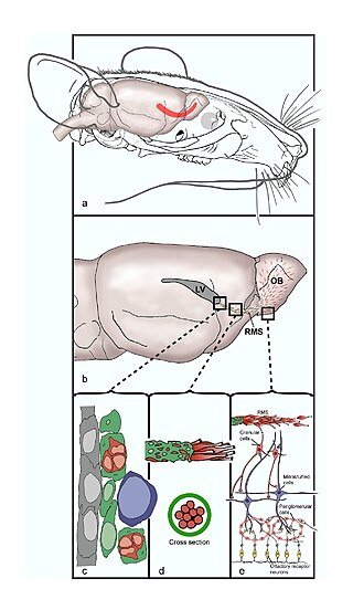
The rostral migratory stream (RMS) is a specialized migratory route found in the brain of some animals along which neuronal precursors that originated in the subventricular zone (SVZ) of the brain migrate to reach the main olfactory bulb (OB). The importance of the RMS lies in its ability to refine and even change an animal's sensitivity to smells, which explains its importance and larger size in the rodent brain as compared to the human brain, as our olfactory sense is not as developed. This pathway has been studied in the rodent, rabbit, and both the squirrel monkey and rhesus monkey. When the neurons reach the OB they differentiate into GABAergic interneurons as they are integrated into either the granule cell layer or periglomerular layer.

Neural stem cells (NSCs) are self-renewing, multipotent cells that firstly generate the radial glial progenitor cells that generate the neurons and glia of the nervous system of all animals during embryonic development. Some neural progenitor stem cells persist in highly restricted regions in the adult vertebrate brain and continue to produce neurons throughout life. Differences in the size of the central nervous system are among the most important distinctions between the species and thus mutations in the genes that regulate the size of the neural stem cell compartment are among the most important drivers of vertebrate evolution.

<span class="mw-page-title-main">Radial glial cell</span> Bipolar-shaped progenitor cells of all neurons in the cerebral cortex and some glia

Radial glial cells, or radial glial progenitor cells (RGPs), are bipolar-shaped progenitor cells that are responsible for producing all of the neurons in the cerebral cortex. RGPs also produce certain lineages of glia, including astrocytes and oligodendrocytes. Their cell bodies (somata) reside in the embryonic ventricular zone, which lies next to the developing ventricular system.

Neuropoiesis is the process by which neural stem cells differentiate to form mature neurons, astrocytes, and oligodendrocytes in the adult mammal. This process is also referred to as adult neurogenesis.

<span class="mw-page-title-main">Subventricular zone</span> Region outside each lateral ventricle of the brain

The subventricular zone (SVZ) is a region situated on the outside wall of each lateral ventricle of the vertebrate brain. It is present in both the embryonic and adult brain. In embryonic life, the SVZ refers to a secondary proliferative zone containing neural progenitor cells, which divide to produce neurons in the process of neurogenesis. The primary neural stem cells of the brain and spinal cord, termed radial glial cells, instead reside in the ventricular zone (VZ).

<span class="mw-page-title-main">Subgranular zone</span>

The subgranular zone (SGZ) is a brain region in the hippocampus where adult neurogenesis occurs. The other major site of adult neurogenesis is the subventricular zone (SVZ) in the brain.

Neurogenins, often abbreviated as Ngn, are a family of bHLH transcription factors involved in specifying neuronal differentiation. The family consisting of Neurogenin-1, Neurogenin-2, and Neurogenin-3, plays a fundamental role in specifying neural precursor cells and regulating the differentiation of neurons during embryonic development. It is one of many gene families related to the atonal gene in Drosophila. Other positive regulators of neuronal differentiation also expressed during early neural development include NeuroD and ASCL1.

<span class="mw-page-title-main">Ganglion mother cell</span>

Ganglion mother cells (GMCs) are cells involved in neurogenesis, in non-mammals, that divide only once to give rise to two neurons, or one neuron and one glial cell or two glial cells, and are present only in the central nervous system. They are also responsible for transcription factor expression. While each ganglion mother cell necessarily gives rise to two neurons, a neuroblast can asymmetrically divide multiple times. GMCs are the progeny of type I neuroblasts. Neuroblasts asymmetrically divide during embryogenesis to create GMCs. GMCs are only present in certain species and only during the embryonic and larval stages of life. Recent research has shown that there is an intermediate stage between a GMC and two neurons. The GMC forms two ganglion cells which then develop into neurons or glial cells. Embryonic neurogenesis has been extensively studied in Drosophila melanogaster embryos and larvae.

Gliogenesis is the generation of non-neuronal glia populations derived from multipotent neural stem cells.

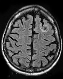
Endogenous regeneration in the brain is the ability of cells to engage in the repair and regeneration process. While the brain has a limited capacity for regeneration, endogenous neural stem cells, as well as numerous pro-regenerative molecules, can participate in replacing and repairing damaged or diseased neurons and glial cells. Another benefit that can be achieved by using endogenous regeneration could be avoiding an immune response from the host.

The development of the cerebral cortex, known as corticogenesis is the process during which the cerebral cortex of the brain is formed as part of the development of the nervous system of mammals including its development in humans. The cortex is the outer layer of the brain and is composed of up to six layers. Neurons formed in the ventricular zone migrate to their final locations in one of the six layers of the cortex. The process occurs from embryonic day 10 to 17 in mice and between gestational weeks seven to 18 in humans.

Epigenetic regulation of neurogenesis is the role that epigenetics plays in the regulation of neurogenesis.

<span class="mw-page-title-main">Neuronal lineage marker</span> Endogenous tag expressed in different cells along neurogenesis and differentiated cells

A neuronal lineage marker is an endogenous tag that is expressed in different cells along neurogenesis and differentiated cells such as neurons. It allows detection and identification of cells by using different techniques. A neuronal lineage marker can be either DNA, mRNA or RNA expressed in a cell of interest. It can also be a protein tag, as a partial protein, a protein or an epitope that discriminates between different cell types or different states of a common cell. An ideal marker is specific to a given cell type in normal conditions and/or during injury. Cell markers are very valuable tools for examining the function of cells in normal conditions as well as during disease. The discovery of various proteins specific to certain cells led to the production of cell-type-specific antibodies that have been used to identify cells.

<span class="mw-page-title-main">Ventricular zone</span> Transient embryonic layer of tissue containing neural stem cells

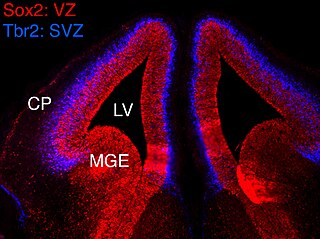
In vertebrates, the ventricular zone (VZ) is a transient embryonic layer of tissue containing neural stem cells, principally radial glial cells, of the central nervous system (CNS). The VZ is so named because it lines the ventricular system, which contains cerebrospinal fluid (CSF). The embryonic ventricular system contains growth factors and other nutrients needed for the proper function of neural stem cells. Neurogenesis, or the generation of neurons, occurs in the VZ during embryonic and fetal development as a function of the Notch pathway, and the newborn neurons must migrate substantial distances to their final destination in the developing brain or spinal cord where they will establish neural circuits. A secondary proliferative zone, the subventricular zone (SVZ), lies adjacent to the VZ. In the embryonic cerebral cortex, the SVZ contains intermediate neuronal progenitors that continue to divide into post-mitotic neurons. Through the process of neurogenesis, the parent neural stem cell pool is depleted and the VZ disappears. The balance between the rates of stem cell proliferation and neurogenesis changes during development, and species from mouse to human show large differences in the number of cell cycles, cell cycle length, and other parameters, which is thought to give rise to the large diversity in brain size and structure.

Neurogenesis is the process by which nervous system cells, the neurons, are produced by neural stem cells (NSCs). In short, it is brain growth in relation to its organization. This occurs in all species of animals except the porifera (sponges) and placozoans. Types of NSCs include neuroepithelial cells (NECs), radial glial cells (RGCs), basal progenitors (BPs), intermediate neuronal precursors (INPs), subventricular zone astrocytes, and subgranular zone radial astrocytes, among others.

<span class="mw-page-title-main">Brain vesicle</span>

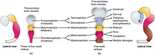
Brain vesicles are the bulge-like enlargements of the early development of the neural tube in vertebrates, which eventually give rise to the brain.

Intermediate progenitor cells (IPCs) are a type of progenitor cell in the developing cerebral cortex. They are multipolar cells produced by radial glial cells who have undergone asymmetric division. IPCs can produce neuron cells via neurogenesis and are responsible for ensuring the proper quantity of cortical neurons are produced. In mammals, neural stem cells are the primary progenitors during embryogenesis whereas intermediate progenitor cells are the secondary progenitors.

References

  1. 1 2 Sadler, T (2006). Langman's medical embryology (11th. ed.). Lippincott William & Wilkins. pp. 295–299. ISBN   9780781790697.
  2. 1 2 3 4 McDonald, A. (2007). Prenatal Development - The Dana Guide. The Dana Foundation. ISBN   978-1-932594-10-2 . Retrieved 7 December 2011.
  3. 1 2 3 Zolessi, F. R. (2009). "Vertebrate Neurogenesis: Cell Polarity". Encyclopedia of Life Sciences. doi:10.1002/9780470015902.a0000826.pub2. ISBN   978-0470016176.
  4. 1 2 3 4 5 6 7 Götz, M.; Huttner, W. B. (2005). "The cell biology of neurogenesis". Nature Reviews Molecular Cell Biology. 6 (10): 777–788. doi:10.1038/nrm1739. PMID   16314867. S2CID   16955231.
  5. Clarke, D. L. (2003). "Neural stem cells". Bone Marrow Transplantation. 32: S13–S17. doi:10.1038/sj.bmt.1703937. PMID   12931233.
  6. Kempermann, G. (2011). "Seven principles in the regulation of adult neurogenesis". European Journal of Neuroscience. 33 (6): 1018–1024. doi:10.1111/j.1460-9568.2011.07599.x. PMID   21395844. S2CID   14149058.
  7. 1 2 Taupin, P. (2008). "Adult neurogenesis, neuroinflammation and therapeutic potential of adult neural stem cells". International Journal of Medical Sciences. 5 (3): 127–132. doi:10.7150/ijms.5.127. PMC   2424180 . PMID   18566676.
  8. Winner, Beate; Zacharias Kohl; Fred H. Gage (2011). "Neurodegenerative disease and adult neurogenesis" (PDF). European Journal of Neuroscience. 33 (6): 1139–1151. doi:10.1111/j.1460-9568.2011.07613.x. PMID   21395858. S2CID   6610255 . Retrieved 2011-11-28.
  9. "Dysembryoplastic Neuroepithelial Tumor". Children's Hospital Boston. Archived from the original on 26 September 2011. Retrieved 1 November 2011.
  10. Chin, L. S.; Jayarao, M. "Colloid Cysts". Medscape. Retrieved 7 December 2011.
  11. "Oligodendroglioma". Macmillan. Retrieved 7 December 2011.
  12. Brüstle, O. (1999). "Building brains: Neural chimeras in the study of nervous system development and repair". Brain Pathology. Zurich, Switzerland. 9 (3): 527–545. doi:10.1111/j.1750-3639.1999.tb00540.x. PMC   8098370 . PMID   10416992. S2CID   14847541.
  13. Jacobs, B. L.; Praag, H.; Gage, F. H. (May 2000). "Adult brain neurogenesis and psychiatry: a novel theory of depression". Molecular Psychiatry. 5 (3): 262–269. doi: 10.1038/sj.mp.4000712 . PMID   10889528. S2CID   24913141.