Neural fold

Last updated
Neural fold
Gray18.png
Chick embryo of thirty-three hours’ incubation, viewed from the dorsal aspect. 30x. (Neural fold labeled at center left, third from the bottom.)
Details
Carnegie stage 9
Precursor neural plate
Gives rise to neural tube
Identifiers
Latin plica neuralis
TE fold_by_E5.13.1.0.1.0.2 E5.13.1.0.1.0.2
Anatomical terminology

The neural fold is a structure that arises during neurulation in the embryonic development of both birds and mammals among other organisms. [1] [2] This structure is associated with primary neurulation, meaning that it forms by the coming together of tissue layers, rather than a clustering, and subsequent hollowing out, of individual cells (known as secondary neurulation). In humans, the neural folds are responsible for the formation of the anterior end of the neural tube. The neural folds are derived from the neural plate, a preliminary structure consisting of elongated ectoderm cells. The folds give rise to neural crest cells, as well as bringing about the formation of the neural tube. [1] [3]

Contents

Development

In the embryo, the formation of the neural folds originates from the area where the neural plate and the surrounding ectoderm converge. This region of the embryo is formed after gastrulation, and consists of epithelial tissue. Here, the epithelial cells elongate by means of microtubule polymerization, increasing their height. The thumbnail below shows this process, as well as the subsequent formation of the neural crest cells and the neural tube, which arise from the joining of the neural folds. [4]

Folding

A strip of specialized cells called the notochord (A) induces the cells of the ectoderm directly above it to become the primitive nervous system (i.e., neuroepithelium). The neuroepithelium then folds over (B). As the tips of the folds fuse together, a hollow tube (the neural tube) forms (C)--the precursor of the brain and spinal cord. Meanwhile, the ectoderm and endoderm continue to curve around and fuse to create the body cavity, completing the transformation of the embryo from a flattened disk to a three-dimensional body. Cells originating from the fused tips of the neuroectoderm (neural crest cells) migrate to various locations throughout the embryo, where they will initiate the development of diverse body structures (D). Embryonic Development CNS.png
A strip of specialized cells called the notochord (A) induces the cells of the ectoderm directly above it to become the primitive nervous system (i.e., neuroepithelium). The neuroepithelium then folds over (B). As the tips of the folds fuse together, a hollow tube (the neural tube) forms (C)—the precursor of the brain and spinal cord. Meanwhile, the ectoderm and endoderm continue to curve around and fuse to create the body cavity, completing the transformation of the embryo from a flattened disk to a three–dimensional body. Cells originating from the fused tips of the neuroectoderm (neural crest cells) migrate to various locations throughout the embryo, where they will initiate the development of diverse body structures (D).

The formation of the neural fold is initiated by the release of calcium from within the cells. The released calcium interacts with proteins that can modify the actin filaments in the outer epithelial tissue, or ectoderm, in order to induce the dynamic cell movements necessary to create the fold. [6] These cells are held together by cadherins (specifically E and N-cadherin), types of intercellular binding protein. When the cells at the peaks of the neural folds come in proximity with each other, it is the affinity for similar cadherin molecules (N-cadherins) that allows these cells to bind to each other. Thus, when the neural tube precursor cells begin expressing N-cadherin in the place of E-cadherin, this causes the neural tube to form and separate from the ectoderm and settle inside the embryo. [1] When the cells fail to associate in a manner that is not part of the normal course of development, severe diseases can occur.

Process overview

The process of folding begins when the cells in the central region of the neural plate, the medial hinge point cells, bind to the notochord beneath them. This creates a central anchoring point for the process of folding to occur, and subsequently creates the neural groove. As the neural folds continue to extend, dorsolateral hinge points form, allowing the folds to curve into a tube-like structure. When the peaks of the folds (known as the neural crest regions) touch, they merge and involute, creating the neural tube beneath the newly formed epidermal layer. [7]

Mechanism

Cross section through the embryonic disc showing the fold. Gray16.png
Cross section through the embryonic disc showing the fold.

The molecular mechanism behind this process lies in the expression and repression of bone morphogenetic proteins (BMPs). BMPs are a wide family of proteins that perform many functions throughout the growing embryo, including stimulating the growth of cartilage and bone. In order to allow for the growth of precursor neural tissues, as opposed to precursor bone or cartilage tissues, BMP expression is decreased in the neural plate, specifically along the medial line, where the neural groove will soon form. The proteins produced from the genes Noggin and Chordin inhibit these BMPs, and subsequently allow neural commitment genes, like SOX , to be expressed. These genes encode transcription factors, which alter the genomic expression of these cells, furthering them along the path of neural cell commitment. [8] This process of BMP inhibition allows for the anchoring of the medial hinge point cells, providing the neural folds with the foundation necessary for folding and closure to occur. Noggin and Chordin have other roles in the neurulation process, including stimulating the neural crest cells to emigrate from the newly formed neural tube. [9] [10] The Sonic hedgehog gene also plays a role in attenuating BMP expression, forming the medial hinge point while inhibiting the formation of the dorsolateral hinge points, and in ensuring the proper closure of the neural folds. [11] The prechordal plate, notochord, and non-neural ectoderm are believed to be important inducer tissues that release these chemical signals, in order to trigger neural plate folding. [8]

The final adhesion of the converging neural folds is due to several different types of intercellular binding proteins. Cadherins and their CAM receptor molecules, for example, are present in two types in the neural precursor tissue: E-cadherin keeps the cells of the neural plate and surrounding ectoderm adhered to each other, while N-cadherin does the same for the cells of the neural fold. Only cells expressing the same kind of cadherin can bind to each other; since the peaks of the neural folds both express N-cadherin, they are able to merge into a continuous sheet of cells. Likewise, it is this diminished affinity between cells expressing different types of cadherin that allows the neural tube precursor cells to separate from the ectoderm, forming the neural tube on the interior of the embryo and the true epidermis on the exterior. [1] Another set of molecules involved with the merging of the neural folds are the ephrin molecules and their Eph receptors, which adhere in a similar manner to the cadherin molecules discussed above. [8]

Derivative structures

The merging of the neural folds gives rise to many structures including the neural tube (the precursor to the central nervous system), neural crest cells(which give rise to a variety of diverse mesenchymal cells), and to the true epidermal layer. [1] The neural fold is an extremely important structure in that this mechanism is needed to produce these diverse kinds of cells in the right places.

Clinical significance

A side view of an anencephalic fetus Anencephaly side.jpg
A side view of an anencephalic fetus

There are many potential diseases that can arise from the improper adhesion or merging of the neural folds. During folding, the openings that are formed at the cranial and caudal regions are termed the cranial and caudal neuropores. [12] If the caudal neuropore fails to close, a condition called spina bifida can occur, in which the bottom of the spinal cord remains exposed. Often this condition can be detected during prenatal examinations and be treated before birth, though in more severe cases the individual may cope with the condition for the rest of his or her life. [13] Depending on the severity and the affected area, individuals can experience a variety of symptoms, including a varying motor function and mobility, bladder control, and/or sexual function. [14]

If the failure is instead in the cranial neuropore, anencephaly occurs. In this condition, the brain tissue is directly exposed to the amniotic fluid, and is subsequently degraded. [15] If the entire neural tube fails to close, the condition is referred to as craniorachischisis.

See also

Related Research Articles

Ontogeny Origination and development of an organism

Ontogeny is the origination and development of an organism, usually from the time of fertilization of the egg to adult. The term can also be used to refer to the study of the entirety of an organism's lifespan.

The development of the nervous system, or neural development (neurodevelopment), refers to the processes that generate, shape, and reshape the nervous system of animals, from the earliest stages of embryonic development to adulthood. The field of neural development draws on both neuroscience and developmental biology to describe and provide insight into the cellular and molecular mechanisms by which complex nervous systems develop, from nematodes and fruit flies to mammals.

Neural tube Developmental precursor to the central nervous system

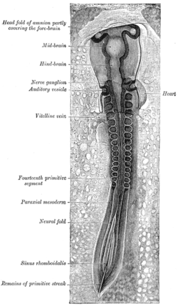
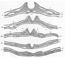
In the developing chordate, the neural tube is the embryonic precursor to the central nervous system, which is made up of the brain and spinal cord. The neural groove gradually deepens as the neural fold become elevated, and ultimately the folds meet and coalesce in the middle line and convert the groove into the closed neural tube. In humans, neural tube closure usually occurs by the fourth week of pregnancy. The ectodermal wall of the tube forms the rudiment of the nervous system. The centre of the tube is the neural canal.

Ectoderm Outer germ layer that forms the neurons of brain, skin, and more

The ectoderm is one of the three primary germ layers formed in early embryonic development. It is the outermost layer, and is superficial to the mesoderm and endoderm. It emerges and originates from the outer layer of germ cells. The word ectoderm comes from the Greek ektos meaning "outside", and derma meaning "skin".

Neurulation Embryological process forming the neural tube

Neurulation refers to the folding process in vertebrate embryos, which includes the transformation of the neural plate into the neural tube. The embryo at this stage is termed the neurula.

Somite Each of several blocks of mesoderm that flank the neural tube on either side in embryogenesis

The somites are a set of bilaterally paired blocks of paraxial mesoderm that form in the embryonic stage of somitogenesis, along the head-to-tail axis in segmented animals. In vertebrates, somites subdivide into the sclerotomes, myotomes, syndetomes and dermatomes that give rise to the vertebrae of the vertebral column, rib cage and part of the occipital bone; skeletal muscle, cartilage, tendons, and skin.

A germ layer is a primary layer of cells that forms during embryonic development. The three germ layers in vertebrates are particularly pronounced; however, all eumetazoans produce two or three primary germ layers. Some animals, like cnidarians, produce two germ layers making them diploblastic. Other animals such as bilaterians produce a third layer between these two layers, making them triploblastic. Germ layers eventually give rise to all of an animal’s tissues and organs through the process of organogenesis.

Organogenesis is the phase of embryonic development that starts at the end of gastrulation and continues until birth. During organogenesis, the three germ layers formed from gastrulation form the internal organs of the organism.

Neural plate

The neural plate is a key developmental structure that serves as the basis for the nervous system. Cranial to the primitive node of the embryonic primitive streak, ectodermal tissue thickens and flattens to become the neural plate. The region anterior to the primitive node can be generally referred to as the neural plate. Cells take on a columnar appearance in the process as they continue to lengthen and narrow. The ends of the neural plate, known as the neural folds, push the ends of the plate up and together, folding into the neural tube, a structure critical to brain and spinal cord development. This process as a whole is termed primary neurulation.

Neurula Embryo at the early stage of development in which neurulation occurs

A neurula is a vertebrate embryo at the early stage of development in which neurulation occurs. The neurula stage is preceded by the gastrula stage; consequentially, neurulation is preceded by gastrulation. Neurulation marks the beginning of the process of organogenesis.

Neural crest They are pluripotent embyronic group of cells giving rise to diverse cell lineages

Neural crest cells are a temporary group of cells unique to vertebrates that arise from the embryonic ectoderm germ layer, and in turn give rise to a diverse cell lineage—including melanocytes, craniofacial cartilage and bone, smooth muscle, peripheral and enteric neurons and glia.

The primitive node is the organizer for gastrulation in most amniote embryos. In birds it is known as Hensen's node, and in amphibians it is known as the Spemann-Mangold organizer. It is induced by the Nieuwkoop center in amphibians, or by the posterior marginal zone in amniotes including birds.

Neural groove Shallow median groove of the neural plate between the neural folds of an embryo.

The neural groove is a shallow median groove of the neural plate between the neural folds of an embryo. The neural plate is a thick sheet of ectoderm surrounded on either side by the neural folds, two longitudinal ridges in front of the primitive streak of the developing embryo.

Neuroectoderm Ectoderm that goes on to form the neural plate

Neuroectoderm consists of cells derived from ectoderm. Formation of the neuroectoderm is first step in the development of the nervous system. The neuroectoderm receives bone morphogenetic protein-inhibiting signals from proteins such as noggin, which leads to the development of the nervous system from this tissue. Histologically, these cells are classified as pseudostratified columnar cells.

Mesenchyme Type of animal embryonic connective tissue

Mesenchyme is a type of loosely organised animal embryonic connective tissue of undifferentiated cells that gives rise to blood and lymph vessels, bone, and muscle.

Eye development Formation of the eye during embryonic development

Eye formation in the human embryo begins at approximately three weeks into embryonic development and continues through the tenth week. Cells from both the mesodermal and the ectodermal tissues contribute to the formation of the eye. Specifically, the eye is derived from the neuroepithelium, surface ectoderm, and the extracellular mesenchyme which consists of both the neural crest and mesoderm.

Fish development

The development of fishes is unique in some specific aspects compared to the development of other animals.

Human embryonic development Development and formation of the human embryo

Human embryonic development, or human embryogenesis, is the development and formation of the human embryo. It is characterised by the processes of cell division and cellular differentiation of the embryo that occurs during the early stages of development. In biological terms, the development of the human body entails growth from a one-celled zygote to an adult human being. Fertilisation occurs when the sperm cell successfully enters and fuses with an egg cell (ovum). The genetic material of the sperm and egg then combine to form a single cell called a zygote and the germinal stage of development commences. Embryonic development in the human, covers the first eight weeks of development; at the beginning of the ninth week the embryo is termed a fetus. Human embryology is the study of this development during the first eight weeks after fertilisation. The normal period of gestation (pregnancy) is about nine months or 40 weeks.

The development of the nervous system in humans, or neural development or neurodevelopment involves the studies of embryology, developmental biology, and neuroscience to describe the cellular and molecular mechanisms by which the complex nervous system forms in humans, develops during prenatal development, and continues to develop postnatally.

Neural crest cells are multipotent cells required for the development of cells, tissues and organ systems. A subpopulation of neural crest cells are the cardiac neural crest complex. This complex refers to the cells found amongst the midotic placode and somite 3 destined to undergo epithelial-mesenchymal transformation and migration to the heart via pharyngeal arches 3, 4 and 6.

References

  1. 1 2 3 4 5 Gilbert, Scott F. (2010). Developmental biology (9th ed.). Sunderland, Mass.: Sinauer Associates. ISBN   978-0878933846.[ page needed ]
  2. Colas JF, Schoenwolf GC (June 2001). "Towards a cellular and molecular understanding of neurulation". Developmental Dynamics. 221 (2): 117–45. doi:10.1002/dvdy.1144. PMID   11376482. S2CID   43666547.
  3. Yamaguchi Y, Miura M (September 2013). "How to form and close the brain: insight into the mechanism of cranial neural tube closure in mammals". Cellular and Molecular Life Sciences. 70 (17): 3171–86. doi:10.1007/s00018-012-1227-7. PMC   3742426 . PMID   23242429.
  4. Lawson A, Anderson H, Schoenwolf GC (February 2001). "Cellular mechanisms of neural fold formation and morphogenesis in the chick embryo". The Anatomical Record. 262 (2): 153–68. doi: 10.1002/1097-0185(20010201)262:2<153::AID-AR1021>3.0.CO;2-W . PMID   11169910.
  5. "File:Embryonic Development CNS.gif". Wikimedia Commons. 2012-04-04. Retrieved 1 April 2013.
  6. Ferreira MC, Hilfer SR (October 1993). "Calcium regulation of neural fold formation: visualization of the actin cytoskeleton in living chick embryos". Developmental Biology. 159 (2): 427–40. doi:10.1006/dbio.1993.1253. PMID   8405669.
  7. Rocky S. Tuan; Cecilia W. Lo, eds. (2000). "15". Developmental biology protocols, Volume 136. Humama. pp.  125–134. ISBN   9781592590650 . Retrieved 1 April 2013.
  8. 1 2 3 Khong, Hrsg. Jean W. Keeling; Hrsg. T. Yee (2007). Fetal and Neonatal Pathology (4th ed.). Godalming: Springer London. pp. 702–704. ISBN   978-1846285240.
  9. Anderson RM, Stottmann RW, Choi M, Klingensmith J (September 2006). "Endogenous bone morphogenetic protein antagonists regulate mammalian neural crest generation and survival". Developmental Dynamics. 235 (9): 2507–20. doi:10.1002/dvdy.20891. PMC   6626635 . PMID   16894609.
  10. Stottmann RW, Berrong M, Matta K, Choi M, Klingensmith J (July 2006). "The BMP antagonist Noggin promotes cranial and spinal neurulation by distinct mechanisms". Developmental Biology. 295 (2): 647–63. doi:10.1016/j.ydbio.2006.03.051. PMC   3001110 . PMID   16712836.
  11. Kirillova I, Novikova I, Augé J, et al. (May 2000). "Expression of the sonic hedgehog gene in human embryos with neural tube defects". Teratology. 61 (5): 347–54. doi:10.1002/(SICI)1096-9926(200005)61:5<347::AID-TERA6>3.0.CO;2-#. PMID   10777830.
  12. Gilbert, SF (2000). "12: Formation of the Neural Tube". Developmental Biology (6th ed.). Sunderland, MA: Sinauer Associates. ISBN   978-0-87893-243-6 . Retrieved 30 November 2011.
  13. "Spina Bifida" . Retrieved 1 April 2013.
  14. "SB and the Spine". Learn about Spina Bifida. Archived from the original on 23 April 2013. Retrieved 1 April 2013.
  15. "7.2: The trilaminar germ disk (3rd week)". Human Embryology: Embryogenesis. Retrieved 22 March 2013.