Cervical artery dissection

Last updated
Cervical artery dissection
Vertebral artery.png
Arteries of the neck.
Types Carotid artery dissection, vertebral artery dissection [1]

Cervical artery dissection is dissection of one of the layers that compose the carotid and vertebral artery in the neck (cervix). [1] [2] They include: [1]

Contents

Cervical dissections can be broadly classified as either "spontaneous" or traumatic. Cervical artery dissections are a significant cause of strokes in young adults. [5]

A dissection typically results in a tear in one of the layers of the arterial wall. [2] The result of this tear is often an intramural hematoma and/or aneurysmal dilation [2] [6] in the arteries leading to the intracranial area. [2] [7]

Signs and symptoms of a cervical artery dissection are often non-specific and can be localized or generalized. [1] [2] There is no specific treatment, although most patients are either given an anti-platelet or anti-coagulation agent to prevent or treat strokes. [2]

Epidemiology

Cervical artery dissection has been noted to be a common cause of young adult strokes, with some sources indicating a prevalence of up to 20% in this young adult population with annual incidence rates between 2.6 and 2.9 per 100,000, although these incidences may be misleading with true incidences being higher because clinical presentations can vary, many being minor or self-limited, and thus these dissections can go undiagnosed. [5] In population-based studies, the peak age of presentation is approximately 45 years with a slight gender predisposition towards males (53-57%). [8] [9]

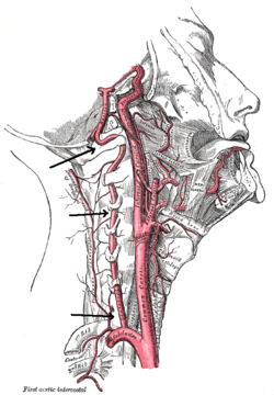
Cervical arteries, as mentioned above, consist of two pairs of arteries: vertebral and carotid. As such, cervical artery dissection can be further categorized based on the involvement of artery: carotid vs. vertebral, and the location of the dissection: intracranial vs. extracranial. [10] [11]

Causes

The two main causes of cervical artery dissection can be broadly categorized as either spontaneous or traumatic. [2] Arterial walls are composed of three layers: an intima (the innermost layer), media (the middle muscular layer), and adventitia (the outermost layer). [12] A tear in one of the layers of the arterial walls can result in blood collecting within a pocket between the layers of the artery, which can result in an intramural hematoma and/or aneurysmal dilation. [2] [6] [10] At the moment, there is no definitive location for the dissection, with some believing dissection initially occurs within the connective tissue and vasa vasorum of the media while others believe dissection begins with an intimal tear. [10] [13]

Spontaneous

Spontaneous cervical artery dissections are dissections that occur without any trauma to the neck. [2] More specifically, this can include potential causes such as hereditary connective tissue diseases, family history of strokes, infections and other miscellaneous causes such as smoking, hypertension, migraines and contraceptive use. [10] [14] Hereditary connective tissue diseases include autosomal polycystic kidney disease, Ehlers-Danlos syndrome, Marfan syndrome, fibromuscular dysplasia, and osteogenesis imperfecta type 1 although studies have shown that the link between hereditary connective tissue diseases and cervical artery dissection is low, ranging from 0-0.6% in one study and 5-18% in another study. [15]

Traumatic

Traumatic cervical artery dissections are dissections that occur after any sort of trauma to the neck, ranging from minor trauma such as nose-blowing or sneezing to severe trauma such as roadside accidents. [2] [10] Most traumatic dissections are secondary to some form of rapid decelerations injuries resulting in hyperextension or hyperflexion with rotational injury of the neck. [6]

Pathophysiology

Cervical artery dissections begin initially with a small tear in the innermost layer of the arterial wall, the tunica intima, or rupture of the vasa vasorum with bleeding within the media.[ citation needed ] As the arterial wall begins to tear, blood begins to enter this newly formed false lumen and the resulting hematoma formation can either narrow (stenosis) or occlude the artery, decreasing or completely blocking blood flow through the artery. A complete occlusion of the artery can result in cerebral ischemia as the brain is depleted of oxygen-rich blood. [16] Because the brain has a pair of carotid and vertebral arteries on each side of the neck, a unilateral occlusion can be asymptomatic, as the bilateral circulation continues perfusing the brain. [17] Thrombus formation is the natural physiologic response to the vascular injury to prevent significant blood loss. [18] However, parts of the thrombus can break apart and result in emboli that can lodge themselves in distal cerebral arteries causing ischemic stroke, otherwise known as a cerebral infarction. [19] [20]

Signs and symptoms

The signs and symptoms of cervical artery dissection are often non-specific or generalized and can either develop acutely or over several days.

Imaging

Various imaging modalities can be used for the diagnosis of cervical artery dissection. The diagnosis of cervical artery dissection can be confirmed with the presence of a mural hematoma on ultrasound (US) or magnetic resonance imaging (MRI) and will also guide therapeutic decision making. [7] MRI, with a fat-suppressed T1 sequence, is often first-line imaging. [7] However, other modalities exist with helical computed topographic angiography (CTA) becoming the new gold-standard. Magnetic resonance angiography (MRA) and doppler US can also be utilized as additional non-invasive imaging techniques. [2] The purpose of these imaging techniques is often multi-functional. These imaging modalities can detect the direction and changes of blood flow within the arteries and whether the dissection has resulted in any damage to brain tissue. [2] [7]

Treatment

The primary goal of treatment in cervical artery dissection is preventing or treating a stroke. Treatment guidelines also depend on the presence of underlying connective tissue disorders, dissection secondary to trauma, and underlying medical conditions. Management is often using either antiplatelet agents (i.e. aspirin) or anti-coagulation to prevent development of thrombus. Antiplatelet drugs can be given as individual drugs, or in combination (i.e. aspirin alone, or aspiring and clopidogrel). Anticoagulation such as heparin, intravenously (IV) or injectable (shot) can be given while inpatient and followed by heparin. [2] A recent trial in 2015, the Cervical Artery Dissection in Stroke Study (CADISS), examined the efficacy of antiplatelet and anticoagulation treatment with the primary endpoint of ipsilateral stroke or death in individuals with symptomatic dissections. The CADISS trial revealed no significant difference in efficacy of antiplatelet and anticoagulant drugs in preventing strokes or death, but did note that strokes were rare in either group, and rarer than what has been reported in observational studies. [21]

Related Research Articles

<span class="mw-page-title-main">Thrombus</span> Blood clot

A thrombus, colloquially called a blood clot, is the final product of the blood coagulation step in hemostasis. There are two components to a thrombus: aggregated platelets and red blood cells that form a plug, and a mesh of cross-linked fibrin protein. The substance making up a thrombus is sometimes called cruor. A thrombus is a healthy response to injury intended to stop and prevent further bleeding, but can be harmful in thrombosis, when a clot obstructs blood flow through healthy blood vessels in the circulatory system.

<span class="mw-page-title-main">Brachiocephalic artery</span> Artery of the mediastinum

The brachiocephalic artery is an artery of the mediastinum that supplies blood to the right arm and the head and neck. It was previously known as the innominate artery, meaning unnamed artery.

<span class="mw-page-title-main">Cerebrovascular disease</span> Condition that affects the arteries that supply the brain

Cerebrovascular disease includes a variety of medical conditions that affect the blood vessels of the brain and the cerebral circulation. Arteries supplying oxygen and nutrients to the brain are often damaged or deformed in these disorders. The most common presentation of cerebrovascular disease is an ischemic stroke or mini-stroke and sometimes a hemorrhagic stroke. Hypertension is the most important contributing risk factor for stroke and cerebrovascular diseases as it can change the structure of blood vessels and result in atherosclerosis. Atherosclerosis narrows blood vessels in the brain, resulting in decreased cerebral perfusion. Other risk factors that contribute to stroke include smoking and diabetes. Narrowed cerebral arteries can lead to ischemic stroke, but continually elevated blood pressure can also cause tearing of vessels, leading to a hemorrhagic stroke.

<span class="mw-page-title-main">Interventional radiology</span> Medical subspecialty

Interventional radiology (IR) is a medical specialty that performs various minimally-invasive procedures using medical imaging guidance, such as x-ray fluoroscopy, computed tomography, magnetic resonance imaging, or ultrasound. IR performs both diagnostic and therapeutic procedures through very small incisions or body orifices. Diagnostic IR procedures are those intended to help make a diagnosis or guide further medical treatment, and include image-guided biopsy of a tumor or injection of an imaging contrast agent into a hollow structure, such as a blood vessel or a duct. By contrast, therapeutic IR procedures provide direct treatment—they include catheter-based medicine delivery, medical device placement, and angioplasty of narrowed structures.

<span class="mw-page-title-main">Ischemia</span> Restriction in blood supply to tissues

Ischemia or ischaemia is a restriction in blood supply to any tissue, muscle group, or organ of the body, causing a shortage of oxygen that is needed for cellular metabolism. Ischemia is generally caused by problems with blood vessels, with resultant damage to or dysfunction of tissue i.e. hypoxia and microvascular dysfunction. It also implies local hypoxia in a part of a body resulting from constriction. Ischemia causes not only insufficiency of oxygen, but also reduced availability of nutrients and inadequate removal of metabolic wastes. Ischemia can be partial or total blockage. The inadequate delivery of oxygenated blood to the organs must be resolved either by treating the cause of the inadequate delivery or reducing the oxygen demand of the system that needs it. For example, patients with myocardial ischemia have a decreased blood flow to the heart and are prescribed with medications that reduce chronotrophy and ionotrophy to meet the new level of blood delivery supplied by the stenosed vasculature so that it is adequate.

<span class="mw-page-title-main">Vascular surgery</span> Medical specialty, operative procedures for the treatment of vascular disorders

Vascular surgery is a surgical subspecialty in which vascular diseases involving the arteries, veins, or lymphatic vessels, are managed by medical therapy, minimally-invasive catheter procedures and surgical reconstruction. The specialty evolved from general and cardiovascular surgery where it refined the management of just the vessels, no longer treating the heart or other organs. Modern vascular surgery includes open surgery techniques, endovascular techniques and medical management of vascular diseases - unlike the parent specialities. The vascular surgeon is trained in the diagnosis and management of diseases affecting all parts of the vascular system excluding the coronaries and intracranial vasculature. Vascular surgeons also are called to assist other physicians to carry out surgery near vessels, or to salvage vascular injuries that include hemorrhage control, dissection, occlusion or simply for safe exposure of vascular structures.

<span class="mw-page-title-main">Stroke</span> Death of a region of brain cells due to poor blood flow

A stroke is a medical condition in which poor blood flow to the brain causes cell death. There are two main types of stroke: ischemic, due to lack of blood flow, and hemorrhagic, due to bleeding. Both cause parts of the brain to stop functioning properly.

<span class="mw-page-title-main">Dura mater</span> Outermost layer of the protective tissues around the central nervous system (meninges)

In neuroanatomy, dura mater is a thick membrane made of dense irregular connective tissue that surrounds the brain and spinal cord. It is the outermost of the three layers of membrane called the meninges that protect the central nervous system. The other two meningeal layers are the arachnoid mater and the pia mater. It envelops the arachnoid mater, which is responsible for keeping in the cerebrospinal fluid. It is derived primarily from the neural crest cell population, with postnatal contributions of the paraxial mesoderm.

<span class="mw-page-title-main">Intracranial hemorrhage</span> Hemorrhage, or bleeding, within the skull

Intracranial hemorrhage (ICH), also known as intracranial bleed, is bleeding within the skull. Subtypes are intracerebral bleeds, subarachnoid bleeds, epidural bleeds, and subdural bleeds. More often than not it ends in a lethal outcome.

<span class="mw-page-title-main">Cerebral angiography</span> Angiography that produces images of blood vessels in and around the brain

Cerebral angiography is a form of angiography which provides images of blood vessels in and around the brain, thereby allowing detection of abnormalities such as arteriovenous malformations and aneurysms. It was pioneered in 1927 by the Portuguese neurologist Egas Moniz at the University of Lisbon, who also helped develop thorotrast for use in the procedure.

A thunderclap headache is a headache that is severe and has a sudden onset. It is defined as a severe headache that takes seconds to minutes to reach maximum intensity. Although approximately 75% are attributed to "primary" headaches—headache disorder, non-specific headache, idiopathic thunderclap headache, or uncertain headache disorder—the remainder are secondary to other causes, which can include some extremely dangerous acute conditions, as well as infections and other conditions. Usually, further investigations are performed to identify the underlying cause.

<span class="mw-page-title-main">Vertebral artery</span> Major arteries of the neck

The vertebral arteries are major arteries of the neck. Typically, the vertebral arteries originate from the subclavian arteries. Each vessel courses superiorly along each side of the neck, merging within the skull to form the single, midline basilar artery. As the supplying component of the vertebrobasilar vascular system, the vertebral arteries supply blood to the upper spinal cord, brainstem, cerebellum, and posterior part of brain.

<span class="mw-page-title-main">Carotid endarterectomy</span> Surgical procedure

Carotid endarterectomy is a surgical procedure used to reduce the risk of stroke from carotid artery stenosis. In endarterectomy, the surgeon opens the artery and removes the plaque. The plaque forms and thickens the inner layer of the artery, or intima, hence the name of the procedure which simply means removal of part of the internal layers of the artery.

Vertebrobasilar insufficiency (VBI) describes a temporary set of symptoms due to decreased blood flow (ischemia) in the posterior circulation of the brain. The posterior circulation supplies the medulla, pons, midbrain, cerebellum and supplies the posterior cerebellar artery to the thalamus and occipital cortex. As a result, symptoms vary widely depending which brain region is predominantly affected.

<span class="mw-page-title-main">Watershed stroke</span> Medical condition

A watershed stroke is defined as a brain ischemia that is localized to the vulnerable border zones between the tissues supplied by the anterior, posterior and middle cerebral arteries. The actual blood stream blockage/restriction site can be located far away from the infarcts. Watershed locations are those border-zone regions in the brain supplied by the major cerebral arteries where blood supply is decreased. Watershed strokes are a concern because they comprise approximately 10% of all ischemic stroke cases. The watershed zones themselves are particularly susceptible to infarction from global ischemia as the distal nature of the vasculature predisposes these areas to be most sensitive to profound hypoperfusion.

<span class="mw-page-title-main">Fibromuscular dysplasia</span> Human arterial disease

Fibromuscular dysplasia (FMD) is a non-atherosclerotic, non-inflammatory disease of the blood vessels that causes abnormal growth within the wall of an artery. FMD has been found in nearly every arterial bed in the body although the most common arteries affected are the renal and carotid arteries.

<span class="mw-page-title-main">Carotid artery dissection</span> Human disease

Carotid artery dissection is a separation of the layers of the artery wall supplying oxygen-bearing blood to the head and brain and is the most common cause of stroke in young adults.

<span class="mw-page-title-main">Arterial dissections</span> Medical condition

An arterial dissection is a tear within the wall of an artery, which allows blood to separate the wall layers. There are several types. Usually, a tear is in an arterial wall, but a vein wall tear has been documented.

<span class="mw-page-title-main">Vertebral artery dissection</span> Tear of the inner lining of the vertebral artery

Vertebral artery dissection (VAD) is a flap-like tear of the inner lining of the vertebral artery, which is located in the neck and supplies blood to the brain. After the tear, blood enters the arterial wall and forms a blood clot, thickening the artery wall and often impeding blood flow. The symptoms of vertebral artery dissection include head and neck pain and intermittent or permanent stroke symptoms such as difficulty speaking, impaired coordination and visual loss. It is usually diagnosed with a contrast-enhanced CT or MRI scan.

<span class="mw-page-title-main">Spontaneous coronary artery dissection</span> Uncommon cause of heart attacks mostly affecting younger, healthy women

Spontaneous coronary artery dissection (SCAD) is an uncommon but potentially lethal condition in which one of the coronary arteries that supply the heart, spontaneously develops a blood collection, or hematoma, within the artery wall due to a tear in the wall. SCAD is one of the arterial dissections that can occur.

References

  1. 1 2 3 4 "When a pain in the neck is serious". Harvard Health. Harvard Publishing. August 2017. Retrieved 8 January 2020.
  2. 1 2 3 4 5 6 7 8 9 10 11 12 13 14 "Cervical (Carotid or Vertebral) Artery Dissection". Cleveland Clinic. 2019-05-24. Archived from the original on 2019-11-29. Retrieved 2021-11-03.
  3. Jasmin L. "Carotid Dissection". Cedars-Sinai. Archived from the original on 2019-09-02. Retrieved 2021-11-03.
  4. "Vertebral Artery: What Is It, Location, Anatomy and Function". Cleveland Clinic. 2021-07-23. Archived from the original on 2021-11-06. Retrieved 2021-11-03.
  5. 1 2 Lee VH, Brown RD, Mandrekar JN, Mokri B (November 2006). "Incidence and outcome of cervical artery dissection: a population-based study". Neurology. 67 (10): 1809–1812. doi:10.1212/01.wnl.0000244486.30455.71. PMID   17130413. S2CID   72123054.
  6. 1 2 3 Chaibi A, Russell MB (March 2019). "A risk-benefit assessment strategy to exclude cervical artery dissection in spinal manual-therapy: a comprehensive review". Annals of Medicine. 51 (2): 118–127. doi:10.1080/07853890.2019.1590627. PMC   7857472 . PMID   30889367.
  7. 1 2 3 4 Ben Hassen W, Machet A, Edjlali-Goujon M, Legrand L, Ladoux A, Mellerio C, et al. (December 2014). "Imaging of cervical artery dissection". Diagnostic and Interventional Imaging. FMC: Cerebrovascular diseases. 95 (12): 1151–1161. doi: 10.1016/j.diii.2014.10.003 . PMID   25632417.
  8. Arnold M, Kappeler L, Georgiadis D, Berthet K, Keserue B, Bousser MG, Baumgartner RW (September 2006). "Gender differences in spontaneous cervical artery dissection". Neurology. 67 (6): 1050–1052. doi:10.1212/01.wnl.0000237341.30854.6a. PMID   17000975. S2CID   12969343.
  9. Touzé E, Gauvrit JY, Moulin T, Meder JF, Bracard S, Mas JL (November 2003). "Risk of stroke and recurrent dissection after a cervical artery dissection: a multicenter study". Neurology. 61 (10): 1347–1351. doi:10.1212/01.wnl.0000094325.95097.86. PMID   14638953. S2CID   9715826.
  10. 1 2 3 4 5 6 Thanvi B, Munshi SK, Dawson SL, Robinson TG (June 2005). "Carotid and vertebral artery dissection syndromes". Postgraduate Medical Journal. 81 (956): 383–388. doi:10.1136/pgmj.2003.016774. PMC   1743284 . PMID   15937204.
  11. Kwon JY, Kim NY, Suh DC, Kang DW, Kwon SU, Kim JS (November 2015). "Intracranial and extracranial arterial dissection presenting with ischemic stroke: Lesion location and stroke mechanism". Journal of the Neurological Sciences. 358 (1–2): 371–376. doi:10.1016/j.jns.2015.09.368. PMID   26434614. S2CID   206289781.
  12. Tucker WD, Arora Y, Mahajan K (2021). "Anatomy, Blood Vessels". StatPearls. Treasure Island (FL): StatPearls Publishing. PMID   29262226 . Retrieved 2021-11-03.
  13. Schievink WI (March 2001). "Spontaneous dissection of the carotid and vertebral arteries". The New England Journal of Medicine. 344 (12): 898–906. doi:10.1056/NEJM200103223441206. PMID   11259724.
  14. Campos-Herrera CR, Scaff M, Yamamoto FI, Conforto AB (December 2008). "Spontaneous cervical artery dissection: an update on clinical and diagnostic aspects". Arquivos de Neuro-Psiquiatria. 66 (4): 922–927. doi: 10.1590/s0004-282x2008000600036 . PMID   19099146.
  15. de Bray JM, Baumgartner RW (July 2005). "History of spontaneous dissection of the cervical carotid artery". Archives of Neurology. 62 (7): 1168–1170. doi:10.1001/archneur.62.7.1168. PMID   16009782.
  16. DeSai C, Hays Shapshak A (2021). "Cerebral Ischemia". StatPearls. Treasure Island (FL): StatPearls Publishing. PMID   32809345 . Retrieved 2021-11-05.
  17. Liebeskind DS (September 2003). "Collateral circulation". Stroke. 34 (9): 2279–2284. doi: 10.1161/01.STR.0000086465.41263.06 . PMID   12881609.
  18. Yazdani A, Li H, Bersi MR, Di Achille P, Insley J, Humphrey JD, Karniadakis GE (February 2018). "Data-driven Modeling of Hemodynamics and its Role on Thrombus Size and Shape in Aortic Dissections". Scientific Reports. 8 (1): 2515. Bibcode:2018NatSR...8.2515Y. doi:10.1038/s41598-018-20603-x. PMC   5802786 . PMID   29410467.
  19. Ibrahim F, Murr N (2021). "Embolic Stroke". StatPearls. Treasure Island (FL): StatPearls Publishing. PMID   33232021 . Retrieved 2021-11-05.
  20. Lucas C, Moulin T, Deplanque D, Tatu L, Chavot D (December 1998). "Stroke patterns of internal carotid artery dissection in 40 patients". Stroke. 29 (12): 2646–2648. doi: 10.1161/01.STR.29.12.2646 . PMID   9836779. S2CID   9500008.
  21. Markus HS, Hayter E, Levi C, Feldman A, Venables G, Norris J (April 2015). "Antiplatelet treatment compared with anticoagulation treatment for cervical artery dissection (CADISS): a randomised trial". The Lancet. Neurology. 14 (4): 361–367. doi: 10.1016/S1474-4422(15)70018-9 . hdl: 2440/102964 . PMID   25684164. S2CID   6985697.