Conoscopy

Last updated

Conoscopy is an optical technique to make observations of a transparent specimen in a cone of converging rays of light. The various directions of light propagation are observable simultaneously.

Contents

A conoscope is an apparatus to carry out conoscopic observations and measurements, often realized by a microscope with a Bertrand lens for observation of the direction's image. The earliest reference to the use of conoscopy (i.e., observation in convergent light with a polarization microscope with a Bertrand lens) for evaluation of the optical properties of liquid crystalline phases (i.e., orientation of the optical axes) is in 1911 when it was used by Charles-Victor Mauguin to investigate the alignment of nematic and chiral-nematic phases. [1]

A beam of convergent (or divergent) light is known to be a linear superposition of many plane waves over a cone of solid angles. The raytracing of Figure 1 illustrates the basic concept of conoscopy: transformation of a directional distribution of rays of light in the front focal plane into a lateral distribution (directions image) appearing in the back focal plane (which is more or less curved). The incoming elementary parallel beams (illustrated by the colors blue, green and red) are converging in the back focal plane of the lens with the distance of their focal point from the optical axis being a (monotonous) function of the angle of beam inclination.

raytracing to illustrate the formation of a directions image in the back focal plane of a positive thin lens Cono-1.png
raytracing to illustrate the formation of a directions image in the back focal plane of a positive thin lens
Figure 1: Imaging of bundles of elementary parallel rays to form a directions image in the back focal plane of a positive thin lens.

This transformation can easily be deduced from two simples rules for the thin positive lens:

The object of measurement is usually located in the front focal plane of the lens. In order to select a specific area of interest on the object (i.e., definition of a measuring spot, or field of measurement) an aperture can be placed on top of the object. In this configuration only rays from the measuring spot (aperture) hit the lens.

The image of the aperture is projected to infinity while the image of the directional distribution of the light passing through the aperture (i. e. directions image) is generated in the back focal plane of the lens. When it is not considered appropriate to place an aperture into the front focal plane of the lens, i.e., on the object, the selection of the measuring spot (field of measurement) can also be achieved by using a second lens. An image of the object (located in the front focal plane of the first lens) is generated in the back focal plane of the second lens. The magnification, M, of this imaging is given by the ratio of the focal lengths of the lenses L1 and L2, M = f2 / f1.

formation of an image of the object (aperture) by addition of a second lens. The field of measurement is determined by the aperture located in the image of the object. Cono-2.png
formation of an image of the object (aperture) by addition of a second lens. The field of measurement is determined by the aperture located in the image of the object.
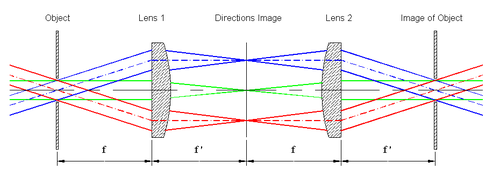
Figure 2: Formation of an image of the object (aperture) by addition of a second lens. The field of measurement is determined by the aperture located in the image of the object.

A third lens transforms the rays passing through the aperture (located in the plane of the image of the object) into a second directions image which may be analyzed by an image sensor (e.g., electronic camera).

schematic raytracing of a complete conoscope: formation of the directions image and imaging of the object Cono-3.png
schematic raytracing of a complete conoscope: formation of the directions image and imaging of the object
Figure 3: Schematic raytracing of a complete conoscope: formation of the directions image and imaging of the object.

The functional sequence is as follows:

This simple arrangement is the basis for all conoscopic devices (conoscopes). It is not straight forward however to design and manufacture lens systems that combine the following features:

Design and manufacturing of this type of complex lens system requires assistance by numerical modelling and a sophisticated manufacturing process.

Modern advanced conoscopic devices are used for rapid measurement and evaluation of the electro-optical properties of LCD-screens (e.g., variation of luminance, contrast and chromaticity with viewing direction).[ citation needed ]

Related Research Articles

<span class="mw-page-title-main">Optical aberration</span> Deviation from perfect paraxial optical behavior

In optics, aberration is a property of optical systems, such as lenses, that causes light to be spread out over some region of space rather than focused to a point. Aberrations cause the image formed by a lens to be blurred or distorted, with the nature of the distortion depending on the type of aberration. Aberration can be defined as a departure of the performance of an optical system from the predictions of paraxial optics. In an imaging system, it occurs when light from one point of an object does not converge into a single point after transmission through the system. Aberrations occur because the simple paraxial theory is not a completely accurate model of the effect of an optical system on light, rather than due to flaws in the optical elements.

<span class="mw-page-title-main">Diffraction</span> Phenomenon of the motion of waves

Diffraction is the interference or bending of waves around the corners of an obstacle or through an aperture into the region of geometrical shadow of the obstacle/aperture. The diffracting object or aperture effectively becomes a secondary source of the propagating wave. Italian scientist Francesco Maria Grimaldi coined the word diffraction and was the first to record accurate observations of the phenomenon in 1660.

<span class="mw-page-title-main">Lens</span> Optical device which transmits and refracts light

A lens is a transmissive optical device that focuses or disperses a light beam by means of refraction. A simple lens consists of a single piece of transparent material, while a compound lens consists of several simple lenses (elements), usually arranged along a common axis. Lenses are made from materials such as glass or plastic and are ground, polished, or molded to the required shape. A lens can focus light to form an image, unlike a prism, which refracts light without focusing. Devices that similarly focus or disperse waves and radiation other than visible light are also called "lenses", such as microwave lenses, electron lenses, acoustic lenses, or explosive lenses.

<span class="mw-page-title-main">Optics</span> Branch of physics that studies light

Optics is the branch of physics that studies the behaviour and properties of light, including its interactions with matter and the construction of instruments that use or detect it. Optics usually describes the behaviour of visible, ultraviolet, and infrared light. Light is a type of electromagnetic radiation, and other forms of electromagnetic radiation such as X-rays, microwaves, and radio waves exhibit similar properties.

<span class="mw-page-title-main">Numerical aperture</span> Characteristic of an optical system

In optics, the numerical aperture (NA) of an optical system is a dimensionless number that characterizes the range of angles over which the system can accept or emit light. By incorporating index of refraction in its definition, NA has the property that it is constant for a beam as it goes from one material to another, provided there is no refractive power at the interface. The exact definition of the term varies slightly between different areas of optics. Numerical aperture is commonly used in microscopy to describe the acceptance cone of an objective, and in fiber optics, in which it describes the range of angles within which light that is incident on the fiber will be transmitted along it.

<span class="mw-page-title-main">Aperture</span> Hole or opening through which light travels

In optics, the aperture of an optical system is a hole or an opening that primarily limits light propagated through the system. More specifically, the entrance pupil as the front side image of the aperture and focal length of an optical system determine the cone angle of a bundle of rays that comes to a focus in the image plane.

The focal length of an optical system is a measure of how strongly the system converges or diverges light; it is the inverse of the system's optical power. A positive focal length indicates that a system converges light, while a negative focal length indicates that the system diverges light. A system with a shorter focal length bends the rays more sharply, bringing them to a focus in a shorter distance or diverging them more quickly. For the special case of a thin lens in air, a positive focal length is the distance over which initially collimated (parallel) rays are brought to a focus, or alternatively a negative focal length indicates how far in front of the lens a point source must be located to form a collimated beam. For more general optical systems, the focal length has no intuitive meaning; it is simply the inverse of the system's optical power.

<span class="mw-page-title-main">Camera lens</span> Optical lens or assembly of lenses used with a camera to create images

A camera lens is an optical lens or assembly of lenses used in conjunction with a camera body and mechanism to make images of objects either on photographic film or on other media capable of storing an image chemically or electronically.

<span class="mw-page-title-main">Transmission electron microscopy</span> Imaging and diffraction using electrons that pass through samples

Transmission electron microscopy (TEM) is a microscopy technique in which a beam of electrons is transmitted through a specimen to form an image. The specimen is most often an ultrathin section less than 100 nm thick or a suspension on a grid. An image is formed from the interaction of the electrons with the sample as the beam is transmitted through the specimen. The image is then magnified and focused onto an imaging device, such as a fluorescent screen, a layer of photographic film, or a detector such as a scintillator attached to a charge-coupled device or a direct electron detector.

<span class="mw-page-title-main">Angular resolution</span> Ability of any image-forming device to distinguish small details of an object

Angular resolution describes the ability of any image-forming device such as an optical or radio telescope, a microscope, a camera, or an eye, to distinguish small details of an object, thereby making it a major determinant of image resolution. It is used in optics applied to light waves, in antenna theory applied to radio waves, and in acoustics applied to sound waves. The colloquial use of the term "resolution" sometimes causes confusion; when an optical system is said to have a high resolution or high angular resolution, it means that the perceived distance, or actual angular distance, between resolved neighboring objects is small. The value that quantifies this property, θ, which is given by the Rayleigh criterion, is low for a system with a high resolution. The closely related term spatial resolution refers to the precision of a measurement with respect to space, which is directly connected to angular resolution in imaging instruments. The Rayleigh criterion shows that the minimum angular spread that can be resolved by an image-forming system is limited by diffraction to the ratio of the wavelength of the waves to the aperture width. For this reason, high-resolution imaging systems such as astronomical telescopes, long distance telephoto camera lenses and radio telescopes have large apertures.

<span class="mw-page-title-main">Diffraction-limited system</span> Optical system with resolution performance at the instruments theoretical limit

In optics, any optical instrument or system – a microscope, telescope, or camera – has a principal limit to its resolution due to the physics of diffraction. An optical instrument is said to be diffraction-limited if it has reached this limit of resolution performance. Other factors may affect an optical system's performance, such as lens imperfections or aberrations, but these are caused by errors in the manufacture or calculation of a lens, whereas the diffraction limit is the maximum resolution possible for a theoretically perfect, or ideal, optical system.

<span class="mw-page-title-main">Airy disk</span> Diffraction pattern in optics

In optics, the Airy disk and Airy pattern are descriptions of the best-focused spot of light that a perfect lens with a circular aperture can make, limited by the diffraction of light. The Airy disk is of importance in physics, optics, and astronomy.

<span class="mw-page-title-main">Magnification</span> Process of enlarging the apparent size of something

Magnification is the process of enlarging the apparent size, not physical size, of something. This enlargement is quantified by a size ratio called optical magnification. When this number is less than one, it refers to a reduction in size, sometimes called de-magnification.

<span class="mw-page-title-main">Point spread function</span> Response in an optical imaging system

The point spread function (PSF) describes the response of a focused optical imaging system to a point source or point object. A more general term for the PSF is the system's impulse response; the PSF is the impulse response or impulse response function (IRF) of a focused optical imaging system. The PSF in many contexts can be thought of as the extended blob in an image that represents a single point object, that is considered as a spatial impulse. In functional terms, it is the spatial domain version of the optical transfer function (OTF) of an imaging system. It is a useful concept in Fourier optics, astronomical imaging, medical imaging, electron microscopy and other imaging techniques such as 3D microscopy and fluorescence microscopy.

<span class="mw-page-title-main">Telecentric lens</span> Optical lens

A telecentric lens is a special optical lens that has its entrance or exit pupil, or both, at infinity. The size of images produced by a telecentric lens is insensitive to either the distance between an object being imaged and the lens, or the distance between the image plane and the lens, or both, and such an optical property is called telecentricity. Telecentric lenses are used for precision optical two-dimensional measurements, reproduction, and other applications that are sensitive to the image magnification or the angle of incidence of light.

<span class="mw-page-title-main">Selected area diffraction</span> Crystallographic electron diffraction technique

Selected area (electron) diffraction is a crystallographic experimental technique typically performed using a transmission electron microscope (TEM). It is a specific case of electron diffraction used primarily in material science and solid state physics as one of the most common experimental techniques. Especially with appropriate analytical software, SAD patterns (SADP) can be used to determine crystal orientation, measure lattice constants or examine its defects.

In Gaussian optics, the cardinal points consist of three pairs of points located on the optical axis of a rotationally symmetric, focal, optical system. These are the focal points, the principal points, and the nodal points; there are two of each. For ideal systems, the basic imaging properties such as image size, location, and orientation are completely determined by the locations of the cardinal points; in fact, only four points are necessary: the two focal points and either the principal points or the nodal points. The only ideal system that has been achieved in practice is a plane mirror, however the cardinal points are widely used to approximate the behavior of real optical systems. Cardinal points provide a way to analytically simplify an optical system with many components, allowing the imaging characteristics of the system to be approximately determined with simple calculations.

Autocollimation is an optical setup where a collimated beam leaves an optical system and is reflected back into the same system by a plane mirror.

The viewing cone refers to the effective viewing directions of an LCD display, as seen from the eye. This collection of angles resembles a cone. The concept has been introduced as an international standard ISO 13406-2, which defines it as the range of viewing directions that can safely be used for the intended task without "reduced visual performance". This standard describes a complex procedure which evaluates the viewing cone from measurements of luminance and chromaticity versus direction of observation. ISO 13406-2 introduces 4 viewing direction range classes, from a wide viewing cone, for many simultaneous observers, to the so-called "privacy display", with a severely limited viewing cone. Compliance routes for different display applications can now be found in the successor standard ISO 9241-300.

The optical properties of all liquid and solid materials change as a function of the wavelength of light used to measure them. This change as a function of wavelength is called the dispersion of the optical properties. The graph created by plotting the optical property of interest by the wavelength at which it is measured is called a dispersion curve.

References

  1. Mauguin, C.: Sur les cristaux liquides de Lehmann. Bull. Soc. Fr. Miner. 34, 71–117 (1911)

Literature