Related Research Articles

Tracheal intubation, usually simply referred to as intubation, is the placement of a flexible plastic tube into the trachea (windpipe) to maintain an open airway or to serve as a conduit through which to administer certain drugs. It is frequently performed in critically injured, ill, or anesthetized patients to facilitate ventilation of the lungs, including mechanical ventilation, and to prevent the possibility of asphyxiation or airway obstruction.

Sevoflurane, sold under the brand name Sevorane, among others, is a sweet-smelling, nonflammable, highly fluorinated methyl isopropyl ether used as an inhalational anaesthetic for induction and maintenance of general anesthesia. After desflurane, it is the volatile anesthetic with the fastest onset. While its offset may be faster than agents other than desflurane in a few circumstances, its offset is more often similar to that of the much older agent isoflurane. While sevoflurane is only half as soluble as isoflurane in blood, the tissue blood partition coefficients of isoflurane and sevoflurane are quite similar. For example, in the muscle group: isoflurane 2.62 vs. sevoflurane 2.57. In the fat group: isoflurane 52 vs. sevoflurane 50. As a result, the longer the case, the more similar will be the emergence times for sevoflurane and isoflurane.

Laryngoscopy is endoscopy of the larynx, a part of the throat. It is a medical procedure that is used to obtain a view, for example, of the vocal folds and the glottis. Laryngoscopy may be performed to facilitate tracheal intubation during general anaesthesia or cardiopulmonary resuscitation or for surgical procedures on the larynx or other parts of the upper tracheobronchial tree.

General anaesthesia (UK) or general anesthesia (US) is a method of medically inducing loss of consciousness that renders a patient unarousable even with painful stimuli. This effect is achieved by administering either intravenous or inhalational general anaesthetic medications, which often act in combination with an analgesic and neuromuscular blocking agent. Spontaneous ventilation is often inadequate during the procedure and intervention is often necessary to protect the airway. General anaesthesia is generally performed in an operating theater to allow surgical procedures that would otherwise be intolerably painful for a patient, or in an intensive care unit or emergency department to facilitate endotracheal intubation and mechanical ventilation in critically ill patients.
A tracheal tube is a catheter that is inserted into the trachea for the primary purpose of establishing and maintaining a patent airway and to ensure the adequate exchange of oxygen and carbon dioxide.
Airway management includes a set of maneuvers and medical procedures performed to prevent and relieve airway obstruction. This ensures an open pathway for gas exchange between a patient's lungs and the atmosphere. This is accomplished by either clearing a previously obstructed airway; or by preventing airway obstruction in cases such as anaphylaxis, the obtunded patient, or medical sedation. Airway obstruction can be caused by the tongue, foreign objects, the tissues of the airway itself, and bodily fluids such as blood and gastric contents (aspiration).
In anaesthesia and advanced airway management, rapid sequence induction (RSI) – also referred to as rapid sequence intubation or as rapid sequence induction and intubation (RSII) or as crash induction – is a special process for endotracheal intubation that is used where the patient is at a high risk of pulmonary aspiration. It differs from other techniques for inducing general anesthesia in that several extra precautions are taken to minimize the time between giving the induction drugs and securing the tube, during which period the patient's airway is essentially unprotected.

The cricoid cartilage, or simply cricoid or cricoid ring, is the only complete ring of cartilage around the trachea. It forms the back part of the voice box and functions as an attachment site for muscles, cartilages, and ligaments involved in opening and closing the airway and in producing speech.

The arytenoid cartilages are a pair of small three-sided pyramids which form part of the larynx. They are the site of attachment of the vocal cords. Each is pyramidal or ladle-shaped and has three surfaces, a base, and an apex. The arytenoid cartilages allow for movement of the vocal cords by articulating with the cricoid cartilage. They may be affected by arthritis, dislocations, or sclerosis.

In anesthesia, the Mallampati score or Mallampati classification, named after the Indian anaesthesiologist Seshagiri Mallampati, is used to predict the ease of endotracheal intubation. The test comprises a visual assessment of the distance from the tongue base to the roof of the mouth, and therefore the amount of space in which there is to work. It is an indirect way of assessing how difficult an intubation will be; this is more definitively scored using the Cormack–Lehane classification system, which describes what is actually seen using direct laryngoscopy during the intubation process itself. A high Mallampati score is associated with more difficult intubation as well as a higher incidence of sleep apnea.

The Combitube—also known as the esophageal tracheal airway or esophageal tracheal double-lumen airway—is a blind insertion airway device (BIAD) used in the pre-hospital and emergency setting. It is designed to provide an airway to facilitate the mechanical ventilation of a patient in respiratory distress.

Throughout recorded history, attempts at producing a state of general anesthesia can be traced back to the writings of ancient Sumerians, Babylonians, Assyrians, Egyptians, Indians, and Chinese. Despite significant advances in anatomy and surgical technique during the Renaissance, surgery remained a last-resort treatment largely due to the pain associated with it. However, scientific discoveries in the late 18th and early 19th centuries paved the way for the development of modern anesthetic techniques.
Tracheal intubation, an invasive medical procedure, is the placement of a flexible plastic catheter into the trachea. For millennia, tracheotomy was considered the most reliable method of tracheal intubation. By the late 19th century, advances in the sciences of anatomy and physiology, as well as the beginnings of an appreciation of the germ theory of disease, had reduced the morbidity and mortality of this operation to a more acceptable rate. Also in the late 19th century, advances in endoscopic instrumentation had improved to such a degree that direct laryngoscopy had finally become a viable means to secure the airway by the non-surgical orotracheal route. Nasotracheal intubation was not widely practiced until the early 20th century. The 20th century saw the transformation of the practices of tracheotomy, endoscopy and non-surgical tracheal intubation from rarely employed procedures to essential components of the practices of anesthesia, critical care medicine, emergency medicine, gastroenterology, pulmonology and surgery.
The following outline is provided as an overview of and topical guide to anesthesia:

The laryngeal tube is an airway management device designed as an alternative to other airway management techniques such as mask ventilation, laryngeal mask airway, and tracheal intubation. This device can be inserted blindly through the oropharynx into the hypopharynx to create an airway during anaesthesia and cardiopulmonary resuscitation so as to enable mechanical ventilation of the lungs.

Airtraq is a fibreoptic intubation device used for indirect tracheal intubation in difficult airway situations. It is designed to enable a view of the glottic opening without aligning the oral with the pharyngeal, and laryngeal axes as an advantage over direct endotracheal intubation and allows for intubation with minimal head manipulation and positioning.

A double-lumen endotracheal tube is a type of endotracheal tube which is used in tracheal intubation during thoracic surgery and other medical conditions to achieve selective, one-sided ventilation of either the right or the left lung.
Advanced airway management is the subset of airway management that involves advanced training, skill, and invasiveness. It encompasses various techniques performed to create an open or patent airway – a clear path between a patient's lungs and the outside world.
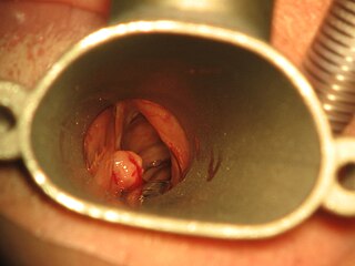
Intubation granuloma is a benign growth of granulation tissue in the larynx or trachea, which arises from tissue trauma due to endotracheal intubation. This medical condition is described as a common late complication of tracheal intubation, specifically caused by irritation to the mucosal tissue of the airway during insertion or removal of the patient's intubation tube.

Suction Assisted Laryngoscopy Airway Decontamination (SALAD) is incremental step-wise approach to the management of a massively contaminated airway.
References
- ↑ "Sellick's maneuver".
- ↑ Takahata, O; Kubota, M; Mamiya, K; Akama, Y; Nozaka, T; Matsumoto, H; Ogawa, H (1997). "The efficacy of the "BURP" maneuver during a difficult laryngoscopy" (PDF). Anesthesia & Analgesia. 84 (2): 419–21. doi:10.1097/00000539-199702000-00033. PMID 9024040. S2CID 16579238.
- ↑ Knill, RL (1993). "Difficult laryngoscopy made easy with a "BURP"". Canadian Journal of Anesthesia. 40 (3): 279–82. doi: 10.1007/BF03037041 . PMID 8467551.
- ↑ Sellick, BA (1961). "Cricoid pressure to control regurgitation of stomach contents during induction of anaesthesia". The Lancet. 2(7199) (7199): 404–406. doi:10.1016/s0140-6736(61)92485-0. PMID 13749923.
- ↑ Barash, Paul (2009). Clinical Anesthesia (6th ed.). Lippencott Williams & Wilkins. p. 1223.
- ↑ Moied, AS; Jyotishka, P (2010). "Cricoid pressure – A misnomer in pediatric anaesthesia". J Emerg Trauma Shock. 3 (1): 96–97. doi: 10.4103/0974-2700.58650 . PMC 2823158 . PMID 20165735.
- ↑ Salem, MR; Sellick, BA; Elam, JO (1974). "The historical background of cricoid pressure in anesthesia and resuscitation". Anesthesia & Analgesia. 53 (2): 230–2. doi: 10.1213/00000539-197403000-00011 . PMID 4593092.
- ↑ Maltby, JR; Beriault, MT (2002). "Science, pseudoscience and Sellick". Canadian Journal of Anesthesia. 49 (5): 443–7. doi: 10.1007/BF03017917 . PMID 11983655.
- ↑ Smith KJ, Dobranowski J, Yip G, Dauphin A, Choi PT (2003). "Cricoid pressure displaces the esophagus: an observational study using magnetic resonance imaging". Anesthesiology. 99 (1): 60–4. doi: 10.1097/00000542-200307000-00013 . PMID 12826843. S2CID 18535821.
- ↑ Haslam, N; Parker, L; Duggan, JE (2005). "Effect of cricoid pressure on the view at laryngoscopy". Anaesthesia. 60 (1): 41–7. doi: 10.1111/j.1365-2044.2004.04010.x . PMID 15601271. S2CID 42387260.
- ↑ "Cricolol by John Hinds". 28 May 2014.
- ↑ Moynihan, RJ; Brock – Utne, JG; Archer, JH; Feld, LH; Kreitzman, TR (Apr 1993). "The effect of cricoid pressure on preventing gastric insufflation in infants and children". Anesthesiology. 78 (4): 652–656. doi: 10.1097/00000542-199304000-00007 . PMID 8466065. S2CID 38041710.
- ↑ Salem, MR; Sellick, BA; Elam, JO (Mar–Apr 1974). "The historical background of cricoid pressure in anesthesia and resuscitation". Anesthesia and Analgesia. 53 (2): 230–2. doi: 10.1213/00000539-197403000-00011 . PMID 4593092.
- ↑ American Heart Association (2006). Textbook of Advanced Cardiac Life Support. Dallas, TX: American Heart Association.
- ↑ American Heart Association's BLS (Basic Life Support) Provider training, as of 2013-05-19
- ↑ Escott MEA, Owen H, Strahan AD, Plummer JL. Cricoid pressure training: how useful are descriptions of force? Anaesth Intensive Care 2003;31:388–391
- ↑ Owen H, Follows V, Reynolds KJ, Burgess G, Plummer J. Learning to apply effective cricoid pressure using a part task trainer. Anaesthesia 2002;57(11):1098–1101
- ↑ Walton S, Pearce A. Auditing the application of cricoid pressure. Anaesthesia 2000;55:1028–1029
- ↑ Koziol CA, Cuddleford JD, Moos DD. Assessing the force generated with the application of cricoid pressure. AORN J 2000;72:1018–1030
- ↑ Meek T, Gittins N, Duggan JE. Cricoid pressure: knowledge and performance amongst anaesthetic assistants. Anaesthesia 1999;54(1):59–62.
- ↑ Smith, K. J., Dobranowski, J., Yip, G., Dauphin, A., & Choi, P. T. (2003). Cricoid pressure displaces the esophagus: an observational study using magnetic resonance imaging. Anesthesiology, 99(1), 60–64;
- ↑ Smith, K. J., Ladak, S., Choi, Pt L., & Dobranowski, J. (2002). The cricoid cartilage and the oesophagus are not aligned in close to half of adult patients. Canadian Journal of Anesthesia, 49(5), 503–507.
- ↑ Palmer, JHM, Ball, D.R. The effect of cricoids pressure on the cricoids cartilage and vocal cords: An endoscopic study in anaesthetized patients. Anaesthesia (2000): 55; 260–287
- ↑ Hartsilver, E. L., Vanner, R. G. Airway obstruction with cricoids pressure. Anesthesia (2000): 55: 208–211
- ↑ Haslam, N., Parker, L., and Duggan, J.E. Effect of cricoid pressure on the view at laryngoscopy. Anesthesia (2005): 60: 41–47
- ↑ Hocking, G., Roberts, F.L., Thew, M.E. Airway obstruction with cricoids pressure and lateral tilt. Anesthesia (2001), 56; 825–828
- ↑ Ovessapian, A; Salem, MR (Nov 2009). "Sellick's Maneuver: To Do or Not Do". Anesthesia & Analgesia. 109 (5): 1360–1362. doi: 10.1213/ane.0b013e3181b763c0 . PMID 19843769 . Retrieved 2012-05-12.