Cutaneous reflex in human locomotion

Last updated
Tripping Activates Cutaneous Reflex PN Ostrzezenie przed niebezpieczenstwem potkniecia sie.svg
Tripping Activates Cutaneous Reflex

Cutaneous, or skin reflexes, are activated by skin receptors and play a valuable role in locomotion, providing quick responses to unexpected environmental challenges. They have been shown to be important in responses to obstacles or stumbling, in preparing for visually challenging terrain, and for assistance in making adjustments when instability is introduced. In addition to the role in normal locomotion, cutaneous reflexes are being studied for their potential in enhancing rehabilitation therapy (physiotherapy) for people with gait abnormalities.

Contents

Quick responses to obstacles Obstacle Course 2 (6452150563).jpg
Quick responses to obstacles

Background of reflex pathway

Reflexes are a vital part of our everyday activities. We have all experienced how fast and automatic these responses to environmental interactions can be. They can protect us from potentially dangerous situations such as touching a hot stove or stepping on a tack. There are also reflex pathways involved in more dynamic activities such as walking and running, helping to ensure a smooth gait and allowing us to respond quickly to obstacles or unexpected perturbations or disturbances.

Pathway of a Monosynaptic Reflex Afferent and efferent neurons en.svg
Pathway of a Monosynaptic Reflex

The reflex pathway (reflex arc) is a sequence of neurons connecting the sensory input (afferent neuron) to the motor output (efferent neuron), resulting in a behavioral response. The general pathway of a spinal reflex is one which involves neurons contained within the spinal cord. However, the brain may also provide additional (supraspinal) contributions, which can modulate the responsiveness of the reflex to the sensory input.

General reflex pathways

Simple Reflex Flowchart.jpg

Reflexes can be very simple, as in the monosynaptic reflex, which only contains one synapse, or more complicated, as in the polysynaptic reflex, which involves more than one synapse. The knee jerk reflex is a common example of a monosynaptic reflex when one is looking at the quadriceps motor response of kicking your leg out. It can also be used as an example of a polysynaptic reflex when looking at the involvement of inhibitory interneurons to relax the hamstrings. The complexity of the reflex can be estimated by examining the time delay, or latency, between the electrical stimulation of the sensory neuron and the corresponding motor response, as measured by EMG (electromyography). Most reflexes can be categorized in one of three groups depending on the latency of EMG response. The short-latency reflex (SLR) is the fastest (~40-50 ms) and involves a mono-synaptic pathway. The medium-latency reflex (MLR) utilizes interneurons within the spinal cord and is typically ~80-90 ms. The long-latency reflex (LLR) is ~120-140 ms, suggesting that it is mediated by additional supraspinal input from the brain. [1]

Cutaneous receptors and reflexes

Cutaneous receptors are a type of sensory receptor, which respond to stimuli (touch, pressure, pain, temperature) that provide information regarding contact with the external environment. A common reflex involving cutaneous receptors is the crossed extensor reflex. This reflex is recruited when we experience a painful stimulus on the bottom of our foot, such as stepping on a tack. The response is to quickly pull the foot that stepped on the tack away, and at the same time, shift our weight to the opposite (contralateral) leg for balance.

It has been noted that the muscle activation response to cutaneous receptor stimulation can be modulated by a variety of factors. These include the:

In addition to modulation, the cutaneous reflex has been shown to evoke both a MLR (medium latency response) and LLR (long latency response) EMG response, indicating that it is a polysynaptic reflex, involving spinal interneurons or supraspinal pathways [2]

Functional role

Cutaneous nerves
Plantar Cutaneous Nerves.png
General surface location for activation of three cutaneous receptors

The cutaneous reflex has been attributed to functional responses [3] to disturbances encountered during locomotion and is, therefore, dependent on which cutaneous nerve is stimulated. Examples of nerves whose cutaneous branches have been examined are the:

1. Superficial fibular nerve or peroneal nerve (innervating the dorsal aspect of the foot) 2. Tibial nerve (innervating the plantar surface of the foot)3. Sural nerve (innervating the lateral portion of the foot)4. Superficial branch of radial nerve (innervating forearm and hand on the thumb side)

Stance and swing phase of a gait cycle Gait Cycle.png
Stance and swing phase of a gait cycle

Human locomotion is often examined from the perspective of the gait cycle. Cutaneous reflexes demonstrate variations in the muscles activated and the timing at which they are activated depending on which portion of the gait cycle the stimulation occurs. This variation suggests a functional role for the reflex to provide us with a smooth gait alteration when encountering or anticipating obstacles and challenging terrain. The major muscles impacted involve four (4) motions important to locomotion:

Thigh muscles responding to cutaneous reflex
Gray430.png
Vastus lateralis (knee extensor)
Gray434.png
Biceps femoris (knee flexor)
Lower leg muscles responding to cutaneous reflex
Gray437.png
Tibialis anterior (dorsiflexor)
Gray438.png
Gastrocnemius (plantar flexor)
Leg MotionMuscle GroupTypical muscle measured

for EMG activity

Knee flexionHamstringsBiceps femoris
Knee extensionQuadricepsVastus lateralis
Ankle plantar flexion (pointing toes)Calf musclesGastrocnemius or

Soleus

Ankle dorsiflexion (raising toes up)Shin muscleTibialis anterior

Superficial fibular nerve (SF)

Cutaneous stimulation of the superficial fibular nerve (SF) would typically occur when the top of the foot encounters an obstacle. The reflexive response to this activation is to pull the leg up and over the obstacle, while also preparing for a possible stumble or fall. Observed EMG muscle responses to SF stimulation help to explain how this reflexive response is accomplished:

Tibial nerve (TN)

Tibial nerve (TN) stimulation demonstrates a phase dependent response, in which the muscle activation varies depending on what part of the gait cycle a person is in when the stimulation occurs.

1. In normal, unimpeded walking, TN activation during the transition from stance to swing should decrease as the foot begins to leave the ground. In this case, increased TN stimulation would suggest scuffing of the bottom of the foot along the ground. The reflexive response will bring the foot up, pulling it off the ground. EMG responses to stimulation, again, help to explain how this is accomplished.

2. TN activation during late swing in normal walking should begin to increase as the foot begins to contact the ground. TN stimulation during this phase is, therefore, similar to a normal gait pattern sensory input. In this situation, the foot will plantar flex to allow smooth placement for the transition to the stance phase. Here, the opposite EMG effect is observed.

Sural nerve (SN)

Sural nerve (SN) stimulation results in a reflex that is both phase and intensity dependent. The sural nerve innervates the lateral (outside) portion of the foot and could be activated during either the swing or stance phase when encountering uneven terrain. The intensity dependent response is indicative of the level of activation and, therefore, the potential for harm.

1. During the swing phase, SN stimulation indicates encountering an obstacle on the lateral side of the foot. The reaction is to move the foot inward and the leg up, out of the way. Observed EMG responses are:

2. During the stance phase, a moderate intensity SN stimulation is indicative of uneven terrain and will result in a response that provides stiffness and stability to the ankle. The mechanism for this response is accomplished through the simultaneous activation of two antagonistic ankle muscles

3. During the stance phase, a high intensity SN stimulation is indicative of an obstacle that could potentially cause physical damage to the foot. In this case, the goal of the reflex would be to remove the foot completely away from the stimulus.

Superficial radial nerve (SR)

AnimatedAvatar.gif

Cutaneous stimulation of the superficial radial nerve (located on the thumb side of the forearm) will evoke a cutaneous reflex in arm muscles during rhythmic arm movements in a manner similar to that observed with leg muscles during locomotion. In addition to its role in reflex responses to the arm itself, it will modulate leg muscle cutaneous reflexes during locomotion. [7] This reflexive response may be a vestigial remnant from our quadruped ancestry, connecting forelimb movement with hindlimb movement. [8] This connection can be seen with normal walking, when the arm swings in rhythm with the opposite leg. The crossed synchrony may be partially due to a shared neural pathway between upper and lower limbs. While the function of this coordinated movement may be involved in maintaining balance while shifting our center of mass, it is also involved in locomotive reflexes. Stimulation of the radial nerve during walking is unusual and often indicative of an obstacle that is high enough to make contact with the swinging arm. Concurrent, or simultaneous stimulation of both the superficial fibular nerve (top of the foot) and the superficial radial nerve of the opposite (contralateral) arm suggests that the obstacle is large enough to cause a major stumble or fall. The response is, therefore, one which activates the leg muscles in a way as to prepare for that possibility. EMG responses to this type of simultaneous stimulation involves significant changes in the leg opposite or (contralateral) to the side receiving the radial simulation:

Additional variables modulating cutaneous reflex response

The magnitude of the cutaneous reflex in leg muscles can be altered by multiple variables. The alterations are movement dependent, gait phase dependent, and can be either excitatory or inhibitory to the normal cutaneous reflex pattern.

Complexity or challenge of the task

Cutaneous reflexes are modulated in conditions which present instability or challenging locomotive movements. In unstable conditions, such as disturbances occurring when the arms are crossed, there is facilitation or amplification in both the ipsilateral and contralateral reflexes which are muscle specific. [9] With superficial fibular nerve stimulation, the ipsilateral biceps femoris (knee flexor) and ipsilateral soleus (plantar flexor) reflexes are amplified during swing to allow the leg to clear the obstacle. In contrast, the contralateral, opposite, leg demonstrates amplification of both the tibialis anterior (dorsiflexor) and the gastrocnemius (plantar flexor) providing additional leg stiffness for stability of the stance leg.

Similar amplification results are seen in both visually challenging environments, such as horizontal ladder stepping, [10] and backward walking. [11] These situations demonstrate facilitation of the contralateral reflexes during the stance phase to in a manner which would provide stability and ipsilateral reflex modulation in a way to allow smooth transition past the perceived obstacle.

Potential clinical applications of cutaneous reflex modulation

The ability to modulate the magnitude of cutaneous reflexes via rhythmic arm movements or challenging environments has potential implications in rehabilitation for patients with motor weakness. Some stroke patients with lower limb complications demonstrate reduced or blunted cutaneous reflexes. These lower limb reflexes can be amplified with superficial radial nerve stimulation by taking advantage of the interlimb reflex observed during rhythmic arm locomotion between the upper and lower limbs. The amplification of the contralateral tibialis anterior seen during the swing to stance transition may be clinically useful in preventing foot drop during the swing phase for stroke patients. [12] Future research is needed to further explore the efficacy of cutaneous reflex amplification for as a tool for enhancing motor receptivity with stroke rehabilitation.

Related Research Articles

In neuroscience, an F wave is one of several motor responses which may follow the direct motor response (M) evoked by electrical stimulation of peripheral motor or mixed nerves. F-waves are the second of two late voltage changes observed after stimulation is applied to the skin surface above the distal region of a nerve, in addition to the H-reflex which is a muscle reaction in response to electrical stimulation of innervating sensory fibers. Traversal of F-waves along the entire length of peripheral nerves between the spinal cord and muscle, allows for assessment of motor nerve conduction between distal stimulation sites in the arm and leg, and related motoneurons (MN's) in the cervical and lumbosacral cord. F-waves are able to assess both afferent and efferent loops of the alpha motor neuron in its entirety. As such, various properties of F-wave motor nerve conduction are analyzed in nerve conduction studies (NCS), and often used to assess polyneuropathies, resulting from states of neuronal demyelination and loss of peripheral axonal integrity.

Foot Anatomical structure found in vertebrates

The foot is an anatomical structure found in many vertebrates. It is the terminal portion of a limb which bears weight and allows locomotion. In many animals with feet, the foot is a separate organ at the terminal part of the leg made up of one or more segments or bones, generally including claws or nails.

Human leg lower extremity or limb of the human body (foot, lower leg, thigh and hip)

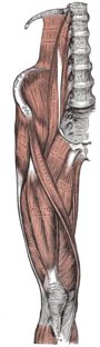
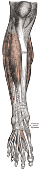
The human leg, in the general word sense, is the entire lower limb of the human body, including the foot, thigh and even the hip or gluteal region. However, the definition in human anatomy refers only to the section of the lower limb extending from the knee to the ankle, also known as the crus. Legs are used for standing, and all forms of locomotion including recreational such as dancing, and constitute a significant portion of a person's mass. Female legs generally have greater hip anteversion and tibiofemoral angles, but shorter femur and tibial lengths than those in males.

Plantar reflex reflex elicited when the sole of the foot is stimulated with a blunt instrument

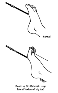
The plantar reflex is a reflex elicited when the sole of the foot is stimulated with a blunt instrument. The reflex can take one of two forms. In healthy adults, the plantar reflex causes a downward response of the hallux (flexion). An upward response (extension) of the hallux is known as the Babinski response or Babinski sign, named after the neurologist Joseph Babinski. The presence of the Babinski sign can identify disease of the spinal cord and brain in adults, and also exists as a primitive reflex in infants.

Gait (human) Locomotion achieved through the movement of human limbs

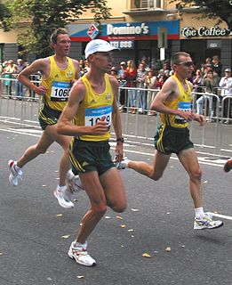
Human gait refers to locomotion achieved through the movement of human limbs. Human gait is defined as bipedal, biphasic forward propulsion of center of gravity of the human body, in which there are alternate sinuous movements of different segments of the body with least expenditure of energy. Different gait patterns are characterized by differences in limb-movement patterns, overall velocity, forces, kinetic and potential energy cycles, and changes in the contact with the surface. Human gaits are the various ways in which a human can move, either naturally or as a result of specialized training. Gait also refers to a religious approach to life, one can walk with a narrow or wide gait.

The withdrawal reflex is a spinal reflex intended to protect the body from damaging stimuli. The reflex rapidly coordinates the contractions of all the flexor muscles and the relaxations of the extensors in that limb causing sudden withdrawal from the potentially damaging stimulus. Spinal reflexes are often monosynaptic and are mediated by a simple reflex arc. A withdrawal reflex is mediated by a polysynaptic reflex resulting in the stimulation of many motor neurons in order to give a quick response.

The crossed extensor reflex or crossed extensor response or crossed extension reflex is a reflex in which the contralateral limb compensates for loss of support when the ipsilateral limb withdraws from painful stimulus in a withdrawal reflex. During a withdrawal reflex, the flexors in the withdrawing limb contract and the extensors relax, while in the other limb, the opposite occurs as part of the crossed extensor reflex. Besides shifting the body weight to the other side, the reflex pathway is also associated with leg coordination when walking by flexing muscle on one side, while extending muscle on the other side. This crossed extensor response is properly part of the withdrawal reflex.

Tibialis anterior muscle

The tibialis anterior is a muscle in humans that originates along the upper two-thirds of the lateral (outside) surface of the tibia and inserts into the medial cuneiform and first metatarsal bones of the foot. It acts to dorsiflex and invert the foot. This muscle is mostly located near the shin.

Tibial nerve

The tibial nerve is a branch of the sciatic nerve. The tibial nerve passes through the popliteal fossa to pass below the arch of soleus.

Plantaris muscle muscle

The plantaris is one of the superficial muscles of the superficial posterior compartment of the leg, one of the fascial compartments of the leg.

Foot drop Gait abnormality

Foot drop is a gait abnormality in which the dropping of the forefoot happens due to weakness, irritation or damage to the common fibular nerve including the sciatic nerve, or paralysis of the muscles in the anterior portion of the lower leg. It is usually a symptom of a greater problem, not a disease in itself. Foot drop is characterized by inability or impaired ability to raise the toes or raise the foot from the ankle (dorsiflexion). Foot drop may be temporary or permanent, depending on the extent of muscle weakness or paralysis and it can occur in one or both feet. In walking, the raised leg is slightly bent at the knee to prevent the foot from dragging along the ground.

Vestibulospinal tract

The vestibulospinal tract is a neural tract in the central nervous system. Specifically, it is a component of the extrapyramidal system and is classified as a component of the medial pathway. Like other descending motor pathways, the vestibulospinal fibers of the tract relay information from nuclei to motor neurons. The vestibular nuclei receive information through the vestibulocochlear nerve about changes in the orientation of the head. The nuclei relay motor commands through the vestibulospinal tract. The function of these motor commands is to alter muscle tone, extend, and change the position of the limbs and head with the goal of supporting posture and maintaining balance of the body and head.

Sole (foot) bottom part of foot

The sole is the bottom of the foot.

Locomotor effects of shoes

Locomotor effects of shoes are the way in which the physical characteristics or components of shoes influence the locomotion neuromechanics of a person. Depending on the characteristics of the shoes, the effects are various, ranging from alteration in balance and posture, muscle activity of different muscles as measured by electromyography (EMG), and the impact force. There are many different types of shoes that exist, such as running, walking, loafers, high heels, sandals, slippers, work boots, dress shoes, and many more. However, a typical shoe will be composed of an insole, midsole, outsole, and heels, if any. In an unshod condition, where one is without any shoes, the locomotor effects are primarily observed in the heel strike patterns and resulting impact forces generated on the ground.

Spinal locomotion

Spinal locomotion results from intricate dynamic interactions between a central program in lower thoracolumbar spine and proprioceptive feedback from body in the absence of central control by brain as in complete spinal cord injury (SCI). Following SCI, the spinal circuitry below the lesion site does not become silent rather it continues to maintain active and functional neuronal properties although in a modified manner.

Terrestrial locomotion by means of a running gait can be accomplished on level surfaces. However, in most outdoor environments an individual will experience terrain undulations requiring uphill running. Similar conditions can be mimicked in a controlled environment on a treadmill also. Additionally, running on inclines is used by runners, both distance and sprinter, to improve cardiovascular conditioning and lower limb strength.

Neural substrate of locomotor central pattern generators in mammals

Central pattern generators are biological neural networks organized to produce any rhythmic output without requiring a rhythmic input. In mammals, locomotor CPGs are organized in the lumbar and cervical segments of the spinal cord, and are used to control rhythmic muscle output in the arms and legs. Certain areas of the brain initiate the descending neural pathways that ultimately control and modulate the CPG signals. In addition to this direct control, there exist different feedback loops that coordinate the limbs for efficient locomotion and allow for the switching of gaits under appropriate circumstances.

Neuromechanics field of study

As originally proposed by Enoka, neuromechanics is a field of study that combines concepts from biomechanics and neurophysiology to study human movement. Neuromechanics examines the combined roles of the skeletal, muscular, and nervous systems and how they interact to produce the motion required to complete a motor task.

Proportional myoelectric control can be used to activate robotic lower limb exoskeletons

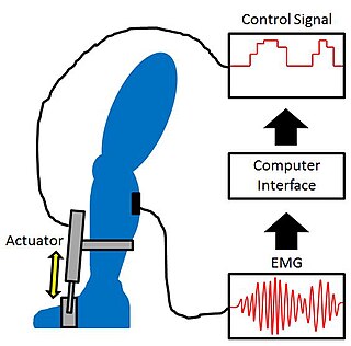
Proportional myoelectric control can be used to activate robotic lower limb exoskeletons. A proportional myoelectric control system utilizes a microcontroller or computer that inputs electromyography (EMG) signals from sensors on the leg muscle(s) and then activates the corresponding joint actuator(s) proportionally to the EMG signal.

References

  1. Scholz, E., Diener, H. C., Noth, J., Friedemann, H., Dichgans, J., & Bacher, M. (1987). Medium and long latency EMG responses in leg muscles: Parkinson's disease. Journal of Neurology, Neurosurgery, and Psychiatry, 50(1), 66-70.
  2. Nakajima, T., Barss, T., Klarner, T., Komiyama, T., & Zehr, E. P. (2013). Amplification of interlimb reflexes evoked by stimulating the hand simultaneously with conditioning from the foot during locomotion. Bmc Neuroscience, 14.
  3. Zehr, E. P., Komiyama, T., & Stein, R. B. (1997). Cutaneous reflexes during human gait: Electromyographic and kinematic responses to electrical stimulation. Journal of Neurophysiology, 77(6), 3311-3325.
  4. Zehr, E. P., Komiyama, T., & Stein, R. B. (1997). Cutaneous reflexes during human gait: Electromyographic and kinematic responses to electrical stimulation. Journal of Neurophysiology, 77(6), 3311-3325.
  5. Zehr, E. P., Komiyama, T., & Stein, R. B. (1997). Cutaneous reflexes during human gait: Electromyographic and kinematic responses to electrical stimulation. Journal of Neurophysiology, 77(6), 3311-3325.
  6. Zehr, E. P., Stein, R. B., & Komiyama, T. (1998). Function of sural nerve reflexes during human walking. Journal of Physiology-London, 507(1), 305-314.
  7. de Kam, D., Rijken, H., Manintveld, T., Nienhuis, B., Dietz, V., & Duysens, J. (2013). Arm movements can increase leg muscle activity during submaximal recumbent stepping in neurologically intact individuals. Journal of Applied Physiology, 115(1), 34-42.
  8. Nakajima
  9. Lamont, E. V., & Zehr, E. P. (2007). Earth-referenced handrail contact facilitates interlimb cutaneous reflexes during locomotion. Journal of Neurophysiology, 98(1), 433-442.
  10. Ruff, C. R., Miller, A. B., Delva, M. L., Lajoie, K., & Marigold, D. S. (2014). Modification of cutaneous reflexes during visually guided walking. Journal of Neurophysiology, 111(2), 379-393.
  11. Hoogkamer, W., Massaad, F., Jansen, K., Bruijn, S. M., & Duysens, J. (2012). Selective bilateral activation of leg muscles after cutaneous nerve stimulation during backward walking. Journal of Neurophysiology, 108(7), 1933-1941.
  12. Zehr, E. P., & Loadman, P. M. (2012). Persistence of locomotor-related interlimb reflex networks during walking after stroke. Clinical Neurophysiology, 123(4), 796-807.