Dibucaine number

Last updated

Dibucaine, also known as cinchocaine, is an amino amide local anesthetic. When administered to humans intravenously, it is capable of inhibiting the plasma cholinesterase (butyrylcholinesterase) enzyme. The dibucaine number is used to differentiate individuals who have substitution mutations (point mutations) of the enzyme's gene, resulting in decreased enzyme function.

Contents

Metabolism

Plasma cholinesterase is also known as butyrylcholinesterase, in part because once an individual is given butyrylcholine intravenously, the enzyme converts it to the products butyric acid and choline. This tetrameric enzyme is responsible for the metabolism of a number of substances, including amino ester local anesthetics and succinylcholine, which it hydrolyses in two stages to succinyl monocholine and choline, then to succinic acid and a second molecule of choline. Dibucaine inhibits normal butyrylcholinesterase activity, reducing the ability to convert butyrylcholine to its byproducts. The extent of the catalysis can be determined by measuring the percentage of butyrylcholine that remains unchanged in the blood of individuals administered a standard dose after dibucaine inhibition challenge in what has been established as the dibucaine number test. Kalow and Genest [1] first described this means of determining butyrylcholinestersase activity in 1957. Typical measurement of dibucaine number in the United States yields values of 80 and above for wild type homozygotes (normal), 40–60 for heterozygotes (atypical), and 20 or less for atypical homozygotes. [2]

Dibucaine number

The dibucaine number is used to differentiate individuals who have substitution mutations of the butyrylcholinesterase enzyme resulting in decreased enzyme function. At least one substitution mutation has been characterized that is capable of altering the efficiency of enzymatic catalysis. Reduced butyrylcholinesterase activity may occur as a result of inherited or acquired causes. Inherited reductions in butyrylcholinesterase activity occur because of mutations at a single autosomal location on the long arm of chromosome 3. Physiologic reductions may occur with extremes of age and during pregnancy. Other acquired causes of decreased activity include kidney and liver disease, malignancy (cancer), malnutrition, and burns. In the inherited type, an individual receives a gene from each parent, one of which may be the wild type butyrylcholinesterase, or the mutant. Thus, there may be individuals who are homozygous for the wild type butyrylcholinesterase (normal) or the mutant butyrylcholinesterase (incidence 1/3200), and there is the group of heterozygotes with one of each (incidence 1/480).

Point mutation

Miller's Anesthesia [2] notes that a point mutation in the gene for human serum cholinesterase has been identified that changes Asp-70 to Gly in the atypical form of serum cholinesterase. The mutation in nucleotide 209, which changes codon 70 from GAT to GGT, was found by sequencing a genomic clone and sequencing selected regions of DNA amplified by the polymerase chain reaction. McGuire et al. [3] compared the entire coding sequences for usual and atypical cholinesterases, and found no other consistent base differences. They described a polymorphic site near the C terminus of the coded region, but neither allele at this locus segregated consistently with the atypical trait. They conclude that the Asp-70 to Gly mutation (acidic to neutral amino acid substitution) accounts for reduced affinity of atypical cholinesterase for choline esters and that Asp-70 must be an important component of the anionic site. Heterogeneity in atypical alleles may exist, but the Asp-70 point mutation may represent an appreciable portion of the atypical gene pool.

More recently, Gaffney and Campbell [4] have described a PCR-based method to identify the Kalow allele for butyrylcholinesterase. A quantitative variant of the usual gene and was shown to result from a single base pair change in the DNA as described above. A new method based on the polymerase chain reaction to distinguish Kalow alleles of the cholinesterase gene was developed. Using the amplification refractory mutagenesis system, two different reactions distinguished the presence of a guanine (normal E1u allele) from that of an adenine (Kalow E1k allele) at nucleotide 1615 within the coding sequences of the gene. The frequency of the Kalow allele in their sample of 51 individuals was determined to be 20%. The mean total cholinesterase activity in heterozygotes was 90% of that in persons who typed as E1uE1u homozygotes. Two E1kE1k homozygotes were identified and their cholinesterase activities were the two lowest measured.

Distinction

The distinctive quality of dibucaine is that its enzyme inhibition of the wild type butyrylcholinesterase (Typical) is substantially greater than that of the mutant butyrylcholinesterase (Atypical). Thus, the atypical enzyme is said to be resistant to dibucaine inhibition. This can be used to distinguish individuals in the aforementioned genetic classes. Lockridge and La Du [5] measured atypical and usual human serum cholinesterases with the fluorescent probe, N-methyl-(7-dimethylcarbamoxy)quinolinium iodide. Four active sites per tetramer were found in each enzyme. The turnover numbers of usual and atypical cholinesterases were the same: 15,000 mumol of benzoylcholine hydrolyzed/min/mumol of active site; 48,000 min-1 for o-nitrophenylbutyrate; and 0.0025 min-1 for N-methyl-(7-dimethylcarbamoxy)quinolinium iodide. They had identical rate constants for carbamylation, (5.0 min-1) and for decarbamylation (0.15 h-1). The major difference between the two genetically determined forms of the enzyme was substrate affinity, KD being 0.16 mM for usual and 5.4 mM for atypical cholinesterase, for the fluorescent probe substrate. Km for the uncharged ester, o-nitrophenylbutyrate, was 0.14 mM for both enzymes, whereas Km for benzoylcholine was 0.005 mM for usual and 0.024 mM for atypical cholinesterase. We interpret these data to mean that the two enzymes differ only in the structure of their anionic site.

Neuromuscular blocking

When given succinylcholine, a commonly used neuromuscular-blocking drug administered for general anesthesia during surgery, the heterozygous and mutant homozygous individual will experience a prolonged duration of action of neuromuscular blockade. This results in unexpected and unwanted postoperative respiratory muscle paralysis requiring mechanical ventilation in such patients. The duration of such paralysis may last from hours to days. To identify susceptible individuals, the dibucaine number can be determined so as to alert the care team to the risks of use of butyrylcholinesterase substrates. Pestel et al. [6] measured 24,830 Dibucaine numbers over a period of four years in a European trial. Numbers below 30 (atypical homozygous) were found in 0.07% (n=18) giving an incidence of 1:1,400. Dibucaine numbers from 30 to 70 (atypical heterozygous) were found in 1.23% (n=306). On the basis of identification of the Dibucaine numbers we could avoid the administration of succinylcholine resulting in a cost reduction of 12,280 Euro offset against the total laboratory costs amounting to 10,470 Euro.

Cost effect

This incidence is higher than documented in the literature. [2] Pestel et al. conclude that routine measurement of dibucaine number is a cost-effective method of identifying patients at increased risk of prolonged neuromuscular blockade due to atypical cholinesterase. It is currently not standard practice to obtain such testing prior to surgery. Today, dibucaine number is typically determined after an episode of prolonged paralysis following administration of succinylcholine in order to explain the cause of the incident. Succinylcholine duration is usually on the order of 7–15 minutes and the extent of blockade is monitored with a neuromuscular stimulator. If activity at the motor endplate is not reestablished, as determined by nerve stimulator testing, an anesthesiologist will grow concerned that the patient may have a mutant form of the plasma cholinesterase enzyme and will withhold subsequent dosing of neuromuscular blocking agents until return of function.

Related Research Articles

An allele, or allelomorph, is a variant of the sequence of nucleotides at a particular location, or locus, on a DNA molecule.

<span class="mw-page-title-main">Tay–Sachs disease</span> Human medical condition

Tay–Sachs disease is a genetic disorder that results in the destruction of nerve cells in the brain and spinal cord. The most common form is infantile Tay–Sachs disease, which becomes apparent around the age of three to six months of age, with the baby losing the ability to turn over, sit, or crawl. This is then followed by seizures, hearing loss, and inability to move, with death usually occurring by the age of three to five. Less commonly, the disease may occur later in childhood, adolescence, or adulthood. These forms tend to be less severe, but the juvenile form typically results in death by age 15.

<span class="mw-page-title-main">Dominance (genetics)</span> One gene variant masking the effect of another in the other copy of the gene

In genetics, dominance is the phenomenon of one variant (allele) of a gene on a chromosome masking or overriding the effect of a different variant of the same gene on the other copy of the chromosome. The first variant is termed dominant and the second is called recessive. This state of having two different variants of the same gene on each chromosome is originally caused by a mutation in one of the genes, either new or inherited. The terms autosomal dominant or autosomal recessive are used to describe gene variants on non-sex chromosomes (autosomes) and their associated traits, while those on sex chromosomes (allosomes) are termed X-linked dominant, X-linked recessive or Y-linked; these have an inheritance and presentation pattern that depends on the sex of both the parent and the child. Since there is only one copy of the Y chromosome, Y-linked traits cannot be dominant or recessive. Additionally, there are other forms of dominance, such as incomplete dominance, in which a gene variant has a partial effect compared to when it is present on both chromosomes and co-dominance, in which different variants on each chromosome both show their associated traits.
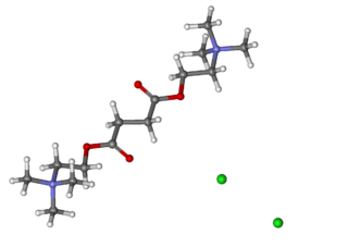
<span class="mw-page-title-main">Suxamethonium chloride</span> Chemical compound

Suxamethonium chloride, [Scoline, Sucostrin] also known as suxamethonium or succinylcholine, or simply sux by medical abbreviation, is a medication used to cause short-term paralysis as part of general anesthesia. This is done to help with tracheal intubation or electroconvulsive therapy. It is administered by injection, either into a vein or into a muscle. When used in a vein, onset of action is generally within one minute and effects last for up to 10 minutes.

<span class="mw-page-title-main">ENU</span> Chemical compound

ENU, also known as N-ethyl-N-nitrosourea (chemical formula C3H7N3O2), is a highly potent mutagen. For a given gene in mice, ENU can induce 1 new mutation in every 700 loci. It is also toxic at high doses.

<span class="mw-page-title-main">Cholinesterase</span> Esterase that lyses choline-based esters

The enzyme cholinesterase (EC 3.1.1.8, choline esterase; systematic name acylcholine acylhydrolase) catalyses the hydrolysis of choline-based esters:

Balancing selection refers to a number of selective processes by which multiple alleles are actively maintained in the gene pool of a population at frequencies larger than expected from genetic drift alone. Balancing selection is rare compared to purifying selection. It can occur by various mechanisms, in particular, when the heterozygotes for the alleles under consideration have a higher fitness than the homozygote. In this way genetic polymorphism is conserved.

A heterozygote advantage describes the case in which the heterozygous genotype has a higher relative fitness than either the homozygous dominant or homozygous recessive genotype. Loci exhibiting heterozygote advantage are a small minority of loci. The specific case of heterozygote advantage due to a single locus is known as overdominance. Overdominance is a rare condition in genetics where the phenotype of the heterozygote lies outside of the phenotypical range of both homozygote parents, and heterozygous individuals have a higher fitness than homozygous individuals.

<span class="mw-page-title-main">Overdominance</span>

Overdominance is a phenomenon in genetics where the phenotype of the heterozygote lies outside the phenotypical range of both homozygous parents. Overdominance can also be described as heterozygote advantage regulated by a single genomic locus, wherein heterozygous individuals have a higher fitness than homozygous individuals. However, not all cases of the heterozygote advantage are considered overdominance, as they may be regulated by multiple genomic regions. Overdominance has been hypothesized as an underlying cause for heterosis.

Pseudocholinesterase deficiency is an autosomal recessive inherited blood plasma enzyme abnormality in which the body's production of butyrylcholinesterase is impaired. People who have this abnormality may be sensitive to certain anesthetic drugs, including the muscle relaxants succinylcholine and mivacurium as well as other ester local anesthetics.

Mutation–selection balance is an equilibrium in the number of deleterious alleles in a population that occurs when the rate at which deleterious alleles are created by mutation equals the rate at which deleterious alleles are eliminated by selection. The majority of genetic mutations are neutral or deleterious; beneficial mutations are relatively rare. The resulting influx of deleterious mutations into a population over time is counteracted by negative selection, which acts to purge deleterious mutations. Setting aside other factors, the equilibrium number of deleterious alleles is then determined by a balance between the deleterious mutation rate and the rate at which selection purges those mutations.

A null allele is a nonfunctional allele caused by a genetic mutation. Such mutations can cause a complete lack of production of the associated gene product or a product that does not function properly; in either case, the allele may be considered nonfunctional. A null allele cannot be distinguished from deletion of the entire locus solely from phenotypic observation.

<span class="mw-page-title-main">Triosephosphate isomerase deficiency</span> Medical condition

Triosephosphate isomerase deficiency is a rare autosomal recessive metabolic disorder which was initially described in 1965.

In population genetics, fixation is the change in a gene pool from a situation where there exists at least two variants of a particular gene (allele) in a given population to a situation where only one of the alleles remains. That is, the allele becomes fixed. In the absence of mutation or heterozygote advantage, any allele must eventually be lost completely from the population or fixed. Whether a gene will ultimately be lost or fixed is dependent on selection coefficients and chance fluctuations in allelic proportions. Fixation can refer to a gene in general or particular nucleotide position in the DNA chain (locus).

In medical genetics, compound heterozygosity is the condition of having two or more heterogeneous recessive alleles at a particular locus that can cause genetic disease in a heterozygous state; that is, an organism is a compound heterozygote when it has two recessive alleles for the same gene, but with those two alleles being different from each other. Compound heterozygosity reflects the diversity of the mutation base for many autosomal recessive genetic disorders; mutations in most disease-causing genes have arisen many times. This means that many cases of disease arise in individuals who have two unrelated alleles, who technically are heterozygotes, but both the alleles are defective.

Lethal alleles are alleles that cause the death of the organism that carries them. They are usually a result of mutations in genes that are essential for growth or development. Lethal alleles may be recessive, dominant, or conditional depending on the gene or genes involved.
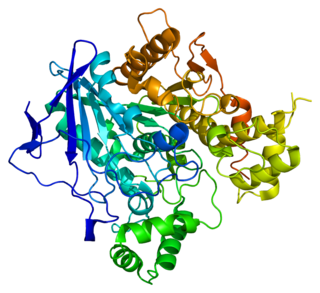
<span class="mw-page-title-main">Butyrylcholinesterase</span> Mammalian protein found in humans

Butyrylcholinesterase, also known as BChE, BuChE, BuChase, pseudocholinesterase, or plasma (cholin)esterase, is a nonspecific cholinesterase enzyme that hydrolyses many different choline-based esters. In humans, it is made in the liver, found mainly in blood plasma, and encoded by the BCHE gene.

The infinite alleles model is a mathematical model for calculating genetic mutations. The Japanese geneticist Motoo Kimura and American geneticist James F. Crow (1964) introduced the infinite alleles model, an attempt to determine for a finite diploid population what proportion of loci would be homozygous. This was, in part, motivated by assertions by other geneticists that more than 50 percent of Drosophila loci were heterozygous, a claim they initially doubted. In order to answer this question they assumed first, that there were a large enough number of alleles so that any mutation would lead to a different allele ; and second, that the mutations would result in a number of different outcomes from neutral to deleterious.

<span class="mw-page-title-main">Acetylcholinesterase</span> Primary cholinesterase in the body

Acetylcholinesterase (HGNC symbol ACHE; EC 3.1.1.7; systematic name acetylcholine acetylhydrolase), also known as AChE, AChase or acetylhydrolase, is the primary cholinesterase in the body. It is an enzyme that catalyzes the breakdown of acetylcholine and some other choline esters that function as neurotransmitters:

<span class="mw-page-title-main">Zygosity</span> Degree of similarity of the alleles in an organism

Zygosity is the degree to which both copies of a chromosome or gene have the same genetic sequence. In other words, it is the degree of similarity of the alleles in an organism.

References

  1. Kalow, W; Genest, K (1957). "A method for the detection of atypical forms of human serum cholinesterase: Determination of dibucaine". Can. J. Biochem. 35 (s): 339–46. doi:10.1139/o57-041.
  2. 1 2 3 Miller, R (2005). Miller's Anesthesia (6th ed.). Philadelphia: Elsevier.
  3. McGuire, MC; Nogueira, CP; Bartels, CF; Lightstone, H; Hajra, A; Van der Spek, AF; Lockridge, O; La Du, BN (February 1989). "Identification of the structural mutation responsible for the dibucaine-resistant (atypical) variant form of human serum cholinesterase". Proc Natl Acad Sci USA. 86 (3): 953–957. Bibcode:1989PNAS...86..953M. doi: 10.1073/pnas.86.3.953 . PMC   286597 . PMID   2915989.
  4. Gaffney, D; Campbell, RA (March 1994). "A PCR based method to determine the Kalow allele of the cholinesterase gene: the E1k allele frequency and its significance in the normal population". J Med Genet. 31 (3): 248–250. doi:10.1136/jmg.31.3.248. PMC   1049753 . PMID   8014977.
  5. Lockridge, O; La Du, BN (25 January 1978). "Comparison of atypical and usual human serum cholinesterase. Purification, number of active sites, substrate affinity, and turnover number". J Biol Chem. 253 (2): 361–6. doi: 10.1016/S0021-9258(17)38214-5 . PMID   618874.
  6. Pestel, G; Sprenger, H; Rothhammer, A (June 2003). "Frequency distribution of dibucaine numbers in 24,830 patients". Der Anaesthesist. 52 (6): 495–9. doi:10.1007/s00101-003-0497-8. PMID   12835869.