External ventricular drain

Last updated
External ventricular drain
EVD-ICH.jpg
Drainage system showing bloody CSF due to intracranial hemorrhage

An external ventricular drain (EVD), also known as a ventriculostomy or extraventricular drain, is a device used in neurosurgery to treat hydrocephalus and relieve elevated intracranial pressure when the normal flow of cerebrospinal fluid (CSF) inside the brain is obstructed. An EVD is a flexible plastic catheter placed by a neurosurgeon or neurointensivist and managed by intensive care unit (ICU) physicians and nurses. The purpose of external ventricular drainage is to divert fluid from the ventricles of the brain and allow for monitoring of intracranial pressure. An EVD must be placed in a center with full neurosurgical capabilities, because immediate neurosurgical intervention can be needed if a complication of EVD placement, such as bleeding, is encountered.

Contents

EVDs are a short-term solution to hydrocephalus, and if the underlying hydrocephalus does not eventually resolve, it may be necessary to convert the EVD to a cerebral shunt, which is a fully internalized, long-term treatment for hydrocephalus. [1]

EVD placement

The EVD catheter is most frequently placed by way of a twist-drill craniostomy placed at Kocher's point, a location in the frontal bone of the skull, with the goal of placing the catheter tip in the frontal horn of the lateral ventricle or in the third ventricle. [2] [3] The catheter is typically inserted on the right side of the brain, but in some cases a left-sided approach is used, and in other situations catheters are needed on both sides. [3] EVDs can be used to monitor intracranial pressure in patients with traumatic brain injury (TBI), [4] subarachnoid hemorrhage (SAH), [5] intracerebral hemorrhage (ICH), or other brain abnormalities that lead to increased CSF build-up. In draining the ventricle, the EVD can also remove blood products from the ventricular spaces. This is important because blood is an irritant to brain tissue and can cause complications such as vasospasm.

Ongoing care

The EVD is leveled to a common reference point that corresponds to the skull base, usually the tragus or external auditory meatus. The EVD is set to drain into a closed, graduated burette at a height corresponding to a particular pressure level, as prescribed by a healthcare professional, usually a neurosurgeon or neurointensivist. Leveling the EVD to a set pressure level is the basis for cerebrospinal fluid (CSF) drainage; hydrostatic pressure dictates CSF drainage. The fluid column pressure must be greater than the weight of the CSF in the system before drainage occurs. It is therefore important that family members and visitors understand the patient's head of bed position cannot be changed without assistance. [6]

An example of a healthcare provider order regarding an EVD is: set EVD to drain CSF for ICP > 15 mm Hg, check and record cerebrospinal fluid drainage and intracranial pressure at least hourly. Continuous CSF drainage is associated with a higher risk of complications. [7] The cerebral perfusion pressure (CPP) can be calculated from data obtained from the EVD and systemic blood pressure. In order to calculate the CPP the intracranial pressure and mean arterial pressure (MAP) must be available. [6]

Other areas that should be monitored are: signs and symptoms of intracranial hypertension, looking for any leaks in the EVD system to prevent infection from entering the brain, and changes in the amount and color of the CSF. A sudden increase in hourly output of CSF may indicate intracranial hypertension, bloody CSF may indicate recurrent aneurysm rupture, and cloudy CSF may indicate brain infection. [8]

Complications

EVD placement is an invasive procedure. It is associated with several potential complications:

Bleeding

Bleeding can occur along the EVD insertion tract or in the several layers of the meninges that prohibit passage into the brain. If drilling or dural puncture is not successful, the surgeon may dissect away dura and create a secondary bleed known as an epidural or subdural hemorrhage. Bleeding from EVD placement can be life-threatening and can require neurosurgical intervention in some cases. The risk of hemorrhage with EVD placement is increased if the patient suffers from coagulopathy.

Mechanical

Mechanical complications from EVD placement can be categorized into:

Malplacement of the EVD tube into the brain tissue instead of ventricles can occur in 10 to 40% of the cases. Therefore, computed tomography (CT), ultrasound, endoscopy, and stereotactic neuronavigation are used to minimize placement errors of EVD tubes. [8]

Obstruction/occlusion of EVD commonly due to fibrinous/clot like material or kinking of the tube. The brain can swell due to pressure build up in the ventricles and permanent brain damage can occur. Physicians or nurses may have to adjust or flush these small diameter catheters to manage medical tube obstructions and occlusions at the intensive-care bedside. [9]

After EVD placement, the drain is tunneled subcutaneously and secured with surgical sutures and/or surgical staples. However, it is possible for the EVD to dislodge or migrate. This will cause the tip of the drain to migrate away from its intended position and provide inaccurate ICP measurement or lead to occlusion of the drain.

Infection

The EVD is a foreign body inserted into the brain, and as such it represents a potential portal for serious infection. Historically, the rate of infections associated with EVDs has been very high, ranging from 5% to > 20%. [10] [11] Infections associated with EVDs can progress to become a severe form of brain infection known as ventriculitis. Protocols designed to reduce the rate of EVD infections have been successful, applying infection control 'bundle' approaches to reduce the rate of infection to well less than 1%. [12] [13] [14]

Neurological

Although neurological deficits from passing the EVD catheter across the brain are uncommon, there can be an association of a patient's poor neurological status with EVD malplacement. [15] In one report, the EVD was inserted too deeply into the fourth ventricle; the authors hypothesized that the patient's coma was due to irritation of the recticular activating system. The patient's level of consciousness improved after the EVD was adjusted. [15]

Related Research Articles

<span class="mw-page-title-main">Cerebrospinal fluid</span> Clear, colorless bodily fluid found in the brain and spinal cord

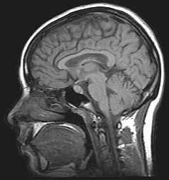
Cerebrospinal fluid (CSF) is a clear, colorless body fluid found within the tissue that surrounds the brain and spinal cord of all vertebrates.

<span class="mw-page-title-main">Idiopathic intracranial hypertension</span> Medical condition

Idiopathic intracranial hypertension (IIH), previously known as pseudotumor cerebri and benign intracranial hypertension, is a condition characterized by increased intracranial pressure without a detectable cause. The main symptoms are headache, vision problems, ringing in the ears, and shoulder pain. Complications may include vision loss.

<span class="mw-page-title-main">Hydrocephalus</span> Abnormal increase in cerebrospinal fluid in the ventricles of the brain

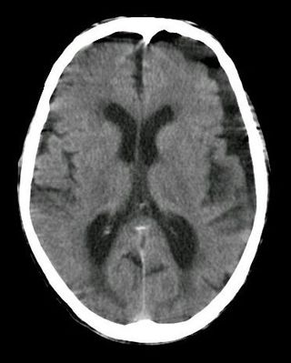
Hydrocephalus is a condition in which an accumulation of cerebrospinal fluid (CSF) occurs within the brain. This typically causes increased pressure inside the skull. Older people may have headaches, double vision, poor balance, urinary incontinence, personality changes, or mental impairment. In babies, it may be seen as a rapid increase in head size. Other symptoms may include vomiting, sleepiness, seizures, and downward pointing of the eyes.

<span class="mw-page-title-main">Intracranial pressure</span> Pressure exerted by fluids inside the skull and on the brain

Intracranial pressure (ICP) is the pressure exerted by fluids such as cerebrospinal fluid (CSF) inside the skull and on the brain tissue. ICP is measured in millimeters of mercury (mmHg) and at rest, is normally 7–15 mmHg for a supine adult. The body has various mechanisms by which it keeps the ICP stable, with CSF pressures varying by about 1 mmHg in normal adults through shifts in production and absorption of CSF.

<span class="mw-page-title-main">Subarachnoid hemorrhage</span> Bleeding into the subarachnoid space

Subarachnoid hemorrhage (SAH) is bleeding into the subarachnoid space—the area between the arachnoid membrane and the pia mater surrounding the brain. Symptoms may include a severe headache of rapid onset, vomiting, decreased level of consciousness, fever, weakness, numbness, and sometimes seizures. Neck stiffness or neck pain are also relatively common. In about a quarter of people a small bleed with resolving symptoms occurs within a month of a larger bleed.

<span class="mw-page-title-main">Intracranial hemorrhage</span> Hemorrhage, or bleeding, within the skull

Intracranial hemorrhage (ICH), also known as intracranial bleed, is bleeding within the skull. Subtypes are intracerebral bleeds, subarachnoid bleeds, epidural bleeds, and subdural bleeds. More often than not it ends in death.

Normal-pressure hydrocephalus (NPH), also called malresorptive hydrocephalus, is a form of communicating hydrocephalus in which excess cerebrospinal fluid (CSF) occurs in the ventricles, and with normal or slightly elevated cerebrospinal fluid pressure. As the fluid builds up, it causes the ventricles to enlarge and the pressure inside the head to increase, compressing surrounding brain tissue and leading to neurological complications. The disease presents in a classic triad of symptoms, which are memory impairment, urinary frequency, and balance problems/gait deviations. The disease was first described by Salomón Hakim and Adams in 1965.

<span class="mw-page-title-main">Intracerebral hemorrhage</span> Type of intracranial bleeding that occurs within the brain tissue itself

Intracerebral hemorrhage (ICH), also known as hemorrhagic stroke, is a sudden bleeding into the tissues of the brain, into its ventricles, or into both. An ICH is a type of bleeding within the skull and one kind of stroke. Symptoms can vary dramatically depending on the severity, acuity, and location (anatomically) but can include headache, one-sided weakness, numbness, tingling, or paralysis, speech problems, vision or hearing problems, memory loss, attention problems, coordination problems, balance problems, dizziness or lightheadedness or vertigo, nausea/vomiting, seizures, decreased level of consciousness or total loss of consciousness, neck stiffness, and fever.

<span class="mw-page-title-main">Ventriculitis</span> Inflammation of the ventricles in the brain

Ventriculitis is the inflammation of the ventricles in the brain. The ventricles are responsible for containing and circulating cerebrospinal fluid throughout the brain. Ventriculitis is caused by infection of the ventricles, leading to swelling and inflammation. This is especially prevalent in patients with external ventricular drains and intraventricular stents. Ventriculitis can cause a wide variety of short-term symptoms and long-term side effects ranging from headaches and dizziness to unconsciousness and death if not treated early. It is treated with some appropriate combination of antibiotics in order to rid the patient of the underlying infection. Much of the current research involving ventriculitis focuses specifically around defining the disease and what causes it. This will allow for much more advancement in the subject. There is also a lot of attention being paid to possible treatments and prevention methods to help make this disease even less prevalent and dangerous.

<span class="mw-page-title-main">Subdural hygroma</span> Collection of cerebrospinal fluid (CSF), without blood, located under the dural membrane

A subdural hygroma (SDG) is a collection of cerebrospinal fluid (CSF), without blood, located under the dural membrane of the brain. Most subdural hygromas are believed to be derived from chronic subdural hematomas. They are commonly seen in elderly people after minor trauma but can also be seen in children following infection or trauma. One of the common causes of subdural hygroma is a sudden decrease in pressure as a result of placing a ventricular shunt. This can lead to leakage of CSF into the subdural space especially in cases with moderate to severe brain atrophy. In these cases the symptoms such as mild fever, headache, drowsiness and confusion can be seen, which are relieved by draining this subdural fluid.

<span class="mw-page-title-main">Colloid cyst</span> Medical condition

A colloid cyst is a non-malignant tumor in the brain. It consists of a gelatinous material contained within a membrane of epithelial tissue. It is almost always found just posterior to the foramen of Monro in the anterior aspect of the third ventricle, originating from the roof of the ventricle. Because of its location, it can cause obstructive hydrocephalus and increased intracranial pressure. Colloid cysts represent 0.5–1.0% of intracranial tumors.

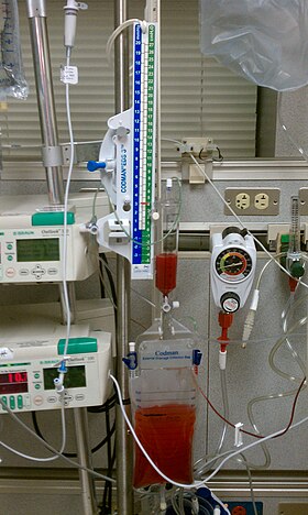
Ventriculostomy is a neurosurgical procedure that involves creating a hole (stoma) within a cerebral ventricle for drainage. It is most commonly performed on those with hydrocephalus. It is done by surgically penetrating the skull, dura mater, and brain such that the ventricular system ventricle of the brain is accessed. When catheter drainage is temporary, it is commonly referred to as an external ventricular drain (EVD). When catheter drainage is permanent, it is usually referred to as a shunt. There are many catheter-based ventricular shunts that are named for where they terminate, for example, a ventriculi-peritoneal shunt terminates in the peritoneal cavity, a ventriculoarterial shunt terminates within the atrium of the heart, etc. The most common entry point on the skull is called Kocher's point, which is measured 11 cm posterior to the nasion and 3 cm lateral to midline. EVD ventriculostomy is done primarily to monitor the intracranial pressure as well as to drain cerebrospinal fluid (CSF), primarily, or blood to relieve pressure from the central nervous system (CNS).

<span class="mw-page-title-main">Cerebral shunt</span> Surgical implant to treat hydrocephalus

A cerebral shunt is a device permanently implanted inside the head and body to drain excess fluid away from the brain. They are commonly used to treat hydrocephalus, the swelling of the brain due to excess buildup of cerebrospinal fluid (CSF). If left unchecked, the excess CSF can lead to an increase in intracranial pressure (ICP), which can cause intracranial hematoma, cerebral edema, crushed brain tissue or herniation. The drainage provided by a shunt can alleviate or prevent these problems in patients with hydrocephalus or related diseases.

<span class="mw-page-title-main">Intraventricular hemorrhage</span> Medical condition

Intraventricular hemorrhage (IVH), also known as intraventricular bleeding, is a bleeding into the brain's ventricular system, where the cerebrospinal fluid is produced and circulates through towards the subarachnoid space. It can result from physical trauma or from hemorrhagic stroke.

<span class="mw-page-title-main">Neurointensive care</span> Branch of medicine that deals with life-threatening diseases of the nervous system

Neurocritical care is a medical field that treats life-threatening diseases of the nervous system and identifies, prevents, and treats secondary brain injury.

Endoscopic third ventriculostomy (ETV) is a surgical procedure for treatment of hydrocephalus in which an opening is created in the floor of the third ventricle using an endoscope placed within the ventricular system through a burr hole. This allows the cerebrospinal fluid to flow directly to the basal cisterns, bypassing the obstruction. Specifically, the opening is created in the translucent tuber cinereum on the third ventricular floor.

A lumbar–peritoneal shunt is a technique to channelise the cerebrospinal fluid (CSF) from the lumbar thecal sac into the peritoneal cavity.

<span class="mw-page-title-main">Low pressure hydrocephalus</span> Medical condition

Low-pressure hydrocephalus (LPH) is a condition whereby ventricles are enlarged and the individual experiences severe dementia, inability to walk, and incontinence – despite very low intracranial pressure (ICP).

The monitoring of intracranial pressure (ICP) is used in the treatment of a number of neurological conditions ranging from severe traumatic brain injury to stroke and brain bleeds. This process is called intracranial pressure monitoring. Monitoring is important as persistent increases in ICP is associated with worse prognosis in brain injuries due to decreased oxygen delivery to the injured area and risk of brain herniation.

<span class="mw-page-title-main">Aqueductal stenosis</span> Narrowing of the aqueduct of Sylvius

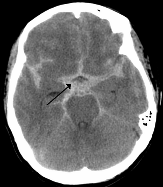
Aqueductal stenosis is a narrowing of the aqueduct of Sylvius which blocks the flow of cerebrospinal fluid (CSF) in the ventricular system. Blockage of the aqueduct can lead to hydrocephalus, specifically as a common cause of congenital and/or obstructive hydrocephalus.

References

  1. Rammos, S; et al. (2008), "Conversion of external ventricular drains to ventriculoperitoneal shunts after aneurysmal subarachnoid hemorrhage: effects of site and protein/red blood cell counts on shunt infection and malfunction", J Neurosurg, 109 (6): 1001–1004, doi:10.3171/JNS.2008.109.12.1001, PMID   19035711, S2CID   34114285.
  2. Monisha Kumar; Joshua Levine; James Schuster; W. Andrew Kofke (20 January 2017). Neurocritical Care Management of the Neurosurgical Patient E-Book. Elsevier Health Sciences. pp. 265–. ISBN   978-0-323-32222-5.
  3. 1 2 Mark R. Proctor (23 November 2007). Minimally Invasive Neurosurgery. Springer Science & Business Media. pp. 402–. ISBN   978-1-59259-899-1.
  4. Terje Sundstrøm; Per-Olof Grände; Niels Juul; Carsten Kock-Jensen, Bertil Romner and Knut Wester (25 June 2012). Management of Severe Traumatic Brain Injury: Evidence, Tricks, and Pitfalls. Springer Science & Business Media. pp. 120–. ISBN   978-3-642-28126-6.
  5. John M. O'Donnell; Flávio E. Nácul (12 July 2016). Surgical Intensive Care Medicine. Springer. pp. 226–. ISBN   978-3-319-19668-8.
  6. 1 2 "Care of the Patient Undergoing Intracranial Pressure Monitoring/ External Ventricular Drainage or Lumbar Drainage". AANN Clinical Practice Guideline Series. American Association of Neuroscience Nurses. 23 October 2012 [accessed].
  7. Olson, DaiWai M.; Zomorodi, Meg; Britz, Gavin W.; Zomorodi, Ali R.; Amato, Anthony; Graffagnino, Carmelo (October 2013). "Continuous cerebral spinal fluid drainage associated with complications in patients admitted with subarachnoid hemorrhage". Journal of Neurosurgery. 119 (4): 974–980. doi: 10.3171/2013.6.JNS122403 . PMID   23957382.
  8. 1 2 Muralidharan R (2015). "External ventricular drains: Management and complications". Surgical Neurology International. 6 (Suppl 6): S271–4. doi:10.4103/2152-7806.157620. PMC   4450504 . PMID   26069848.{{cite journal}}: CS1 maint: unflagged free DOI (link)
  9. Kakarla UK, Kim LJ, Chang SW, Theodore N, Spetzler RF (2008). "Safety and accuracy of bedside external ventricular drain placement". Neurosurgery. 63 (suppl_1): ONS162–6, discussion ONS166–7. doi:10.1227/01.NEU.0000312390.83127.7F. PMID   18728595. S2CID   71985774.
  10. Babu, Maya A.; Patel, Robin; Marsh, W. Richard; Wijdicks, Eelco F. M. (2012-02-01). "Strategies to decrease the risk of ventricular catheter infections: a review of the evidence". Neurocritical Care. 16 (1): 194–202. doi:10.1007/s12028-011-9647-z. ISSN   1556-0961. PMID   22045248. S2CID   43099710.
  11. Beer, R.; Lackner, P.; Pfausler, B.; Schmutzhard, E. (2008-11-01). "Nosocomial ventriculitis and meningitis in neurocritical care patients". Journal of Neurology. 255 (11): 1617–1624. doi: 10.1007/s00415-008-0059-8 . ISSN   0340-5354. PMID   19156484. S2CID   38800504.
  12. Flint, Alexander C.; Rao, Vivek A.; Renda, Natalie C.; Faigeles, Bonnie S.; Lasman, Todd E.; Sheridan, William (2013-06-01). "A simple protocol to prevent external ventricular drain infections". Neurosurgery. 72 (6): 993–999, discussion 999. doi:10.1227/NEU.0b013e31828e8dfd. ISSN   1524-4040. PMID   23467249. S2CID   29117121.
  13. Flint, Alexander C.; Toossi, Shahed; Chan, Sheila L.; Rao, Vivek A.; Sheridan, William (2016-12-21). "A simple infection control protocol durably reduces external ventricular drain infections to near-zero levels". World Neurosurgery. 99: 518–523. doi:10.1016/j.wneu.2016.12.042. ISSN   1878-8769. PMID   28012890.
  14. "EVD Infection Control". www.cleanbrain.org. Retrieved 2017-01-16.
  15. 1 2 Chai FY, Farizal F, Jegan T (2013). "Coma due to malplaced external ventricular drain". Turkish Neurosurgery. 23 (4): 561–563. doi: 10.5137/1019-5149.JTN.5724-12.1 . PMID   24101284.