Related Research Articles

The urethra is a tube that connects the urinary bladder to the urinary meatus for the removal of urine from the body of both females and males. In human females and other primates, the urethra connects to the urinary meatus above the vagina, whereas in marsupials, the female's urethra empties into the urogenital sinus.

The urinary system, also known as the urinary tract or renal system, consists of the kidneys, ureters, bladder, and the urethra. The purpose of the urinary system is to eliminate waste from the body, regulate blood volume and blood pressure, control levels of electrolytes and metabolites, and regulate blood pH. The urinary tract is the body's drainage system for the eventual removal of urine. The kidneys have an extensive blood supply via the renal arteries which leave the kidneys via the renal vein. Each kidney consists of functional units called nephrons. Following filtration of blood and further processing, wastes exit the kidney via the ureters, tubes made of smooth muscle fibres that propel urine towards the urinary bladder, where it is stored and subsequently expelled from the body by urination (voiding). The female and male urinary system are very similar, differing only in the length of the urethra.

The ureters are tubes made of smooth muscle that propel urine from the kidneys to the urinary bladder. In a human adult, the ureters are usually 20–30 cm (8–12 in) long and around 3–4 mm (0.12–0.16 in) in diameter. The ureter is lined by urothelial cells, a type of transitional epithelium, and has an additional smooth muscle layer that assists with peristalsis in its lowest third.

Retrograde ejaculation occurs when semen which would be ejaculated via the urethra is redirected to the urinary bladder. Normally, the sphincter of the bladder contracts before ejaculation, sealing the bladder which besides inhibiting the release of urine also prevents a reflux of seminal fluids into the male bladder during ejaculation. The semen is forced to exit via the urethra, the path of least resistance. When the bladder sphincter does not function properly, retrograde ejaculation may occur. It can also be induced deliberately by a male as a primitive form of male birth control or as part of certain alternative medicine practices. The retrograde-ejaculated semen, which goes into the bladder, is excreted with the next urination.

Hypospadias is a common variation in fetal development of the penis in which the urethra does not open from its usual location on the head of the penis. It is the second-most common birth abnormality of the male reproductive system, affecting about one of every 250 males at birth. Roughly 90% of cases are the less serious distal hypospadias, in which the urethral opening is on or near the head of the penis (glans). The remainder have proximal hypospadias, in which the meatus is all the way back on the shaft of the penis, near or within the scrotum. Shiny tissue that typically forms the urethra instead extends from the meatus to the tip of the glans; this tissue is called the urethral plate.

Chordee is a condition in which the head of the penis curves downward or upward, at the junction of the head and shaft of the penis. The curvature is usually most obvious during erection, but resistance to straightening is often apparent in the flaccid state as well. In many cases but not all, chordee is associated with hypospadias. This is not the same condition as Peyronie's disease, which involves curvature of the shaft of the penis most commonly due to injury during adult life.

The dartos fascia or simply dartos is a layer of connective tissue found in the penile shaft, foreskin, scrotum and labia. The penile portion is referred to as the superficial fascia of penis or the subcutaneous tissue of penis, while the scrotal part is the dartos proper. In addition to being continuous with itself between the scrotum and the penis, it is also continuous with Colles' fascia of the perineum and Scarpa's fascia of the abdomen.

The fascia of Scarpa is the deep membranous layer (stratum membranosum) of the superficial fascia of the abdomen. It is a layer of the anterior abdominal wall. It is found deep to the fascia of Camper and superficial to the external oblique muscle.

A ureteral stent, or ureteric stent, is a thin tube inserted into the ureter to prevent or treat obstruction of the urine flow from the kidney. The length of the stents used in adult patients varies between 24 and 30 cm. Additionally, stents come in differing diameters or gauges, to fit different size ureters. The stent is usually inserted with the aid of a cystoscope. One or both ends of the stent may be coiled to prevent it from moving out of place; this is called a JJ stent, double J stent or pig-tail stent.

The superficial perineal pouch is a compartment of the perineum.

The spongy urethra is the longest part of the male urethra, and is contained in the corpus spongiosum of the penis.

In human male anatomy, the dorsal veins of the penis are blood vessels that drain the shaft, the skin and the glans of the human penis. They are typically located in the midline on the dorsal aspect of the penis and they comprise the superficial dorsal veinof the penis, that lies in the subcutaneous tissue of the shaft, and the deep dorsal veinof the penis, that lies beneath the deep fascia.
Buck's fascia is a layer of deep fascia covering the three erectile bodies of the penis.
Urethroplasty is the surgical repair of an injury or defect within the walls of the urethra. Trauma, iatrogenic injury and infections are the most common causes of urethral injury/defect requiring repair. Urethroplasty is regarded as the gold standard treatment for urethral strictures and offers better outcomes in terms of recurrence rates than dilatations and urethrotomies. It is probably the only useful modality of treatment for long and complex strictures though recurrence rates are higher for this difficult treatment group.
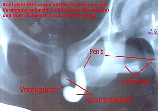
A retrograde urethrography is a routine radiologic procedure used to image the integrity of the urethra. Hence a retrograde urethrogram is essential for diagnosis of urethral injury, or urethral stricture.

In human male anatomy, the radix or root of the penis is the internal and most proximal portion of the human penis that lies in the perineum. Unlike the pendulous body of the penis which is suspended from the pubic symphysis, the root is attached to the pubic arch of the pelvis and is not visible externally. It is triradiate in form, consisting of three masses of erectile tissue; the two diverging crura, one on either side, and the median bulb of the penis or urethral bulb. Approximately one third to one half of the penis is embedded in the pelvis and can be felt through the scrotum and in the perineum.
Bulbar urethral necrosis is a problem that can occur after a pelvic fracture associated urethral distraction defect (PFUDD).

Webbed penis also known as buried or concealed penis is an acquired or congenital condition in which the scrotal skin extends onto the ventral penile shaft. The penile shaft is buried in the scrotum or tethered to the scrotal midline by a fold or web of skin. The urethra and erectile bodies are usually normal. Webbed penis is usually asymptomatic, but the cosmetic appearance is often unacceptable. This condition may be corrected by surgical techniques.
Rupture of the urethra is an uncommon result of penile injury, incorrect catheter insertion, straddle injury, or pelvic girdle fracture. The urethra, the muscular tube that allows for urination, may be damaged by trauma. When urethral rupture occurs, urine may extravasate (escape) into the surrounding tissues. The membranous urethra is most likely to be injured in pelvic fractures, allowing urine and blood to enter the deep perineal space and subperitoneal spaces via the genital hiatus. The spongy urethra is most likely to be injured with a catheter or in a straddle injury, allowing urine and blood to escape into the scrotum, the penis, and the superficial peritoneal space. Urethral rupture may be diagnosed with a cystourethrogram. Due to the tight adherence of the fascia lata, urine from a urethral rupture cannot spread into the thighs.
The genitourinary tract, or simply the urinary tract, consists of the kidneys, ureters, bladder, and the urethra. The kidney is the most frequently injured. Injuries to the kidney commonly occur after automobile or sports-related accidents. A blunt force is involved in 80-85% of injuries. Major decelerations can result in vascular injuries near the kidney's hilum. Gunshots and knife wounds and fractured ribs can result in penetrating injuries to the kidney.
References
- ↑ Gild, Philipp; Kluth, Luis A.; Vetterlein, Malte W.; Engel, Oliver; Chun, Felix K.H.; Fisch, Margit (2018). "Adult iatrogenic ureteral injury and stricture–incidence and treatment strategies". Asian Journal of Urology. 5 (2): 101–106. doi:10.1016/j.ajur.2018.02.003. PMC 5934506 . PMID 29736372.