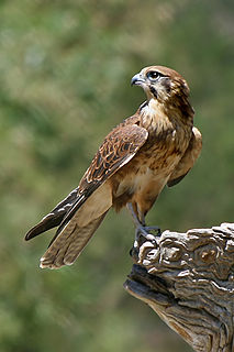
Falcons are birds of prey in the genus Falco, which includes about 40 species. Falcons are widely distributed on all continents of the world except Antarctica, though closely related raptors did occur there in the Eocene.

Rotavirus is a genus of double-stranded RNA viruses in the family Reoviridae. Rotaviruses are the most common cause of diarrhoeal disease among infants and young children. Nearly every child in the world is infected with a rotavirus at least once by the age of five. Immunity develops with each infection, so subsequent infections are less severe; adults are rarely affected. There are nine species of the genus, referred to as A, B, C, D, F, G, H, I and J. Rotavirus A, the most common species, causes more than 90% of rotavirus infections in humans. Rotavirus E, which is seen in pigs, has not been confirmed as a distinct species.
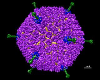
Adenoviruses are medium-sized, nonenveloped viruses with an icosahedral nucleocapsid containing a double stranded DNA genome. Their name derives from their initial isolation from human adenoids in 1953.

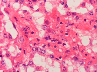
Kaposi's sarcoma-associated herpesvirus (KSHV) is the ninth known human herpesvirus; its formal name according to the International Committee on Taxonomy of Viruses (ICTV) is Human gammaherpesvirus 8, or HHV-8 in short. Like other herpesviruses, its informal names are used interchangeably with its formal ICTV name. This virus causes Kaposi's sarcoma, a cancer commonly occurring in AIDS patients, as well as primary effusion lymphoma, HHV-8-associated multicentric Castleman's disease and KSHV inflammatory cytokine syndrome. It is one of seven currently known human cancer viruses, or oncoviruses. Even after so many years of discovery of KSHV/HHV8, there is no known cure for KSHV associated tumorigenesis.
An oncolytic virus is a virus that preferentially infects and kills cancer cells. As the infected cancer cells are destroyed by oncolysis, they release new infectious virus particles or virions to help destroy the remaining tumour. Oncolytic viruses are thought not only to cause direct destruction of the tumour cells, but also to stimulate host anti-tumour immune system responses.

Carnivore protoparvovirus 1 is a species of parvovirus that infects carnivorans. It causes a highly contagious disease in both dogs and cats. The disease is generally divided into two major genogroups: CPV-1 containing the classical feline panleukopenia virus (FPLV), and CPV-2 containing the canine parvovirus (CPV) which appeared in the 1970s.
Aviadenoviruses are adenoviruses that affect birds—particularly chickens, ducks, geese, turkeys and pheasants. There are 15 species in this genus. Viruses in this genus cause specific disease syndromes such as Quail Bronchitis (QB), Egg Drop Syndrome (EDS), Haemorrhagic Enteritis (HE), Pheasant Marble Spleen Disease (MSD), and Inclusion Body Hepatitis (IBH). Avian adenoviruses have a worldwide distribution and it is common to find multiple species on a single farm. The most common serogroups are serogroup 1, 2 and 3.
Adenovirus infections most commonly cause illness of the respiratory system; however, depending on the infecting serotype, they may also cause various other illnesses and presentations.

Astroviruses are a type of virus that was first discovered in 1975 using electron microscopes following an outbreak of diarrhea in humans. In addition to humans, astroviruses have now been isolated from numerous mammalian animal species and from avian species such as ducks, chickens, and turkey poults. Astroviruses are 28–35 nm diameter, icosahedral viruses that have a characteristic five- or sixpointed star-like surface structure when viewed by electron microscopy. Along with the Picornaviridae and the Caliciviridae, the Astroviridae comprise a third family of nonenveloped viruses whose genome is composed of plus-sense, single-stranded RNA. Astrovirus has a non-segmented, single stranded, positive sense RNA genome within a non-enveloped icosahedral capsid. Human astroviruses have been shown in numerous studies to be an important cause of gastroenteritis in young children worldwide. In animals, Astroviruses also cause infection of the gastrointestinal tract but may also result in encephalitis, hepatitis (avian) and nephritis (avian).
Bovine alphaherpesvirus 1 (BoHV-1) is a virus of the family Herpesviridae and the subfamily Alphaherpesvirinae, known to cause several diseases worldwide in cattle, including rhinotracheitis, vaginitis, balanoposthitis, abortion, conjunctivitis, and enteritis. BoHV-1 is also a contributing factor in shipping fever, also known as bovine respiratory disease (BRD). It is spread horizontally through sexual contact, artificial insemination, and aerosol transmission and it may also be transmitted vertically across the placenta. BoHV-1 can cause both clinical and subclinical infections, depending on the virulence of the strain. Although these symptoms are mainly non-life-threatening it is an economically important disease as infection may cause a drop in production and affect trade restrictions. Like other herpesviruses, BoHV-1 causes a lifelong latent infection and sporadic shedding of the virus. The sciatic nerve and trigeminal nerve are the sites of latency. A reactivated latent carrier is normally the source of infection in a herd. The clinical signs displayed are dependent on the virulence of the strain. There is a vaccine available which reduces the severity and incidence of disease. Some countries in Europe have successfully eradicated the disease by applying a strict culling policy.

Duck plague is a worldwide disease caused by Anatid alphaherpesvirus 1 (AnHV-1) of the family Herpesviridae that causes acute disease with high mortality rates in flocks of ducks, geese, and swans. It is spread both vertically and horizontally—through contaminated water and direct contact. Migratory waterfowl are a major factor in the spread of this disease as they are often asymptomatic carriers of disease. The incubation period is three to seven days. Birds as young as one week old can be infected. DEV is not zoonotic.
A helper dependent virus, also termed a gutless virus, is a synthetic viral vector dependent on the assistance of a helper virus in order to replicate, and can be used for purposes such as gene therapy. Naturally-occurring satellite viruses are also helper virus dependent, and can sometimes be modified to become viral vectors.

A subclinical infection — sometimes called a preinfection or inapparent infection — is an infection that, being subclinical, is nearly or completely asymptomatic. A subclinically infected person is thus an asymptomatic carrier of a microbe, intestinal parasite, or virus that usually is a pathogen causing illness, at least in some individuals. Many pathogens spread by being silently carried in this way by some of their host population. Such infections occur both in humans and animals.

Agamid adenovirus, also called Bearded dragon adenovirus 1, is a type of virus in the Adenoviridae family. The virus is widespread in captive populations of Pogona vitticeps, known commonly as the central bearded dragon, in the United States. Other countries with confirmed cases are Australia, Japan, Germany, The Netherlands, Belgium, the UK, and El Salvador. It is often discovered in association with other infections, and causes increased juvenile mortality and adult deaths.
Gianotti–Crosti syndrome, also known as infantile papular acrodermatitis, papular acrodermatitis of childhood, and papulovesicular acrolocated syndrome, is a reaction of the skin to a viral infection. Hepatitis B virus and Epstein–Barr virus are the most frequently reported pathogens. Other viruses implicated are hepatitis A virus, hepatitis C virus, cytomegalovirus, coxsackievirus, adenovirus, enterovirus, rotavirus, rubella virus, HIV, and parainfluenza virus.
Ehrlichiosis ewingii infection is an infectious disease caused by an intracellular bacteria, Ehrlichia ewingii. The infection is transmitted to humans by the tick, Amblyomma americanum. This tick can also transmit Ehrlichia chaffeensis, the bacteria that causes human monocytic ehrlichiosis (HME).
The epidemiology of herpes simplex is of substantial epidemiologic and public health interest. Worldwide, the rate of infection with herpes simplex virus—counting both HSV-1 and HSV-2—is around 90%. Although many people infected with HSV develop labial or genital lesions, the majority are either undiagnosed or display no physical symptoms—individuals with no symptoms are described as asymptomatic or as having subclinical herpes.
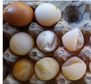
Egg drop syndrome '76 is one of the important viral diseases of birds, notably chickens, ducks, geese and swans. It is characterised by a sudden drop in production of eggs as well as its eggshell quality in apparent healthy laying birds.
KI polyomavirus is a virus of the family Polyomaviridae. It was discovered in 2007 in stored samples of human respiratory secretions collected by the Karolinska Institute, after which the virus is named.
MW polyomavirus is a virus of the polyomavirus family that infects human hosts. It was discovered in 2012 and reported independently by several research groups. It has been identified mostly in stool samples from children and has been detected in a variety of geographic locations.