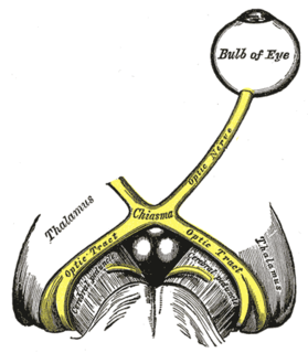
Septo-optic dysplasia (SOD), is a rare congenital malformation syndrome featuring underdevelopment of the optic nerve, pituitary gland dysfunction, and absence of the septum pellucidum. Two of these features need to be present for a clinical diagnosis — only 30% of patients have all three. De Mosier first recognized the relation of a rudimentary or absent septum pellucidum with hypoplasia of the optic nerves and chiasm in 1956.

In dogs, hip dysplasia is an abnormal formation of the hip socket that, in its more severe form, can eventually cause crippling lameness and painful arthritis of the joints. It is a genetic (polygenic) trait that is affected by environmental factors. It is common in many dog breeds, particularly the larger breeds, and is the most common single cause of arthritis of the hips.
An osteotomy is a surgical operation whereby a bone is cut to shorten or lengthen it or to change its alignment. It is sometimes performed to correct a hallux valgus, or to straighten a bone that has healed crookedly following a fracture. It is also used to correct a coxa vara, genu valgum, and genu varum. The operation is done under a general anaesthetic.

Coxa vara is a deformity of the hip, whereby the angle between the head and the shaft of the femur is reduced to less than 120 degrees. This results in the leg being shortened, and the development of a limp. It is commonly caused by injury, such as a fracture. It can also occur when the bone tissue in the neck of the femur is softer than normal, causing it to bend under the weight of the body. This may either be congenital or the result of a bone disorder. The most common cause of coxa vara is either congenital or developmental. Other common causes include metabolic bone diseases, post-Perthes deformity, osteomyelitis, and post traumatic. Shepherd's Crook deformity is a severe form of coxa vara where the proximal femur is severely deformed with a reduction in the neck shaft angle beyond 90 degrees. It is most commonly a sequela of osteogenesis imperfecta, Pagets disease, osteomyelitis, tumour and tumour-like conditions.
Arthroplasty is an orthopedic surgical procedure where the articular surface of a musculoskeletal joint is replaced, remodeled, or realigned by osteotomy or some other procedure. It is an elective procedure that is done to relieve pain and restore function to the joint after damage by arthritis or some other type of trauma.

Dysplasia is a term used in pathology to refer to an abnormality of development or an epithelial anomaly of growth and differentiation.

The gluteal sulcus is an area of the body of humans and great apes, described by a horizontal crease formed by the inferior aspect of the buttocks and the posterior upper thigh. The gluteal sulcus is formed by the posterior horizontal skin crease of the hip joint and overlying fat, and is not formed by the lower border of the gluteus maximus muscle, which crosses the fold obliquely. It is one of the major defining features of the buttocks. Children with developmental dysplasia of the hips are born with uneven gluteal folds and can be diagnosed with physical examination and sonogram.

A hip dislocation a disruption of the joint between the femur and pelvis. Specifically it is when the ball–shaped head of the femur comes out of the cup–shaped acetabulum of the pelvis. Symptoms typically include pain and an inability move the hip. Complications may include avascular necrosis of the hip, injury to the sciatic nerve, or arthritis.
Fairbank's disease or multiple epiphyseal dysplasia (MED) is a rare genetic disorder that affects the growing ends of bones. Long bones normally elongate by expansion of cartilage in the growth plate near their ends. As it expands outward from the growth plate, the cartilage mineralizes and hardens to become bone (ossification). In MED, this process is defective.

Pseudoachondroplasia is an inherited disorder of bone growth. It is a genetic autosomal dominant disorder. It is generally not discovered until 2–3 years of age, since growth is normal at first. Pseudoachondroplasia is usually first detected by a drop of linear growth in contrast to peers, a curious, waddling gait or arising lower limb deformities.

The Ortolani test is part of the physical examination for developmental dysplasia of the hip, along with the Barlow maneuver. Specifically, the Ortolani test is positive when a posterior dislocation of the hip is reducible with this maneuver. This is part of the standard infant exam performed preferably in early infancy.
A GALS screen is an examination used by doctors and other healthcare professionals to detect locomotor abnormalities and functional disability relating to gait, arms, legs and the spine.
The Barlow maneuver is a physical examination performed on infants to screen for developmental dysplasia of the hip. It is named for Dr. Thomas Geoffrey Barlow, an English orthopedic surgeon, who devised this test. It was clinically tested during 1957–1962 at Hope Hospital, Salford, Lancashire.

Hip dysplasia is an abnormality of the hip joint where the socket portion does not fully cover the ball portion, resulting in an increased risk for joint dislocation. congenital dysplasia of the hip (CDH) Hip dysplasia may occur at birth or develop in early life. Regardless, it does not typically produce symptoms in babies less than a year old. Occasionally one leg may be shorter than the other. The left hip is more often affected than the right. Complications without treatment can include arthritis, limping, and low back pain.

Trevor disease, also known as Fairbank's disease and Trevor's disease, is a congenital bone developmental disorder. There is 1 case per million population. The condition is three times more common in males than in females.
The Thomas test is a physical examination test, named after Dr. Hugh Owen Thomas (1834–1891), a British orthopaedic surgeon, used to rule out hip flexion contracture and psoas syndrome. Often associated with runners, dancers, and gymnasts who complain of hip "stiffness" and reported "snapping" feeling when flexing at the waist.
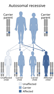
Spondyloepimetaphyseal dysplasia, Pakistani type is a form of spondyloepimetaphyseal dysplasia involving PAPSS2. The condition is rare.
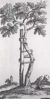
Orthopedic surgery is the branch of surgery concerned with conditions involving the musculoskeletal system. Orthopedic surgeons use both surgical and nonsurgical means to treat musculoskeletal injuries, sports injuries, degenerative diseases, infections, bone tumours, and congenital limb deformities. Trauma surgery and traumatology is a sub-specialty dealing with the operative management of fractures, major trauma and the multiply-injured patient.
X-rays of hip dysplasia are one of the two main methods of medical imaging to diagnose hip dysplasia, the other one being medical ultrasonography.. Ultrasound imaging yields better results defining the anatomy until the cartilage is ossified. When the infant is around 3 months old a clear roentgenographic image can be achieved. Unfortunately the time the joint gives a good x-ray image is also the point at which nonsurgical treatment methods cease to give good results.
