Related Research Articles

In humans and other primates, the knee joins the thigh with the leg and consists of two joints: one between the femur and tibia, and one between the femur and patella. It is the largest joint in the human body. The knee is a modified hinge joint, which permits flexion and extension as well as slight internal and external rotation. The knee is vulnerable to injury and to the development of osteoarthritis.

The posterior cruciate ligament (PCL) is a ligament in each knee of humans and various other animals. It works as a counterpart to the anterior cruciate ligament (ACL). It connects the posterior intercondylar area of the tibia to the medial condyle of the femur. This configuration allows the PCL to resist forces pushing the tibia posteriorly relative to the femur.
A Baker's cyst, also known as a popliteal cyst, is a type of fluid collection behind the knee. Often there are no symptoms. If symptoms do occur these may include swelling and pain behind the knee, or knee stiffness. If the cyst breaks open, pain may significantly increase with swelling of the calf. Rarely complications such as deep vein thrombosis, peripheral neuropathy, ischemia, or compartment syndrome may occur.
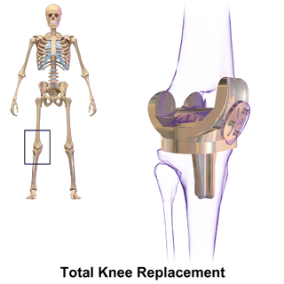
Knee replacement, also known as knee arthroplasty, is a surgical procedure to replace the weight-bearing surfaces of the knee joint to relieve pain and disability, most commonly offered when joint pain is not diminished by conservative sources and also for other knee diseases such as rheumatoid arthritis and psoriatic arthritis. In patients with severe deformity from advanced rheumatoid arthritis, trauma, or long-standing osteoarthritis, the surgery may be more complicated and carry higher risk. Osteoporosis does not typically cause knee pain, deformity, or inflammation and is not a reason to perform knee replacement.

The popliteus muscle in the leg is used for unlocking the knees when walking, by laterally rotating the femur on the tibia during the closed chain portion of the gait cycle. In open chain movements, the popliteus muscle medially rotates the tibia on the femur. It is also used when sitting down and standing up. It is the only muscle in the posterior (back) compartment of the lower leg that acts just on the knee and not on the ankle. The gastrocnemius muscle acts on both joints.

The plantaris is one of the superficial muscles of the superficial posterior compartment of the leg, one of the fascial compartments of the leg.

In medicine, the Ottawa ankle rules are a set of guidelines for clinicians to help decide if a patient with foot or ankle pain should be offered X-rays to diagnose a possible bone fracture. Before the introduction of the rules most patients with ankle injuries would have been imaged. However the vast majority of patients with unclear ankle injuries do not have bone fractures. As a result, many unnecessary X-rays were taken, which was costly, time-consuming and a slight health risk due to radiation exposure.
The Apley grind test or Apley test is used to evaluate individuals for problems in the meniscus of the knee. The Apley grind test has a reported sensitivity of 97% and a specificity of 87%.
A meniscus transplant or meniscal transplant is a transplant of the meniscus of the knee, which separates the thigh bone (femur) from the lower leg bone (tibia). The worn or damaged meniscus is removed and is replaced with a new one from a donor. The meniscus to be transplanted is taken from a cadaver, and, as such, is known as an allograft. Meniscal transplantation is technically difficult, as it must be sized accurately for each person, positioned properly and secured to the tibial plateau. As of 2012, only a few surgeons have significant volume of experience in meniscus transplantation worldwide.
The lateral meniscus is a fibrocartilaginous band that spans the lateral side of the interior of the knee joint. It is one of two menisci of the knee, the other being the medial meniscus. It is nearly circular and covers a larger portion of the articular surface than the medial. It can occasionally be injured or torn by twisting the knee or applying direct force, as seen in contact sports.
The knee examination, in medicine and physiotherapy, is performed as part of a physical examination, or when a patient presents with knee pain or a history that suggests a pathology of the knee joint.
An anterior cruciate ligament injury occurs when the anterior cruciate ligament (ACL) is either stretched, partially torn, or completely torn. The most common injury is a complete tear. Symptoms include pain, an audible cracking sound during injury, instability of the knee, and joint swelling. Swelling generally appears within a couple of hours. In approximately 50% of cases, other structures of the knee such as surrounding ligaments, cartilage, or meniscus are damaged.
The unhappy triad, also known as a blown knee among other names, is an injury to the anterior cruciate ligament, medial collateral ligament, and meniscus. Analysis during the 1990s indicated that this 'classic' O'Donoghue triad is actually an unusual clinical entity among athletes with knee injuries. Some authors mistakenly believe that in this type of injury, "combined anterior cruciate and medial collateral ligament disruptions that were incurred during athletic endeavors" always present with concomitant medial meniscus injury. However, the 1990 analysis showed that lateral meniscus tears are more common than medial meniscus tears in conjunction with sprains of the ACL.

The straight leg raise is a test that can be performed during a physical examination, with the leg being lifted actively by the patient or passively by the clinician. If the straight leg raise is done actively by the patient it is a test of functional leg strength, particularly the rectus femoris element of the quadriceps. If carried out passively, it is used to determine whether a patient with low back pain has an underlying nerve root sensitivity, often located at L5. The rest of this article relates to the passive version of the test.

A tear of a meniscus is a rupturing of one or more of the fibrocartilage strips in the knee called menisci. When doctors and patients refer to "torn cartilage" in the knee, they actually may be referring to an injury to a meniscus at the top of one of the tibiae. Menisci can be torn during innocuous activities such as walking or squatting. They can also be torn by traumatic force encountered in sports or other forms of physical exertion. The traumatic action is most often a twisting movement at the knee while the leg is bent. In older adults, the meniscus can be damaged following prolonged 'wear and tear'. Especially acute injuries can lead to displaced tears which can cause mechanical symptoms such as clicking, catching, or locking during motion of the joint. The joint will be in pain when in use, but when there is no load, the pain goes away.
The pivot-shift test is one of the three major tests for assessing anterior cruciate injury or laxity, the other two being the anterior drawer and Lachman test. However, unlike the other two, it tests for instability, an important determinant as to how the knee will function. In fact, it is instability, not simply the injury to the anterior cruciate ligament itself, that places the menisci at future risk, and gives rise to the feeling that the "knee is not secure" or "may give out".
In medicine, joint locking is a symptom of pathology in a joint. It is a complaint by a person when they are unable to fully flex or fully extend a joint.
Knee pain is pain in or around the knee.
Medial knee injuries are the most common type of knee injury. The medial ligament complex of the knee is composed of the superficial medial collateral ligament (sMCL), deep medial collateral ligament (dMCL), and the posterior oblique ligament (POL). These ligaments have also been called the medial collateral ligament (MCL), tibial collateral ligament, mid-third capsular ligament, and oblique fibers of the sMCL, respectively. This complex is the major stabilizer of the medial knee. Injuries to the medial side of the knee are most commonly isolated to these ligaments. A thorough understanding of the anatomy and function of the medial knee structures, along with a detailed history and physical exam, are imperative to diagnosing and treating these injuries.

The function of the posterior cruciate ligament (PCL) is to prevent the femur from sliding off the anterior edge of the tibia and to prevent the tibia from displacing posterior to the femur. Common causes of PCL injuries are direct blows to the flexed knee, such as the knee hitting the dashboard in a car accident or falling hard on the knee, both instances displacing the tibia posterior to the femur.
References
- ↑ Solomon, D. H.; Simel, D. L.; Bates, D. W.; Katz, J. N.; Schaffer, J. L. (2001). "The rational clinical examination. Does this patient have a torn meniscus or ligament of the knee? Value of the physical examination". JAMA: The Journal of the American Medical Association. 286 (13): 1610–1620. doi:10.1001/jama.286.13.1610. PMID 11585485. S2CID 111298239.
- 1 2 McMurray, T.P. (1942). "The semilunar cartilages". British Journal of Surgery. 29 (116): 407–414. doi:10.1002/bjs.18002911612. S2CID 72803852.
- ↑ Stratford, Paul W.; Binkley, Jill (1995). "A Review of the McMurray Test: Definition, Interpretation, and Clinical Usefulness". Journal of Orthopaedic & Sports Physical Therapy. 22 (3): 116–120. doi:10.2519/jospt.1995.22.3.116. PMID 8535469.
- ↑ Corea JR, Moussa M, al Othman A (1994). "McMurray's test tested". Knee Surg Sports Traumatol Arthrosc . 2 (2): 70–72. doi:10.1007/BF01476474. PMID 7584186. S2CID 34039963.
- ↑ Jackson JL, O'Malley PG, Kroenke K (2003). "Evaluation of acute knee pain in primary care". Ann Intern Med. 139 (7): 575–88. doi:10.7326/0003-4819-139-7-200310070-00010. PMID 14530229. S2CID 46705071.
{{cite journal}}: CS1 maint: multiple names: authors list (link)