Bone healing, or fracture healing, is a proliferative physiological process in which the body facilitates the repair of a bone fracture.

A sarcoma is a malignant tumor, a type of cancer that arises from cells of mesenchymal origin. Connective tissue is a broad term that includes bone, cartilage, muscle, fat, vascular, or other structural tissues, and sarcomas can arise in any of these types of tissues. As a result, there are many subtypes of sarcoma, which are classified based on the specific tissue and type of cell from which the tumor originates.

A bone tumor is an abnormal growth of tissue in bone, traditionally classified as noncancerous (benign) or cancerous (malignant). Cancerous bone tumors usually originate from a cancer in another part of the body such as from lung, breast, thyroid, kidney and prostate. There may be a lump, pain, or neurological signs from pressure. A bone tumor might present with a pathologic fracture. Other symptoms may include fatigue, fever, weight loss, anemia and nausea. Sometimes there are no symptoms and the tumour is found when investigating another problem.

An osteosarcoma (OS) or osteogenic sarcoma (OGS) is a cancerous tumor in a bone. Specifically, it is an aggressive malignant neoplasm that arises from primitive transformed cells of mesenchymal origin and that exhibits osteoblastic differentiation and produces malignant osteoid.

The periosteum is a membrane that covers the outer surface of all bones, except at the articular surfaces of long bones. Endosteum lines the inner surface of the medullary cavity of all long bones.

Hemipelvectomy, also known as a pelvic resection, is a surgical procedure that involves the removal of part of the pelvic girdle. This procedure is most commonly performed to treat oncologic conditions of the pelvis. Hemipelvectomy can be further classified as internal and external hemipelvectomy. An internal hemipelvectomy is a limb-sparing procedure where the innominate bone is resected while preserving the ipsilateral limb. An external hemipelvectomy involves the resection of the innominate bone plus amputation of the ipsilateral limb.
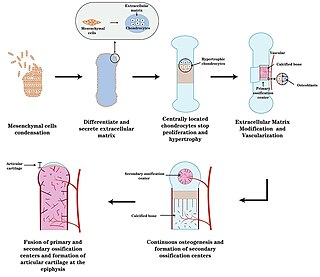
Endochondral ossification is one of the two essential pathways by which bone tissue is produced during fetal development of the mammalian skeletal system, the other pathway being intramembranous ossification. Both endochondral and intramembranous processes initiate from a precursor mesenchymal tissue, but their transformations into bone are different. In intramembranous ossification, mesenchymal tissue is directly converted into bone. On the other hand, endochondral ossification starts with mesenchymal tissue turning into an intermediate cartilage stage, which is eventually substituted by bone.

A chest radiograph, chest X-ray (CXR), or chest film is a projection radiograph of the chest used to diagnose conditions affecting the chest, its contents, and nearby structures. Chest radiographs are the most common film taken in medicine.

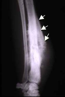
A periosteal reaction is the formation of new bone in response to injury or other stimuli of the periosteum surrounding the bone. It is most often identified on X-ray films of the bones.

Myositis ossificans comprises two syndromes characterized by heterotopic ossification (calcification) of muscle. The World Health Organization, 2020, has grouped myositis ossificans together with fibro-osseous pseudotumor of digits as a single specific entity in the category of fibroblastic and myofibroblastic tumors.

Giant-cell tumor of the bone (GCTOB), is a relatively uncommon bone tumor. It is characterized by the presence of multinucleated giant cells. Malignancy in giant-cell tumor is uncommon and occurs in about 2% of all cases. However, if malignant degeneration does occur, it is likely to metastasize to the lungs. Giant-cell tumors are normally benign, with unpredictable behavior.
Infantile cortical hyperostosis (ICH) is a self-limited inflammatory disorder of infants that causes bone changes, soft tissue swelling and irritability. The disease may be present at birth or occur shortly thereafter. The cause is unknown. Both familial and sporadic forms occur. It is also known as Caffey disease or Caffey's disease.

Chondroblastoma is a rare, benign, locally aggressive bone tumor that typically affects the epiphyses or apophyses of long bones. It is thought to arise from an outgrowth of immature cartilage cells (chondroblasts) from secondary ossification centers, originating from the epiphyseal plate or some remnant of it.

Osteoblastoma is an uncommon osteoid tissue-forming primary neoplasm of the bone.

Ewing sarcoma is a type of pediatric cancer that forms in bone or soft tissue. Symptoms may include swelling and pain at the site of the tumor, fever, and a bone fracture. The most common areas where it begins are the legs, pelvis, and chest wall. In about 25% of cases, the cancer has already spread to other parts of the body at the time of diagnosis. Complications may include a pleural effusion or paraplegia.

Orthopedic surgery is the branch of surgery concerned with conditions involving the musculoskeletal system. Orthopedic surgeons use both surgical and nonsurgical means to treat musculoskeletal injuries, sports injuries, degenerative diseases, infections, bone tumours, and congenital limb deformities. Trauma surgery and traumatology is a sub-specialty dealing with the operative management of fractures, major trauma and the multiply-injured patient.
Miriam Dorothy (Posner) Finkel was a radiobiologist who made significant contributions to molecular biology. Finkel lent her name to the Finkel-Biskis-Jinkins or FBJ virus.
A bone sarcoma is a primary malignant bone tumour, a type of sarcoma that starts in the bones. This is in contrast to most bone cancers that are secondary having developed as a metastasis from another cancer. Bone sarcomas are rare, and mostly affect the legs. The other type of sarcoma is a soft-tissue sarcoma.
Onion skin periosteal reaction also known as multilayered periosteal reaction or lamellated periosteal reaction refers to the multi layered concentric layers of new bone adjacent to the bone cortex. It is called onion skin periosteal reaction because it resembles the layers of an onion. These layers are formed due to any pathological process that leads to the variable, excessive growth of the bone. Onion skin periosteal reaction is seen in osteosarcoma, Ewing sarcoma and Langerhans cell histiocytosis. In radiological images, onion skin periosteal reaction is seen as radiolucent areas along the multiple layers of dense bone. The lucent areas may be occupied by tumors or inflammation.
Extraskeletal Ewing sarcoma (EES), is a cancer of soft tissue, a type of Ewing sarcoma that does not arise from bone.