Ghrelin O-acyltransferase

Last updated
MBOAT4
Identifiers
Aliases MBOAT4 , GOAT, OACT4, FKSG89, membrane bound O-acyltransferase domain containing 4
External IDs OMIM: 611940 MGI: 2685017 HomoloGene: 19272 GeneCards: MBOAT4
Orthologs
SpeciesHumanMouse
Entrez
Ensembl
UniProt
RefSeq (mRNA)

NM_001100916

NM_001126314

RefSeq (protein)

NP_001094386

NP_001119786

Location (UCSC) Chr 8: 30.13 – 30.14 Mb Chr 8: 34.58 – 34.59 Mb
PubMed search [3] [4]
Wikidata
View/Edit Human View/Edit Mouse

Ghrelin O-acyltransferase also known as membrane bound O-acyltransferase domain containing 4 is an enzyme that in humans is encoded by the MBOAT4 gene. [5] It is homologous to other membrane-bound O-acyltransferases. It is a polytopic membrane protein what takes part in lipid signaling reactions. It is the only known enzyme that catalyzes the acylation of ghrelin through the transfer of n-octanoic acid to ghrelin Ser3. [6] Ghrelin O-acyltransferase function is essential in regulation of appetite and the release of growth hormone. Ghrelin O-acyltransferase is a target for scientific research due to promising applications in the treatment of diabetes, eating disorders, and metabolic diseases.

Contents

Consistent with its function relative to ghrelin, ghrelin O-acyltransferase can be found in all vertebrates, including mammals, birds, and fish species. The enzyme is primarily expressed in the stomach and the gastrointestinal system. [6] Other sites where ghrelin O-acyltransferase can be found include tissue from the brain, the pancreas, the pituitary gland, and certain forms of cancer.

Structure

The structure of ghrelin O-acyltransferase has not been fully elucidated or tested experimentally. Using biochemical mapping tools, researchers showed that the ghrelin O-acyltransferase enzyme consists of eleven transmembrane helical domains and one reentrant loop. [7] The C-terminus of ghrelin O-acyltransferase is located on the cytosolic side of the endoplasmic reticulum (ER) while the N-terminus resides in the lumen of the membrane. The topology models of ghrelin O-acyltransferase are similar to those of acetyl-coenzyme A acetyltransferase 1 and glycerol uptake protein 1. This is consistent with ghrelin O-acyltransferase being a member of the membrane bound O-acyl transferase family.

Ghrelin O-acyltransferase contains conserved asparagine (Asn307) and histidine (His338) residues that are found across the membrane bound O-acyl transferase family. [7] These amino acids are positioned on opposite side of the endoplasmic reticulum membrane. His338 resides in the lumen and is necessary for the observed catalytic activity of the enzyme.  

Topology model of ghrelin O-acyltransferase spanning the ER membrane with the cytosolic part depicted below. Ghrelin O-Acyl Transferase Topology Model.jpg
Topology model of ghrelin O-acyltransferase spanning the ER membrane with the cytosolic part depicted below.

History

The first reported discovery of ghrelin O-acyl transferase was in February 2008 by two separate research groups at Eli Lilly and the University of Texas at Austin (UT). [6] The UT group identified orphan acyltransferase enzymes through the genome database and expressed both the enzymes in ghrelin in cells.[7] They found that membrane-bound O-acyltransferase domain 4 specifically acylated ghrelin. The Eli Lilly team confirmed that enzyme is necessary to modify the ghrelin peptide by performing GOAT knock down experiments to prevent expression on the gene. Currently, the major pharmaceutical companies with patents related to ghrelin O-acyl transferase are Eli Lilly, Takeda Pharmaceutical Company, and Boehringer Ingelheim.

Pharmaceutical development

One medical use of ghrelin O-acyl transferase inhibitors has been in the preclinical treatment of Prader–Willi syndrome, a rare genetic disorder resulting in early-onset diabetes. In 2018, Rhythm Pharmaceuticals announced a licensing agreement with Takeda Pharmaceutical to develop an orally administered ghrelin O-acyl transferase inhibitor in order to decrease the amount of active ghrelin in individuals suffering from Prader-Willi Syndrome. [8] Preclinical research has shown that this drug prevented weight gain with positive pharmacological and safety profiles.

Additionally, ghrelin O-acyl transferase inhibition has been used in clinical trials in the United Kingdom in studying appetite regulation of post-surgical bariatric patients. The study measured, among other things, the effect of decreased ghrelin levels on gut hormone, adipokine, and cytokine levels. [9]

Chemical properties

Structure-activity studies have demonstrated that the ‘GSSF’ N-terminal sequence of ghrelin is essential to ghrelin O-acyl transferase substrate recognition. [10] While the enzyme will accept sequences with a threonine reside in place of serine, catalytic activity is reduced in these cases. Ghrelin O-acyl transferase is also poorly stereospecific and is able to recognize and acylate both stereoisomers of serine. The enzymatic activity of GOAT is also altered when treated with cysteine-modifying substances. Interestingly, the enzyme is relatively insensitive to pH changes in the range of 6–9. [11] Kinetic studies also suggest that GOAT activity is consistent with Michaelis-Menten kinetics in the acylation of ghrelin. One paper proposes that bioavailability of the GOAT substrates represent the rate-limiting step of the acyl transfer reaction and not the chemical process itself.

Biochemical assays

Several biochemical assays have been developed to study the activity of the enzyme under in vitro conditions. The most common is the use of an amino acid and radioactive isotope tags to monitor the formation of reaction products. [12] Also widespread is the use of indirect methods to observe ghrelin O-acyl transferase function such as the detection of acylated ghrelin products, which indicate enzyme activity. These assays are based on high-throughput ELISA based techniques. [10] There has also been published literature describing protocols for ghrelin O-acyl transferase function expression and enrichment in insect cells, the use of high performance liquid chromatography assays for acylation activity, and the use of fluorescent protein labels. [13]

Metabolic function

In humans, the ghrelin O-acyl transferase function enzyme post-translationally modifies an inactive form of ghrelin, known as proghrelin, on an N-terminus serine residue. This function is essential in forming an active form of ghrelin, which can then be secreted into the bloodstream. Ghrelin O-acyltransferase also plays a role in preventing hypoglycemia when there is prolonged negative energy balance in the body. [14] GOAT plays a crucial role in the ability to maintain glucose levels during fasting and starvation. Additionally, the enzyme has a key role in modulating the secretion and sensitivity of insulin. These interactions are used in regulating glucose metabolism in the body.

Regulation

Ghrelin O-acyltransferase is highly regulated by energy balance and is upregulated when energy is restricted, such as when fasting. [14] Additionally ghrelin O-acyl transferase function is activated by high bioavailability of lipids and fatty foods. The expression of this enzyme is also regulated by several different growth and appetite related hormones. For example, research has shown that the hormone leptin increases ghrelin O-acyl transferase mRNA levels, and therefore enzyme expression.  

Inhibition

Ghrelin O-acyl transferase inhibitors often result in increased insulin secretion, potentially preventing diabetes and obesity. [7] However, the lack of mechanistic information about the active site of the enzyme prevents rational design of inhibitor molecules. Nonetheless, there have been two general classes of ghrelin O-acyl transferase inhibitors which have been described in scientific literature: substrate mimics and small molecules. [10]

In terms of substrate-memetic inhibitors, it has been determined that ghrelin O-acyl transferase is subject to end product inhibition, also known as feedback inhibition. [12] As such, changing the ester linkage between the n-octanoyl group and ghrelin into an amide group decreases the potency of the enzyme. Replacing the acyl chain entirely with a more biologically stable product inhibits the enzyme through competitive binding with the active site of the enzyme. [10] Many similar substrate mimics use chemical modification on the ghrelin molecule to prevent ghrelin O-acyl transferase from carrying out the acylation reaction that activates ghrelin. Other approaches in the category include altering functional group or amino acid stereochemistry to greatly decrease enzyme binding affinity between ghrelin O-acyl transferase and ghrelin. [15]

There have also been recent advances in small molecule, drug-like inhibition of the ghrelin O-acyl transferase enzyme. One study has shown that a class of triterpenoid related molecules has significant inhibitory activity towards the enzyme. [16] These analyses suggest that there is an essential cystine residue in the active site of ghrelin O-acyl transferase that has a role in the enzyme's catalytic function or inhibitor site binding. This class of inhibitors have been shown to alter ghrelin signaling and reportedly result in weight loss and increased glucose tolerance. [10]

See also

Related Research Articles

In chemistry, acylation is a broad class of chemical reactions in which an acyl group is added to a substrate. The compound providing the acyl group is called the acylating agent. The substrate to be acylated and the product include the following:

Phosphatidic acids are anionic phospholipids important to cell signaling and direct activation of lipid-gated ion channels. Hydrolysis of phosphatidic acid gives rise to one molecule each of glycerol and phosphoric acid and two molecules of fatty acids. They constitute about 0.25% of phospholipids in the bilayer.

The peptidyl transferase is an aminoacyltransferase as well as the primary enzymatic function of the ribosome, which forms peptide bonds between adjacent amino acids using tRNAs during the translation process of protein biosynthesis. The substrates for the peptidyl transferase reaction are two tRNA molecules, one bearing the growing peptide chain and the other bearing the amino acid that will be added to the chain. The peptidyl chain and the amino acids are attached to their respective tRNAs via ester bonds to the O atom at the CCA-3' ends of these tRNAs. Peptidyl transferase is an enzyme that catalyzes the addition of an amino acid residue in order to grow the polypeptide chain in protein synthesis. It is located in the large ribosomal subunit, where it catalyzes the peptide bond formation. It is composed entirely of RNA. The alignment between the CCA ends of the ribosome-bound peptidyl tRNA and aminoacyl tRNA in the peptidyl transferase center contribute to its ability to catalyze these reactions. This reaction occurs via nucleophilic displacement. The amino group of the aminoacyl tRNA attacks the terminal carboxyl group of the peptidyl tRNA. Peptidyl transferase activity is carried out by the ribosome. Peptidyl transferase activity is not mediated by any ribosomal proteins but by ribosomal RNA (rRNA), a ribozyme. Ribozymes are the only enzymes which are not made up of proteins, but ribonucleotides. All other enzymes are made up of proteins. This RNA relic is the most significant piece of evidence supporting the RNA World hypothesis.

<span class="mw-page-title-main">Palmitoylation</span>

Palmitoylation is the covalent attachment of fatty acids, such as palmitic acid, to cysteine (S-palmitoylation) and less frequently to serine and threonine (O-palmitoylation) residues of proteins, which are typically membrane proteins. The precise function of palmitoylation depends on the particular protein being considered. Palmitoylation enhances the hydrophobicity of proteins and contributes to their membrane association. Palmitoylation also appears to play a significant role in subcellular trafficking of proteins between membrane compartments, as well as in modulating protein–protein interactions. In contrast to prenylation and myristoylation, palmitoylation is usually reversible (because the bond between palmitic acid and protein is often a thioester bond). The reverse reaction in mammalian cells is catalyzed by acyl-protein thioesterases (APTs) in the cytosol and palmitoyl protein thioesterases in lysosomes. Because palmitoylation is a dynamic, post-translational process, it is believed to be employed by the cell to alter the subcellular localization, protein–protein interactions, or binding capacities of a protein.

<span class="mw-page-title-main">Long-chain-fatty-acid—CoA ligase</span> Class of enzymes

The long chain fatty acyl-CoA ligase is an enzyme of the ligase family that activates the oxidation of complex fatty acids. Long chain fatty acyl-CoA synthetase catalyzes the formation of fatty acyl-CoA by a two-step process proceeding through an adenylated intermediate. The enzyme catalyzes the following reaction,

<span class="mw-page-title-main">Carnitine palmitoyltransferase I</span> Protein-coding gene in the species Homo sapiens

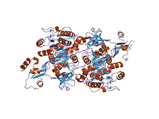
Carnitine palmitoyltransferase I (CPT1) also known as carnitine acyltransferase I, CPTI, CAT1, CoA:carnitine acyl transferase (CCAT), or palmitoylCoA transferase I, is a mitochondrial enzyme responsible for the formation of acyl carnitines by catalyzing the transfer of the acyl group of a long-chain fatty acyl-CoA from coenzyme A to l-carnitine. The product is often Palmitoylcarnitine, but other fatty acids may also be substrates. It is part of a family of enzymes called carnitine acyltransferases. This "preparation" allows for subsequent movement of the acyl carnitine from the cytosol into the intermembrane space of mitochondria.

In enzymology, a 1-acylglycerophosphocholine O-acyltransferase is an enzyme that catalyzes the chemical reaction

<span class="mw-page-title-main">Carnitine O-octanoyltransferase</span>

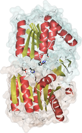
Carnitine O-octanoyltransferase is a member of the transferase family, more specifically a carnitine acyltransferase, a type of enzyme which catalyzes the transfer of acyl groups from acyl-CoAs to carnitine, generating CoA and an acyl-carnitine. The systematic name of this enzyme is octanoyl-CoA:L-carnitine O-octanoyltransferase. Other names in common use include medium-chain/long-chain carnitine acyltransferase, carnitine medium-chain acyltransferase, easily solubilized mitochondrial carnitine palmitoyltransferase, and overt mitochondrial carnitine palmitoyltransferase. Specifically, CROT catalyzes the chemical reaction:

<span class="mw-page-title-main">Glycylpeptide N-tetradecanoyltransferase</span>

In enzymology, a glycylpeptide N-tetradecanoyltransferase is an enzyme that catalyzes the chemical reaction

In enzymology, sphingosine N-acyltransferases (ceramide synthases (CerS), EC 2.3.1.24) are enzymes that catalyze the chemical reaction of synthesis of ceramide:

Sterol O-acyltransferase is an intracellular protein located in the endoplasmic reticulum that forms cholesteryl esters from cholesterol.

<span class="mw-page-title-main">ACOT4</span> Protein-coding gene in the species Homo sapiens

Acyl-coenzyme A thioesterase 4 is an enzyme that in humans is encoded by the ACOT4 gene.

<span class="mw-page-title-main">ACOT11</span> Protein-coding gene in the species Homo sapiens

Acyl-coenzyme A thioesterase 11 also known as StAR-related lipid transfer protein 14 (STARD14) is an enzyme that in humans is encoded by the ACOT11 gene. This gene encodes a protein with acyl-CoA thioesterase activity towards medium (C12) and long-chain (C18) fatty acyl-CoA substrates which relies on its StAR-related lipid transfer domain. Expression of a similar murine protein in brown adipose tissue is induced by cold exposure and repressed by warmth. Expression of the mouse protein has been associated with obesity, with higher expression found in obesity-resistant mice compared with obesity-prone mice. Alternative splicing results in two transcript variants encoding different isoforms.

<span class="mw-page-title-main">DHHC domain</span>

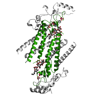
In molecular biology the DHHC domain is a protein domain that acts as an enzyme, which adds a palmitoyl chemical group to proteins in order to anchor them to cell membranes. The DHHC domain was discovered in 1999 and named after a conserved sequence motif found in its protein sequence. Roth and colleagues showed that the yeast Akr1p protein could palmitoylate Yck2p in vitro and inferred that the DHHC domain defined a large family of palmitoyltransferases. In mammals twenty three members of this family have been identified and their substrate specificities investigated. Some members of the family such as ZDHHC3 and ZDHHC7 enhance palmitoylation of proteins such as PSD-95, SNAP-25, GAP43, Gαs. Others such as ZDHHC9 showed specificity only toward the H-Ras protein. However, a recent study questions the involvement of classical enzyme-substrate recognition and specificity in the palmitoylation reaction. Several members of the family have been implicated in human diseases.

<span class="mw-page-title-main">ACOT6</span> Protein-coding gene in the species Homo sapiens

Acyl-CoA thioesterase 6 is a protein that in humans is encoded by the ACOT6 gene. The protein, also known as C14orf42, is an enzyme with thioesterase activity.

<span class="mw-page-title-main">1-Lysophosphatidylcholine</span>

2-acyl-sn-glycero-3-phosphocholines are a class of phospholipids that are intermediates in the metabolism of lipids. Because they result from the hydrolysis of an acyl group from the sn-1 position of phosphatidylcholine, they are also called 1-lysophosphatidylcholine. The synthesis of phosphatidylcholines with specific fatty acids occurs through the synthesis of 1-lysoPC. The formation of various other lipids generates 1-lysoPC as a by-product.

The MBOAT family of membrane proteins is a family of various acyltransferase enzymes. All family members contain multiple transmembrane domains and most carry two conserved residues, a conserved histidine (His) embedded in a hydrophobic stretch of residues and an asparagine (Asn) or histidine within a more hydrophilic region some 30-50 residues upstream.

<span class="mw-page-title-main">ACOT13</span> Protein-coding gene in the species Homo sapiens

Acyl-CoA thioesterase 13 is a protein that in humans is encoded by the ACOT13 gene. This gene encodes a member of the thioesterase superfamily. In humans, the protein co-localizes with microtubules and is essential for sustained cell proliferation.

<span class="mw-page-title-main">ACOT1</span> Protein-coding gene in the species Homo sapiens

Acyl-CoA thioesterase 1 is a protein that in humans is encoded by the ACOT1 gene.

<span class="mw-page-title-main">Acyl-protein thioesterase</span> Enzymes that cleave off lipid modifications on proteins

Acyl-protein thioesterases are enzymes that cleave off lipid modifications on proteins, located on the sulfur atom of cysteine residues linked via a thioester bond. Acyl-protein thioesterases are part of the α/β hydrolase superfamily of proteins and have a conserved catalytic triad. For that reason, acyl-protein thioesterases are also able to hydrolyze oxygen-linked ester bonds.

References

  1. 1 2 3 GRCh38: Ensembl release 89: ENSG00000177669 - Ensembl, May 2017
  2. 1 2 3 GRCm38: Ensembl release 89: ENSMUSG00000071113 - Ensembl, May 2017
  3. "Human PubMed Reference:". National Center for Biotechnology Information, U.S. National Library of Medicine.
  4. "Mouse PubMed Reference:". National Center for Biotechnology Information, U.S. National Library of Medicine.
  5. "Entrez Gene: Membrane bound O-acyltransferase domain containing 4".
  6. 1 2 3 Kojima M, Hamamoto A, Sato T (October 2016). "Ghrelin O-acyltransferase (GOAT), a specific enzyme that modifies ghrelin with a medium-chain fatty acid". Journal of Biochemistry. 160 (4): 189–194. doi: 10.1093/jb/mvw046 . PMID   27489223.
  7. 1 2 3 4 Taylor MS, Ruch TR, Hsiao PY, Hwang Y, Zhang P, Dai L, et al. (November 2013). "Architectural organization of the metabolic regulatory enzyme ghrelin O-acyltransferase". The Journal of Biological Chemistry. 288 (45): 32211–32228. doi: 10.1074/jbc.M113.510313 . PMC   3820860 . PMID   24045953.
  8. "Rhythm Pharmaceuticals Announces Licensing Agreement with Takeda for the Development and Commercialization of Preclinical Treatment for Prader-Willi Syndrome | Rhythm Pharmaceuticals, Inc". ir.rhythmtx.com. Retrieved 2021-11-25.
  9. Clinical trial number NCT03641417 for "BARI-INSIGHT: A Double-blind, Placebo-controlled, Within-subject, Cross-over Mechanistic Study Investigating the Role of Ghrelin in Regulating Appetite and Energy Intake in Patients Following Bariatric Surgery" at ClinicalTrials.gov
  10. 1 2 3 4 5 Hougland JL (February 2019). "Ghrelin octanoylation by ghrelin O-acyltransferase: Unique protein biochemistry underlying metabolic signaling". Biochemical Society Transactions. 47 (1): 169–178. doi:10.1042/BST20180436. PMID   30626708. S2CID   58566712.
  11. Taylor MS, Dempsey DR, Hwang Y, Chen Z, Chu N, Boeke JD, Cole PA (October 2015). "Mechanistic analysis of ghrelin-O-acyltransferase using substrate analogs". Bioorganic Chemistry. 62: 64–73. doi:10.1016/j.bioorg.2015.07.003. PMC   4567917 . PMID   26246082.
  12. 1 2 Yang J, Zhao TJ, Goldstein JL, Brown MS (August 2008). "Inhibition of ghrelin O-acyltransferase (GOAT) by octanoylated pentapeptides". Proceedings of the National Academy of Sciences of the United States of America. 105 (31): 10750–10755. Bibcode:2008PNAS..10510750Y. doi: 10.1073/pnas.0805353105 . PMC   2504781 . PMID   18669668.
  13. Sieburg MA, Cleverdon ER, Hougland JL (2019). "Biochemical Assays for Ghrelin Acylation and Inhibition of Ghrelin O-Acyltransferase". In Linder E (ed.). Protein Lipidation. Methods in Molecular Biology. Vol. 2009. New York, NY: Springer. pp. 227–241. doi:10.1007/978-1-4939-9532-5_18. ISBN   978-1-4939-9532-5. PMID   31152408. S2CID   172136458.
  14. 1 2 Khatib MN, Gaidhane S, Gaidhane AM, Simkhada P, Zahiruddin QS (February 2015). "Ghrelin O Acyl Transferase (GOAT) as a Novel Metabolic Regulatory Enzyme". Journal of Clinical and Diagnostic Research. 9 (2): LE01–LE05. doi:10.7860/JCDR/2015/9787.5514. PMC   4378754 . PMID   25859472.
  15. Cleverdon ER, Davis TR, Hougland JL (September 2018). "Functional group and stereochemical requirements for substrate binding by ghrelin O-acyltransferase revealed by unnatural amino acid incorporation". Bioorganic Chemistry. 79: 98–106. doi:10.1016/j.bioorg.2018.04.009. PMID   29738973. S2CID   13724720.
  16. McGovern-Gooch KR, Mahajani NS, Garagozzo A, Schramm AJ, Hannah LG, Sieburg MA, et al. (February 2017). "Synthetic Triterpenoid Inhibition of Human Ghrelin O-Acyltransferase: The Involvement of a Functionally Required Cysteine Provides Mechanistic Insight into Ghrelin Acylation". Biochemistry. 56 (7): 919–931. doi:10.1021/acs.biochem.6b01008. PMC   5319884 . PMID   28134508.

Further reading