Glutamate-sensitive fluorescent reporter

Last updated

A genetically engineered fluorescent protein that changes its fluorescence when bound to the neurotransmitter glutamate. [1] Glutamate-sensitive fluorescent reporters (iGluSnFR, colloquially pronounced 'glue sniffer') are used to monitor the activity of presynaptic terminals by fluorescence microscopy. GluSnFRs are a class of optogenetic sensors used in neuroscience research. [2] In brain tissue, two-photon microscopy is typically used to monitor GluSnFR fluorescence.

Contents

Design

GluSniffer Scheme.png

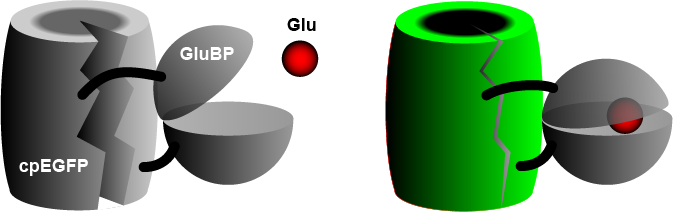
The widely used iGluSnFR consists of a circularly permuted enhanced green fluorescent protein (cpEGFP) fused to a glutamate binding protein (GluBP) from a bacterium. [3] When GluBP binds a glutamate molecule, it changes its shape, pulling the EGFP barrel together, increasing fluorescence. A specific peptide segment (PDGFR) is included to bring the sensor to the outside of the cell membrane. [4] In the more recent version by Aggarwal et al. (2022), [1] researchers introduced iGluSnFR to two additional anchoring domains, a glycosylphostidylinositol (GPI) anchor, and a modified form of the cytosolic -cterminal domain of Stargazin with a PDZ ligand.

History

The first genetically encoded fluorescent glutamate sensors (FLIPE, GluSnFR and SuperGluSnFR) were constructed by attaching cyan-fluorescent protein (CFP) and yellow-fluorescent protein (YFP) to a bacterial glutamate binding protein (GluBP). [5] [6] Glutamate binding changed the distance between CFP and YFP, changing the efficiency of energy transfer (FRET) between the two fluorophores. [7] [8] A breakthrough in visualizing glutamate release was achieved with iGluSnFR, a single-fluorophore glutamate sensor based on EGFP producing a ~5‑fold increase in fluorescence. [3] To measure synaptic transmission at high frequencies, novel iGluSnFR variants with accelerated kinetics have recently been developed. [9] [10]

Related Research Articles

<span class="mw-page-title-main">Green fluorescent protein</span> Protein that exhibits bright green fluorescence when exposed to ultraviolet light

The green fluorescent protein (GFP) is a protein that exhibits bright green fluorescence when exposed to light in the blue to ultraviolet range. The label GFP traditionally refers to the protein first isolated from the jellyfish Aequorea victoria and is sometimes called avGFP. However, GFPs have been found in other organisms including corals, sea anemones, zoanithids, copepods and lancelets.

<span class="mw-page-title-main">Fluorescent tag</span>

In molecular biology and biotechnology, a fluorescent tag, also known as a fluorescent label or fluorescent probe, is a molecule that is attached chemically to aid in the detection of a biomolecule such as a protein, antibody, or amino acid. Generally, fluorescent tagging, or labeling, uses a reactive derivative of a fluorescent molecule known as a fluorophore. The fluorophore selectively binds to a specific region or functional group on the target molecule and can be attached chemically or biologically. Various labeling techniques such as enzymatic labeling, protein labeling, and genetic labeling are widely utilized. Ethidium bromide, fluorescein and green fluorescent protein are common tags. The most commonly labelled molecules are antibodies, proteins, amino acids and peptides which are then used as specific probes for detection of a particular target.

<span class="mw-page-title-main">AMPA receptor</span> Transmembrane protein family

The α-amino-3-hydroxy-5-methyl-4-isoxazolepropionic acid receptor is an ionotropic transmembrane receptor for glutamate (iGluR) that mediates fast synaptic transmission in the central nervous system (CNS). It has been traditionally classified as a non-NMDA-type receptor, along with the kainate receptor. Its name is derived from its ability to be activated by the artificial glutamate analog AMPA. The receptor was first named the "quisqualate receptor" by Watkins and colleagues after a naturally occurring agonist quisqualate and was only later given the label "AMPA receptor" after the selective agonist developed by Tage Honore and colleagues at the Royal Danish School of Pharmacy in Copenhagen. The GRIA2-encoded AMPA receptor ligand binding core was the first glutamate receptor ion channel domain to be crystallized.

<span class="mw-page-title-main">NMDA receptor</span> Glutamate receptor and ion channel protein found in nerve cells

The N-methyl-D-aspartatereceptor (also known as the NMDA receptor or NMDAR), is a glutamate receptor and ion channel found in neurons. The NMDA receptor is one of three types of ionotropic glutamate receptors, the other two being AMPA and kainate receptors. Depending on its subunit composition, its ligands are glutamate and glycine (or D-serine). However, the binding of the ligands is typically not sufficient to open the channel as it may be blocked by Mg2+ ions which are only removed when the neuron is sufficiently depolarized. Thus, the channel acts as a “coincidence detector” and only once both of these conditions are met, the channel opens and it allows positively charged ions (cations) to flow through the cell membrane. The NMDA receptor is thought to be very important for controlling synaptic plasticity and mediating learning and memory functions.

<span class="mw-page-title-main">Immunofluorescence</span> Technique used for light microscopy

Immunofluorescence is a technique used for light microscopy with a fluorescence microscope and is used primarily on biological samples. This technique uses the specificity of antibodies to their antigen to target fluorescent dyes to specific biomolecule targets within a cell, and therefore allows visualization of the distribution of the target molecule through the sample. The specific region an antibody recognizes on an antigen is called an epitope. There have been efforts in epitope mapping since many antibodies can bind the same epitope and levels of binding between antibodies that recognize the same epitope can vary. Additionally, the binding of the fluorophore to the antibody itself cannot interfere with the immunological specificity of the antibody or the binding capacity of its antigen. Immunofluorescence is a widely used example of immunostaining and is a specific example of immunohistochemistry. This technique primarily makes use of fluorophores to visualise the location of the antibodies.

<span class="mw-page-title-main">Förster resonance energy transfer</span> Photochemical energy transfer mechanism

Förster resonance energy transfer (FRET), fluorescence resonance energy transfer, resonance energy transfer (RET) or electronic energy transfer (EET) is a mechanism describing energy transfer between two light-sensitive molecules (chromophores). A donor chromophore, initially in its electronic excited state, may transfer energy to an acceptor chromophore through nonradiative dipole–dipole coupling. The efficiency of this energy transfer is inversely proportional to the sixth power of the distance between donor and acceptor, making FRET extremely sensitive to small changes in distance.

<span class="mw-page-title-main">Bimolecular fluorescence complementation</span>

Bimolecular fluorescence complementation is a technology typically used to validate protein interactions. It is based on the association of fluorescent protein fragments that are attached to components of the same macromolecular complex. Proteins that are postulated to interact are fused to unfolded complementary fragments of a fluorescent reporter protein and expressed in live cells. Interaction of these proteins will bring the fluorescent fragments within proximity, allowing the reporter protein to reform in its native three-dimensional structure and emit its fluorescent signal. This fluorescent signal can be detected and located within the cell using an inverted fluorescence microscope that allows imaging of fluorescence in cells. In addition, the intensity of the fluorescence emitted is proportional to the strength of the interaction, with stronger levels of fluorescence indicating close or direct interactions and lower fluorescence levels suggesting interaction within a complex. Therefore, through the visualisation and analysis of the intensity and distribution of fluorescence in these cells, one can identify both the location and interaction partners of proteins of interest.

<span class="mw-page-title-main">STED microscopy</span>

Stimulated emission depletion (STED) microscopy is one of the techniques that make up super-resolution microscopy. It creates super-resolution images by the selective deactivation of fluorophores, minimizing the area of illumination at the focal point, and thus enhancing the achievable resolution for a given system. It was developed by Stefan W. Hell and Jan Wichmann in 1994, and was first experimentally demonstrated by Hell and Thomas Klar in 1999. Hell was awarded the Nobel Prize in Chemistry in 2014 for its development. In 1986, V.A. Okhonin had patented the STED idea. This patent was unknown to Hell and Wichmann in 1994.

Cameleon is an engineered protein based on variant of green fluorescent protein used to visualize calcium levels in living cells. It is a genetically encoded calcium sensor created by Roger Y. Tsien and coworkers. The name is a conflation of CaM and chameleon to indicate the fact that the sensor protein undergoes a conformation change and radiates at an altered wavelength upon calcium binding to the calmodulin element of the Cameleon. Cameleon was the first genetically encoded calcium sensor that could be used for ratiometric measurements and the first to be used in a transgenic animal to record activity in neurons and muscle cells. Cameleon and other genetically-encoded calcium indicators (GECIs) have found many applications in neuroscience and other fields of biology. It was created by fusing BFP, calmodulin, calmodulin-binding peptide M13 and EGFP.

<span class="mw-page-title-main">Brainbow</span> Neuroimaging technique to differentiate neurons

Brainbow is a process by which individual neurons in the brain can be distinguished from neighboring neurons using fluorescent proteins. By randomly expressing different ratios of red, green, and blue derivatives of green fluorescent protein in individual neurons, it is possible to flag each neuron with a distinctive color. This process has been a major contribution to the field of neural connectomics.

<span class="mw-page-title-main">GRIK1</span> Protein-coding gene in the species Homo sapiens

Glutamate receptor, ionotropic, kainate 1, also known as GRIK1, is a protein that in humans is encoded by the GRIK1 gene.

EosFP is a photoactivatable green to red fluorescent protein. Its green fluorescence (516 nm) switches to red (581 nm) upon UV irradiation of ~390 nm due to a photo-induced modification resulting from a break in the peptide backbone near the chromophore. Eos was first discovered as a tetrameric protein in the stony coral Lobophyllia hemprichii. Like other fluorescent proteins, Eos allows for applications such as the tracking of fusion proteins, multicolour labelling and tracking of cell movement. Several variants of Eos have been engineered for use in specific study systems including mEos2, mEos4 and CaMPARI.

Fluorescent chloride sensors are used for chemical analysis. The discoveries of chloride (Cl) participations in physiological processes stimulates the measurements of intracellular Cl in live cells and the development of fluorescent tools referred below.

Glutamate receptor-interacting protein (GRIP) refers to either a family of proteins that bind to the glutamate receptor or specifically to the GRIP1 protein within this family. Proteins in the glutamate receptor-interacting protein (GRIP) family have been shown to interact with GluR2, a common subunit in the AMPA receptor. This subunit also interacts with other proteins such as protein interacting with C-kinase1 (PICK1) and N-ethylmaleimide-sensitive fusion protein (NSF). Studies have begun to elucidate its function; however, much is still to be learned about these proteins.

Calcium imaging is a microscopy technique to optically measure the calcium (Ca2+) status of an isolated cell, tissue or medium. Calcium imaging takes advantage of calcium indicators, fluorescent molecules that respond to the binding of Ca2+ ions by fluorescence properties. Two main classes of calcium indicators exist: chemical indicators and genetically encoded calcium indicators (GECI). This technique has allowed studies of calcium signalling in a wide variety of cell types. In neurons, electrical activity is always accompanied by an influx of Ca2+ ions. Thus, calcium imaging can be used to monitor the electrical activity in hundreds of neurons in cell culture or in living animals, which has made it possible to dissect the function of neuronal circuits.

PRIME is a molecular biology research tool developed by Alice Y. Ting and the Ting Lab at MIT for site-specific labeling of proteins in living cells with chemical probes. Probes often have useful biophysical properties, such as fluorescence, and allow imaging of proteins. Ultimately, PRIME enables scientists to study functions of specific proteins of interest.

<span class="mw-page-title-main">FMN-binding fluorescent protein</span>

A FMN-binding fluorescent protein (FbFP), also known as a LOV-based fluorescent protein, is a small, oxygen-independent fluorescent protein that binds flavin mononucleotide (FMN) as a chromophore.

<span class="mw-page-title-main">Spinach aptamer</span>

The need for fluorescently tracking RNA rose as its roles in complex cellular functions has grown to not only include mRNA, rRNA, and tRNA, but also RNAi, siRNA, snoRNA, and lncRNA, among others. Spinach is a synthetically derived RNA aptamer born out of the need for a way of studying the role of RNAs at the cellular level. This aptamer was created using Systematic Evolution for Ligands by EXponential enrichment, or SELEX, which is also known as in vitro evolution.

<span class="mw-page-title-main">Red fluorescent protein</span>

Red fluorescent protein (RFP) is a fluorophore that fluoresces red-orange when excited. Several variants have been developed using directed mutagenesis. The original was isolated from Discosoma, and named DsRed. Others are now available that fluoresce orange, red, and far-red.

Optogenetics began with methods to alter neuronal activity with light, using e.g. channelrhodopsins. In a broader sense, optogenetic approaches also include the use of genetically encoded biosensors to monitor the activity of neurons or other cell types by measuring fluorescence or bioluminescence. Genetically encoded calcium indicators (GECIs) are used frequently to monitor neuronal activity, but other cellular parameters such as membrane voltage or second messenger activity can also be recorded optically. The use of optogenetic sensors is not restricted to neuroscience, but plays increasingly important roles in immunology, cardiology and cancer research.

References

  1. 1 2 Aggarwal, Abhi; Liu, Rui; Chen, Yang; Ralowicz, Amelia J.; Bergerson, Samuel J.; Tomaska, Filip; Hanson, Timothy L.; Hasseman, Jeremy P.; Reep, Daniel; Tsegaye, Getahun; Yao, Pantong; Ji, Xiang; Kloos, Marinus; Walpita, Deepika; Patel, Ronak (2022-02-15). "Glutamate indicators with improved activation kinetics and localization for imaging synaptic transmission": 2022.02.13.480251. doi:10.1101/2022.02.13.480251v1.{{cite journal}}: Cite journal requires |journal= (help)
  2. Hefendehl, J. K.; LeDue, J.; Ko, R. W. Y.; Mahler, J.; Murphy, T. H.; MacVicar, B. A. (2016-11-11). "Mapping synaptic glutamate transporter dysfunction in vivo to regions surrounding Aβ plaques by iGluSnFR two-photon imaging". Nature Communications. 7: 13441. Bibcode:2016NatCo...713441H. doi:10.1038/ncomms13441. PMC   5114608 . PMID   27834383.
  3. 1 2 Marvin, Jonathan S; Borghuis, Bart G; Tian, Lin; Cichon, Joseph; Harnett, Mark T; Akerboom, Jasper; Gordus, Andrew; Renninger, Sabine L; Chen, Tsai-Wen (2013). "An optimized fluorescent probe for visualizing glutamate neurotransmission". Nature Methods. 10 (2): 162–170. doi:10.1038/nmeth.2333. ISSN   1548-7105. PMC   4469972 . PMID   23314171.
  4. Marvin, Jonathan S.; Schreiter, Eric R.; Echevarría, Ileabett M.; Looger, Loren L. (2011-11-01). "A genetically encoded, high-signal-to-noise maltose sensor". Proteins: Structure, Function, and Bioinformatics. 79 (11): 3025–3036. doi:10.1002/prot.23118. ISSN   1097-0134. PMC   3265398 . PMID   21989929.
  5. Hu, Yonglin; Fan, Cheng-Peng; Fu, Guangsen; Zhu, Deyu; Jin, Qi; Wang, Da-Cheng (2008). "Crystal Structure of a Glutamate/Aspartate Binding Protein Complexed with a Glutamate Molecule: Structural Basis of Ligand Specificity at Atomic Resolution". Journal of Molecular Biology. 382 (1): 99–111. doi:10.1016/j.jmb.2008.06.091. PMID   18640128.
  6. De Lorimier, Robert M.; Smith, J. Jeff; Dwyer, Mary A.; Looger, Loren L.; Sali, Kevin M.; Paavola, Chad D.; Rizk, Shahir S.; Sadigov, Shamil; Conrad, David W. (2002-11-01). "Construction of a fluorescent biosensor family". Protein Science. 11 (11): 2655–2675. doi:10.1110/ps.021860. ISSN   1469-896X. PMC   2373719 . PMID   12381848.
  7. Okumoto, Sakiko; Looger, Loren L.; Micheva, Kristina D.; Reimer, Richard J.; Smith, Stephen J.; Frommer, Wolf B. (2005-06-14). "Detection of glutamate release from neurons by genetically encoded surface-displayed FRET nanosensors". Proceedings of the National Academy of Sciences of the United States of America. 102 (24): 8740–8745. Bibcode:2005PNAS..102.8740O. doi: 10.1073/pnas.0503274102 . ISSN   0027-8424. PMC   1143584 . PMID   15939876.
  8. Hires, Samuel Andrew; Zhu, Yongling; Tsien, Roger Y. (2008-03-18). "Optical measurement of synaptic glutamate spillover and reuptake by linker optimized glutamate-sensitive fluorescent reporters". Proceedings of the National Academy of Sciences. 105 (11): 4411–4416. Bibcode:2008PNAS..105.4411H. doi: 10.1073/pnas.0712008105 . ISSN   0027-8424. PMC   2393813 . PMID   18332427.
  9. Helassa, Nordine; Dürst, Céline D.; Coates, Catherine; Kerruth, Silke; Arif, Urwa; Schulze, Christian; Wiegert, J. Simon; Geeves, Michael; Oertner, Thomas G.; Török, Katalin (2018-05-22). "Ultrafast glutamate sensors resolve high-frequency release at Schaffer collateral synapses". Proceedings of the National Academy of Sciences. 115 (21): 5594–5599. doi: 10.1073/pnas.1720648115 . PMC   6003469 . PMID   29735711.
  10. Marvin, Jonathan S.; Scholl, Benjamin; Wilson, Daniel E.; Podgorski, Kaspar; Kazemipour, Abbas; Müller, Johannes Alexander; Schoch, Susanne; Quiroz, Francisco José Urra; Rebola, Nelson; Bao, Huan; Little, Justin P. (November 2018). "Stability, affinity, and chromatic variants of the glutamate sensor iGluSnFR". Nature Methods. 15 (11): 936–939. doi:10.1038/s41592-018-0171-3. ISSN   1548-7105. PMC   6394230 . PMID   30377363.