(a) The top view of PR. (b) The side view of PR. In (a,b), the green moiety denotes the chromophore, and the blue moiety is residue 105. (c) The structure of LYR and all hydrogens were removed for simplicity. All atom names are marked for clarity.
Light-activated proteorhodopsin pumps protons outwardly, increasing the proton motive force. Protons can then reenter the cells through ATP-synthase complex, powering the ATP production.
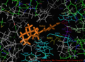
Holoenzyme (Green) with helices A-G labeled (purple) as well as Retinal ligand (orange)
Surface visualization of Proteorhodopsin showing terminals
Visualization of the retinal bound active site of the 2L6X protein structure of pRhodopsin, residues color coded and labeled by activity, ligand is orange.
2L6x In-Active-Site Cartoon Color Coded and Labeled Visualization, D and E Helices hidden for vantage, Retinal ligand binding site
↑ Oesterhelt D, Stoeckenius W (September 1971). "Rhodopsin-like protein from the purple membrane of Halobacterium halobium". Nature. 233 (39): 149–152. doi:10.1038/newbio233149a0. PMID4940442.
This page is based on this Wikipedia article Text is available under the CC BY-SA 4.0 license; additional terms may apply. Images, videos and audio are available under their respective licenses.