Related Research Articles

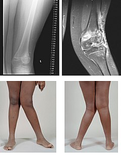
Rickets is a condition that results in weak or soft bones in children. Symptoms include bowed legs, stunted growth, bone pain, large forehead, and trouble sleeping. Complications may include bone fractures, muscle spasms, an abnormally curved spine, or intellectual disability.
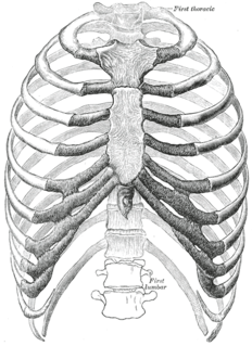
In vertebrate anatomy, ribs are the long curved bones which form the rib cage, part of the axial skeleton. In most tetrapods, ribs surround the chest, enabling the lungs to expand and thus facilitate breathing by expanding the chest cavity. They serve to protect the lungs, heart, and other internal organs of the thorax. In some animals, especially snakes, ribs may provide support and protection for the entire body.
The rib cage is the arrangement of ribs attached to the vertebral column and sternum in the thorax of most vertebrates, that encloses and protects the heart and lungs. In humans, the rib cage, also known as the thoracic cage, is a bony and cartilaginous structure which surrounds the thoracic cavity and supports the shoulder girdle to form the core part of the human skeleton. A typical human rib cage consists of 24 ribs in 12 pairs, the sternum and xiphoid process, the costal cartilages, and the 12 thoracic vertebrae.
A hiatal hernia is a type of hernia in which abdominal organs slip through the diaphragm into the middle compartment of the chest. This may result in gastroesophageal reflux disease (GERD) or laryngopharyngeal reflux (LPR) with symptoms such as a taste of acid in the back of the mouth or heartburn. Other symptoms may include trouble swallowing and chest pains. Complications may include iron deficiency anemia, volvulus, or bowel obstruction.

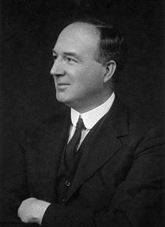
Dame Harriette Chick, was a British microbiologist, protein scientist and nutritionist. She is best remembered for demonstrating the roles of sunlight and cod liver oil in preventing rickets.

The humerus is a long bone in the arm that runs from the shoulder to the elbow. It connects the scapula and the two bones of the lower arm, the radius and ulna, and consists of three sections. The humeral upper extremity consists of a rounded head, a narrow neck, and two short processes. The body is cylindrical in its upper portion, and more prismatic below. The lower extremity consists of 2 epicondyles, 2 processes, and 3 fossae. As well as its true anatomical neck, the constriction below the greater and lesser tubercles of the humerus is referred to as its surgical neck due to its tendency to fracture, thus often becoming the focus of surgeons.

The thoracic diaphragm, or simply the diaphragm, is a sheet of internal skeletal muscle in humans and other mammals that extends across the bottom of the thoracic cavity. The diaphragm separates the thoracic cavity, containing the heart and lungs, from the abdominal cavity and performs an important function in respiration: as the diaphragm contracts, the volume of the thoracic cavity increases, creating a negative pressure there, which draws air into the lungs.
Sir Edward Mellanby discovered vitamin D and its role in preventing rickets in 1919. He also went to Barnard Castle School.

Osteomalacia is a disease characterized by the softening of the bones caused by impaired bone metabolism primarily due to inadequate levels of available phosphate, calcium, and vitamin D, or because of resorption of calcium. The impairment of bone metabolism causes inadequate bone mineralization. Osteomalacia in children is known as rickets, and because of this, use of the term "osteomalacia" is often restricted to the milder, adult form of the disease. Signs and symptoms can include diffuse body pains, muscle weakness, and fragility of the bones. In addition to low systemic levels of circulating mineral ions necessary for bone and tooth mineralization, accumulation of mineralization-inhibiting proteins and peptides occurs in the extracellular matrix of bones and teeth, likely contributing locally to cause matrix hypomineralization (osteomalacia).
Genu valgum, commonly called "knock-knee", is a condition in which the knees angle in and touch each other when the legs are straightened. Individuals with severe valgus deformities are typically unable to touch their feet together while simultaneously straightening the legs. The term originates from the Latin genu, 'knee', and valgus which actually means 'bent outwards', but in this case, it is used to describe the distal portion of the knee joint which bends outwards and thus the proximal portion seems to be bent inwards. For citation and more information on uses of the words Valgus and Varus, please visit the internal link to -varus.
Genu varum , is a varus deformity marked by (outward) bowing at the knee, which means that the lower leg is angled inward (medially) in relation to the thigh's axis, giving the limb overall the appearance of an archer's bow. Usually medial angulation of both lower limb bones is involved.
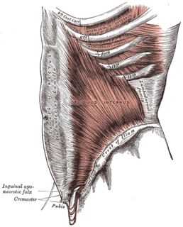
The abdominal internal oblique muscle, also internal oblique muscle or interior oblique, is an abdominal muscle in the abdominal wall that lies below the external oblique muscle and just above the transverse abdominal muscle.

The talus, talus bone, astragalus, or ankle bone is one of the group of foot bones known as the tarsus. The tarsus forms the lower part of the ankle joint. It transmits the entire weight of the body from the lower legs to the foot.
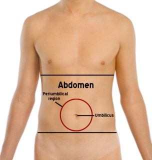
The abdomen is the part of the body between the thorax (chest) and pelvis, in humans and in other vertebrates. The abdomen is the front part of the abdominal segment of the trunk. The region occupied by the abdomen is called the abdominal cavity. In arthropods it is the posterior tagma of the body; it follows the thorax or cephalothorax.

The ischium forms the lower and back part of the hip bone.

X-linked hypophosphatemia (XLH), is an X-linked dominant form of rickets that differs from most cases of rickets in that vitamin D supplementation does not cure it. It can cause bone deformity including short stature and genu varum (bow-leggedness). It is associated with a mutation in the PHEX gene sequence (Xp.22) and subsequent inactivity of the PHEX protein.

Blount's disease is a growth disorder of the tibia that causes the lower leg to angle inward, resembling a bowleg.
Vitamin D deficiency, or hypovitaminosis D, most commonly results from inadequate sunlight exposure. Vitamin D deficiency can also be caused by inadequate nutritional intake of vitamin D, disorders limiting vitamin D absorption, and conditions impairing vitamin D conversion into active metabolites—including certain liver, kidney, and hereditary disorders. Deficiency impairs bone mineralization, leading to bone softening diseases such as rickets in children. It can also worsen osteomalacia and osteoporosis in adults, leading to an increased risk of bone fractures. Muscle weakness is also a common symptom of vitamin D deficiency, further increasing the risk of fall and bone fractures in adults.

Vitamin D is a group of fat-soluble secosteroids responsible for increasing intestinal absorption of calcium, magnesium, and phosphate, and multiple other biological effects. In humans, the most important compounds in this group are vitamin D3 (also known as cholecalciferol) and vitamin D2 (ergocalciferol).
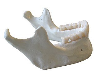
The mandible, lower jaw or jawbone is the largest, strongest and lowest bone in the human face. It forms the lower jaw and holds the lower teeth in place. The mandible sits beneath the maxilla. It is the only movable bone of the skull.
References
- ↑ Naish, J.; Wallis, H. R. E. (20 March 1948). "Significance of Harrison's Grooves". BMJ. 1 (4550): 541–544. doi:10.1136/bmj.1.4550.541. PMC 2090024 . PMID 18909481.