The foot is an anatomical structure found in many vertebrates. It is the terminal portion of a limb which bears weight and allows locomotion. In many animals with feet, the foot is a separate organ at the terminal part of the leg made up of one or more segments or bones, generally including claws and/or nails.

In the human body, the cuboid bone is one of the seven tarsal bones of the foot.

The metatarsal bones or metatarsus are a group of five long bones in the midfoot, located between the tarsal bones and the phalanges (toes). Lacking individual names, the metatarsal bones are numbered from the medial side : the first, second, third, fourth, and fifth metatarsal. The metatarsals are analogous to the metacarpal bones of the hand. The lengths of the metatarsal bones in humans are, in descending order, second, third, fourth, fifth, and first. A bovine hind leg has two metatarsals.

In humans and many other primates, the calcaneus or heel bone is a bone of the tarsus of the foot which constitutes the heel. In some other animals, it is the point of the hock.

The navicular bone is a small bone found in the feet of most mammals.
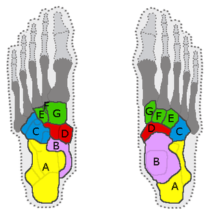
In the human body, the tarsus is a cluster of seven articulating bones in each foot situated between the lower end of the tibia and the fibula of the lower leg and the metatarsus. It is made up of the midfoot and hindfoot.

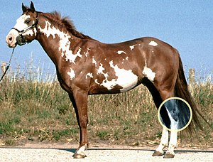
Bog spavin is a swelling of the tibiotarsal joint of the horse's hock which, in itself, does not cause lameness. The joint becomes distended by excess synovial fluid and/or thickened synovial tissue bringing about a soft, fluctuant swelling on the front of the joint, as well as in the medial and lateral plantar pouches. Bog spavin is generally an indication of underlying pathology within the joint.
Bone spavin is a bony growth within the lower hock joint of horse or cattle. It is caused by osteoarthritis, and the degree of lameness that results can be serious enough to end a horse's competitive career.

Fetlock is the common name in horses, large animals, and sometimes dogs for the metacarpophalangeal and metatarsophalangeal joints.
In the anatomy of humans and many other mammals, the tibiotarsal joint is the joint between the tibia and the tarsus.

Equine anatomy encompasses the gross and microscopic anatomy of horses, ponies and other equids, including donkeys, mules and zebras. While all anatomical features of equids are described in the same terms as for other animals by the International Committee on Veterinary Gross Anatomical Nomenclature in the book Nomina Anatomica Veterinaria, there are many horse-specific colloquial terms used by equestrians.
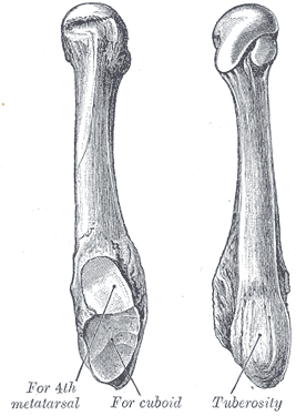
The fifth metatarsal bone is a long bone in the foot, and is palpable along the distal outer edges of the feet. It is the second smallest of the five metatarsal bones. The fifth metatarsal is analogous to the fifth metacarpal bone in the hand.

The fourth metatarsal bone is a long bone in the foot. It is smaller in size than the third metatarsal bone and is the third longest of the five metatarsal bones. The fourth metatarsal is analogous to the fourth metacarpal bone in the hand
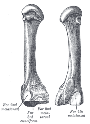
The third metatarsal bone is a long bone in the foot. It is the second longest metatarsal, the longest being the second metatarsal. The third metatarsal is analogous to the third metacarpal bone in the hand
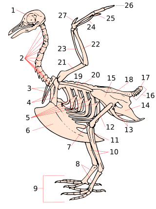
The tarsometatarsus is a bone that is only found in the lower leg of birds and some non-avian dinosaurs. It is formed from the fusion of several bones found in other types of animals, and homologous to the mammalian tarsus and metatarsal bones (foot). Despite this, the tarsometatarsus of birds is often referred to as just the shank, tarsus or metatarsus.

The skeletal system of the horse is a skeletal system of a horse that has three major functions in the body. It protects vital organs, provides framework, and supports soft parts of the body. Horses typically have 205 bones. The pelvic limb typically contains 19 bones, while the thoracic limb contains 20 bones.
Lameness is an abnormal gait or stance of an animal that is the result of dysfunction of the locomotor system. In the horse, it is most commonly caused by pain, but can be due to neurologic or mechanical dysfunction. Lameness is a common veterinary problem in racehorses, sport horses, and pleasure horses. It is one of the most costly health problems for the equine industry, both monetarily for the cost of diagnosis and treatment, and for the cost of time off resulting in loss-of-use.
Plantar ligament refer to ligaments in the sole of the foot:
Curb is defined in older literature as enlargement secondary to inflammation and thickening of the long plantar ligament in horses. However, with the widespread use of diagnostic ultrasonography in equine medicine, curb has been redefined as a collection of soft tissue injuries of the distal plantar hock region. Curb is a useful descriptive term when describing swelling in this area.
The limbs of the horse are structures made of dozens of bones, joints, muscles, tendons, and ligaments that support the weight of the equine body. They include two apparatuses: the suspensory apparatus, which carries much of the weight, prevents overextension of the joint and absorbs shock, and the stay apparatus, which locks major joints in the limbs, allowing horses to remain standing while relaxed or asleep. The limbs play a major part in the movement of the horse, with the legs performing the functions of absorbing impact, bearing weight, and providing thrust. In general, the majority of the weight is borne by the front legs, while the rear legs provide propulsion. The hooves are also important structures, providing support, traction and shock absorption, and containing structures that provide blood flow through the lower leg. As the horse developed as a cursorial animal, with a primary defense mechanism of running over hard ground, its legs evolved to the long, sturdy, light-weight, one-toed form seen today.