Related Research Articles

Transcranial magnetic stimulation (TMS) is a noninvasive form of brain stimulation in which a changing magnetic field is used to induce an electric current at a specific area of the brain through electromagnetic induction. An electric pulse generator, or stimulator, is connected to a magnetic coil connected to the scalp. The stimulator generates a changing electric current within the coil which creates a varying magnetic field, inducing a current within a region in the brain itself.
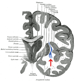
The claustrum is a thin sheet of neurons and supporting glial cells, that connects to the cerebral cortex and subcortical regions including the amygdala, hippocampus and thalamus of the brain. It is located between the insular cortex laterally and the putamen medially, encased by the extreme and external capsules respectively. Blood to the claustrum is supplied by the middle cerebral artery. It is considered to be the most densely connected structure in the brain, and thus hypothesized to allow for the integration of various cortical inputs such as vision, sound and touch, into one experience. Other hypotheses suggest that the claustrum plays a role in salience processing, to direct attention towards the most behaviorally relevant stimuli amongst the background noise. The claustrum is difficult to study given the limited number of individuals with claustral lesions and the poor resolution of neuroimaging.

Brain-derived neurotrophic factor (BDNF), or abrineurin, is a protein that, in humans, is encoded by the BDNF gene. BDNF is a member of the neurotrophin family of growth factors, which are related to the canonical nerve growth factor (NGF), a family which also includes NT-3 and NT-4/NT-5. Neurotrophic factors are found in the brain and the periphery. BDNF was first isolated from a pig brain in 1982 by Yves-Alain Barde and Hans Thoenen.

Interleukin 6 (IL-6) is an interleukin that acts as both a pro-inflammatory cytokine and an anti-inflammatory myokine. In humans, it is encoded by the IL6 gene.
A gamma wave or gamma rhythm is a pattern of neural oscillation in humans with a frequency between 25 and 140 Hz, the 40 Hz point being of particular interest. Gamma rhythms are correlated with large-scale brain network activity and cognitive phenomena such as working memory, attention, and perceptual grouping, and can be increased in amplitude via meditation or neurostimulation. Altered gamma activity has been observed in many mood and cognitive disorders such as Alzheimer's disease, epilepsy, and schizophrenia.

l-Kynurenine is a metabolite of the amino acid l-tryptophan used in the production of niacin.

The posterior cingulate cortex (PCC) is the caudal part of the cingulate cortex, located posterior to the anterior cingulate cortex. This is the upper part of the "limbic lobe". The cingulate cortex is made up of an area around the midline of the brain. Surrounding areas include the retrosplenial cortex and the precuneus.

Transcranial direct current stimulation (tDCS) is a form of neuromodulation that uses constant, low direct current delivered via electrodes on the head. It was originally developed to help patients with brain injuries or neuropsychiatric conditions such as major depressive disorder. It can be contrasted with cranial electrotherapy stimulation, which generally uses alternating current the same way, as well as transcranial magnetic stimulation.

Synaptosomal-Associated Protein, 25kDa (SNAP-25) is a Target Soluble NSF (N-ethylmaleimide-sensitive factor) Attachment Protein Receptor (t-SNARE) protein encoded by the SNAP25 gene found on chromosome 20p12.2 in humans. SNAP-25 is a component of the trans-SNARE complex, which accounts for membrane fusion specificity and directly executes fusion by forming a tight complex that brings the synaptic vesicle and plasma membranes together.
Kynurenic acid is a product of the normal metabolism of amino acid L-tryptophan. It has been shown that kynurenic acid possesses neuroactive activity. It acts as an antiexcitotoxic and anticonvulsant, most likely through acting as an antagonist at excitatory amino acid receptors. Because of this activity, it may influence important neurophysiological and neuropathological processes. As a result, kynurenic acid has been considered for use in therapy in certain neurobiological disorders. Conversely, increased levels of kynurenic acid have also been linked to certain pathological conditions.

Transcription factor 7-like 2 , also known as TCF7L2 or TCF4, is a protein acting as a transcription factor that, in humans, is encoded by the TCF7L2 gene. The TCF7L2 gene is located on chromosome 10q25.2–q25.3, contains 19 exons. As a member of the TCF family, TCF7L2 can form a bipartite transcription factor and influence several biological pathways, including the Wnt signalling pathway.

Kalirin, also known as Huntingtin-associated protein-interacting protein (HAPIP), protein duo (DUO), or serine/threonine-protein kinase with Dbl- and pleckstrin homology domain, is a protein that in humans is encoded by the KALRN gene. Kalirin was first identified in 1997 as a protein interacting with huntingtin-associated protein 1. Is also known to play an important role in nerve growth and axonal development.

The GABAA beta-2 subunit is a protein that in humans is encoded by the GABRB2 gene. It combines with other subunits to form the ionotropic GABAA receptors. GABA system is the major inhibitory system in the brain, and its dominant GABAA receptor subtype is composed of α1, β2, and γ2 subunits with the stoichiometry of 2:2:1, which accounts for 43% of all GABAA receptors. Alternative splicing of the GABRB2 gene leads at least to four isoforms, viz. β2-long (β2L) and β2-short. Alternatively spliced variants displayed similar but non-identical electrophysiological properties. GABRB2 is subjected to positive selection and known to be both an alternative splicing and a recombination hotspot; it is regulated via epigenetic regulation including imprinting and gene and promoter methylation GABRB2 has been associated with a number of neuropsychiatric disorders, and found to display altered expression in cancer.

The Abelson helper integration site 1 (AHI1) is a protein coding gene that is known for the critical role it plays in brain development. Proper cerebellar and cortical development in the human brain depends heavily on AHI1. The AHI1 gene is prominently expressed in the embryonic hindbrain and forebrain. AHI1 specifically encodes the Jouberin protein and mutations in the expression of the gene is known to cause specific forms of Joubert syndrome. Joubert syndrome is autosomal recessive and is characterized by the brain malformations and mental retardation that AHI1 mutations have the potential to induce. AHI1 has also been associated with schizophrenia and autism due to the role it plays in brain development. An AHI1 heterozygous knockout mouse model was studied by Bernard Lerer and his group at Hadassah Medical Center in Jerusalem to elucidate the correlation between alterations in AHI1 expression and the pathogenesis of neuropsychiatric disorders. The core temperatures and corticosterone secretions of the heterozygous knockout mice after exposure to environmental and visceral stress exhibited extreme repression of autonomic nervous system and hypothalamic-pituitary-adrenal responses. The knockout mice demonstrated an increased resilience to different types of stress and these results lead to a correlation between emotional regulation and neuropsychiatric disorders.
Connectomics is the production and study of connectomes: comprehensive maps of connections within an organism's nervous system. More generally, it can be thought of as the study of neuronal wiring diagrams with a focus on how structural connectivity, individual synapses, cellular morphology, and cellular ultrastructure contribute to the make up of a network. The nervous system is a network made of billions of connections and these connections are responsible for our thoughts, emotions, actions, memories, function and dysfunction. Therefore, the study of connectomics aims to advance our understanding of mental health and cognition by understanding how cells in the nervous system are connected and communicate. Because these structures are extremely complex, methods within this field use a high-throughput application of functional and structural neural imaging, most commonly magnetic resonance imaging (MRI), electron microscopy, and histological techniques in order to increase the speed, efficiency, and resolution of these nervous system maps. To date, tens of large scale datasets have been collected spanning the nervous system including the various areas of cortex, cerebellum, the retina, the peripheral nervous system and neuromuscular junctions.
Scientific studies have found that different brain areas show altered activity in humans with major depressive disorder (MDD), and this has encouraged advocates of various theories that seek to identify a biochemical origin of the disease, as opposed to theories that emphasize psychological or situational causes. Factors spanning these causative groups include nutritional deficiencies in magnesium, vitamin D, and tryptophan with situational origin but biological impact. Several theories concerning the biologically based cause of depression have been suggested over the years, including theories revolving around monoamine neurotransmitters, neuroplasticity, neurogenesis, inflammation and the circadian rhythm. Physical illnesses, including hypothyroidism and mitochondrial disease, can also trigger depressive symptoms.
Magnetic seizure therapy (MST) is a proposed form of electrotherapy and electrical brain stimulation. It is currently being investigated for the treatment of major depressive disorder, treatment-resistant depression (TRD), bipolar depression, schizophrenia and obsessive-compulsive disorder. MST is stated to work by inducing seizures via magnetic fields, in contrast to ECT which does so using alternating electric currents. Additionally, MST works in a more concentrated fashion than ECT, thus able to create a seizure with less of a total electric charge. In contrast to (r)TMS, the stimulation rates are higher resulting in more energy transfer. Currently it is thought that MST works in patients with major depressive disorder by activating the connection between the subgenual anterior cingulate cortex and the parietal cortex.

Resting state fMRI is a method of functional magnetic resonance imaging (fMRI) that is used in brain mapping to evaluate regional interactions that occur in a resting or task-negative state, when an explicit task is not being performed. A number of resting-state brain networks have been identified, one of which is the default mode network. These brain networks are observed through changes in blood flow in the brain which creates what is referred to as a blood-oxygen-level dependent (BOLD) signal that can be measured using fMRI.
Heather Clare Whalley is a Scottish scientist. She is a senior research fellow in neuroimaging at the Centre for Clinical Brain Sciences, University of Edinburgh, and is an affiliate member of the Centre for Genomic and Experimental Medicine at the University of Edinburgh. Her main focus of research is on the mechanisms underlying the development of major psychiatric disorders using the latest genomic and neuroimaging approaches.
Network neuroscience is an approach to understanding the structure and function of the human brain through an approach of network science, through the paradigm of graph theory. A network is a connection of many brain regions that interact with each other to give rise to a particular function. Network Neuroscience is a broad field that studies the brain in an integrative way by recording, analyzing, and mapping the brain in various ways. The field studies the brain at multiple scales of analysis to ultimately explain brain systems, behavior, and dysfunction of behavior in psychiatric and neurological diseases. Network neuroscience provides an important theoretical base for understanding neurobiological systems at multiple scales of analysis.
References
- ↑ Mancuso L, Costa T, Nani A, Manuello J, Liloia D, Gelmini G, et al. (March 2019). "The homotopic connectivity of the functional brain: a meta-analytic approach". Scientific Reports. 9 (1): 3346. Bibcode:2019NatSR...9.3346M. doi:10.1038/s41598-019-40188-3. PMC 6399443 . PMID 30833662.
- ↑ Shan X, Cui X, Liu F, Li H, Huang R, Tang Y, et al. (May 2021). "Shared and distinct homotopic connectivity changes in melancholic and non-melancholic depression". Journal of Affective Disorders. 287: 268–275. doi:10.1016/j.jad.2021.03.038. PMID 33799047. S2CID 232775109.
- ↑ Hermesdorf M, Sundermann B, Feder S, Schwindt W, Minnerup J, Arolt V, et al. (March 2016). "Major depressive disorder: Findings of reduced homotopic connectivity and investigation of underlying structural mechanisms". Human Brain Mapping. 37 (3): 1209–1217. doi:10.1002/hbm.23097. PMC 6867499 . PMID 26704348.
- ↑ Li HJ, Xu Y, Zhang KR, Hoptman MJ, Zuo XN (April 2015). "Homotopic connectivity in drug-naïve, first-episode, early-onset schizophrenia". Journal of Child Psychology and Psychiatry, and Allied Disciplines. 56 (4): 432–443. doi:10.1111/jcpp.12307. PMC 4333112 . PMID 25130214.
- ↑ Rossi LF, Wykes RC, Kullmann DM, Carandini M (August 2017). "Focal cortical seizures start as standing waves and propagate respecting homotopic connectivity". Nature Communications. 8 (1): 217. Bibcode:2017NatCo...8..217R. doi:10.1038/s41467-017-00159-6. PMC 5550430 . PMID 28794407.
| | This biology article is a stub. You can help Wikipedia by expanding it. |