Related Research Articles
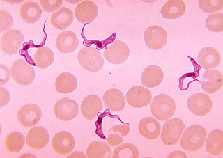
African trypanosomiasis, also known as African sleeping sickness or simply sleeping sickness, is an insect-borne parasitic infection of humans and other animals. It is caused by the species Trypanosoma brucei. Humans are infected by two types, Trypanosoma brucei gambiense (TbG) and Trypanosoma brucei rhodesiense (TbR). TbG causes over 98% of reported cases. Both are usually transmitted by the bite of an infected tsetse fly and are most common in rural areas.

Erythema is redness of the skin or mucous membranes, caused by hyperemia in superficial capillaries. It occurs with any skin injury, infection, or inflammation. Examples of erythema not associated with pathology include nervous blushes.
Panniculitis is a group of diseases whose hallmark is inflammation of subcutaneous adipose tissue. Symptoms include tender skin nodules, and systemic signs such as weight loss and fatigue.

Erythema multiforme (EM) is a skin condition of unknown cause; it is a type of erythema possibly mediated by deposition of immune complexes in the superficial microvasculature of the skin and oral mucous membrane that usually follows an infection or drug exposure. It is an uncommon disorder, with peak incidence in the second and third decades of life. The disorder has various forms or presentations, which its name reflects. Target lesions are a typical manifestation. Two types, one mild to moderate and one severe, are recognized.
Palmar erythema is reddening of the palms at the thenar and hypothenar eminences.

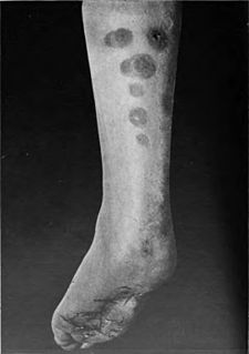
Erythema nodosum (EN), is an inflammatory condition characterized by inflammation of the fat cells under the skin, resulting in tender red nodules or lumps that are usually seen on both shins. It can be caused by a variety of conditions, and typically resolves spontaneously within 30 days. It is common in young people aged 12–20 years.

Erythema migrans or erythema chronicum migrans is an expanding rash often seen in the early stage of Lyme disease, and can also be caused by southern tick-associated rash illness (STARI). It can appear anywhere from one day to one month after a tick bite. This rash does not represent an allergic reaction to the bite, but rather an actual skin infection of one of the Lyme bacteria species from the genus Borrelia. The rash's name comes from New Latin for "migrating redness".

Necrolytic migratory erythema is a red, blistering rash that spreads across the skin. It particularly affects the skin around the mouth and distal extremities; but may also be found on the lower abdomen, buttocks, perineum, and groin. It is strongly associated with glucagonoma, a glucagon-producing tumor of the pancreas, but is also seen in a number of other conditions including liver disease and intestinal malabsorption.

Erythema annulare centrifugum (EAC), is a descriptive term for a class of skin lesion presenting redness (erythema) in a ring form (anulare) that spreads from a center (centrifugum). It was first described by Darier in 1916. Many different terms have been used to classify these types of lesions and it is still controversial on what exactly defines EAC. Some of the types include annular erythema, erythema perstans, erythema gyratum perstans, erythema gyratum repens, darier erythema and erythema figuratum perstans.

Chemotherapy-induced acral erythema is reddening, swelling, numbness and desquamation on palms of the hands and soles of the feet that can occur after chemotherapy in patients with cancer. Hand-foot syndrome is also rarely seen in sickle-cell disease. These skin changes usually are well demarcated. Acral erythema typically disappears within a few weeks after discontinuation of the offending drug.
Dermatophytids are fungus-free disseminated skin lesions resulting from induced sensitization in patients with ringworm infections.
Erythema gyratum repens is a figurate erythema that is rapidly moving and usually a marker of underlying cancer, usually from the lung.
Red lunulae is characterized by a dusky erythema confined to the lunulae, as has been reported in association with alopecia areata.
Generalized erythema is a skin condition that may be caused by medications, bacterial toxins, or viral infections.
Erythema multiforme is usually a reaction of the skin and mucous membranes that occurs suddenly. It appears as a symmetrical rash and may include the mucous membrane lesions. This means that the body is sensitive to something that causes the skin and mucous membranes to react. The more common mild form is refer to as EM minor. It consists of a skin rash that involve no more than one mucosal surface. The sudden onset will progress rapidly as symmetrical lesions with circular color changes in some or all of the lesions. Rash will spread towards center or trunk of the body. Evenly distributed bumps on the skin become classic iris or target lesions. They have bright red borders and small white bumps in the center. The cause of EM appears to be a highly sensitive reaction that can be triggered by a variety of causes. The causes can include bacterial, viral or chemical products, such as antibiotics – specifically penicillins or cephalosporins. This reaction is an allergic reaction and is in no way contagious.

Erythema dyschromicum perstans is an uncommon skin condition with peak age of onset being young adults, but it may also be seen in children or adults of any age. EDP is characterized by hyperpigmented macules that are ash-grey in color and may vary in size and shape. While agents such as certain medications, radiographic contrast, pesticides, infection with parasites, and HIV have been implicated in the occurrence of this disease, the cause of this skin disease remains unknown.
Septal panniculitis is a condition of the subcutaneous fat affecting the layer of adipose tissue that lies between the dermis and underlying fascia, of which there are two forms: acute erythema nodosum and chronic erythema nodosum.
Roseola vaccinia is a cutaneous condition characterized by a prominent rim of erythema surrounding the site of vaccinia injection.
References
- ↑ James, William D.; Berger, Timothy G.; et al. (2006). Andrews' Diseases of the Skin: clinical Dermatology. Saunders Elsevier. ISBN 0-7216-2921-0.
| This infection-related cutaneous condition article is a stub. You can help Wikipedia by expanding it. |