Hypotrichosis with juvenile macular dystrophy

Last updated
Hypotrichosis with juvenile macular dystrophy
Other namesHypotrichosis with juvenile macular degeneration [1]
HJMD.jpg
Hypotrichosis (sparse hair growth) in a 5-year-old boy with HJMD

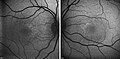
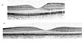
Hypotrichosis with juvenile macular dystrophy (HJMD or CDH3) is an extremely rare congenital disease characterized by sparse hair growth (hypotrichosis) from birth and progressive macular corneal dystrophy.

Contents

Signs and symptoms

Hair growth on the head is noticeably less full than normal, and the hairs are very weak; the rest of the body shows normal hair. The macular degeneration comes on slowly with deterioration of central vision, leading to a loss of reading ability. Those affected may otherwise develop in a completely healthy manner; life expectancy is normal.[ citation needed ]

Cause

Hypotrichosis with juvenile macular dystrophy is an autosomal recessive hereditary disease. [2] It is caused by a combination of mutations (compound heterozygosity) in the CDH3 gene, which codes for Cadherin-3 (also known as P-Cadherin), a calcium-binding protein that is responsible for cellular adhesion in various tissues.[ citation needed ]

Diagnosis

The markedly anomalous hair growth should lead to a retinal examination by school entry at the latest, since weak vision will not necessarily be detected in the course of normal medical check-ups. Confirmation of a diagnosis, which is necessary for any future therapeutic options, is only possible by means of a molecular genetic diagnosis in the context of genetic counseling.[ citation needed ]

Examination method

The extent of retinal damage is assessed by fluorescent angiography, retinal scanning and optical coherence tomography; electrophysiological examinations such as electroretinography (ERG) or multifocal electroretinography (mfERG) may also be used.[ citation needed ]

Differential diagnosis

Anomalies of the hair shaft caused by ectodermal dysplasia should be ruled out. Mutations in the CDH3 gene can also appear in EEM syndrome.

Treatment

There is no treatment for the disorder. A number of studies are looking at gene therapy, exon skipping and CRISPR interference to offer hope for the future. Accurate determination through confirmed diagnosis of the genetic mutation that has occurred also offers potential approaches beyond gene replacement for a specific group, namely in the case of diagnosis of a so-called nonsense mutation, a mutation where a stop codon is produced by the changing of a single base in the DNA sequence. This results in premature termination of protein biosynthesis, resulting in a shortened and either functionless or function-impaired protein. In what is sometimes called "read-through therapy", translational skipping of the stop codon, resulting in a functional protein, can be induced by the introduction of specific substances. However, this approach is only conceivable in the case of narrowly circumscribed mutations, which cause differing diseases.[ citation needed ]

Life planning

A disease that threatens the eyesight and additionally produces a hair anomaly that is apparent to strangers causes harm beyond the physical. It is therefore not surprising that learning the diagnosis is a shock to the patient. This is as true of the affected children as of their parents and relatives. They are confronted with a statement that there are at present no treatment options. They probably have never felt so alone and abandoned in their lives. The question comes to mind, "Why me/my child?" However, there is always hope and especially for affected children, the first priority should be a happy childhood. Too many examinations and doctor appointments take up time and cannot practically solve the problem of a genetic mutation within a few months. It is therefore advisable for parents to treat their child with empathy, but to raise him or her to be independent and self-confident by the teenage years. Openness about the disease and talking with those affected about their experiences, even though its rarity makes it unlikely that others will be personally affected by it, will together assist in managing life.

Epidemiology

It is estimated to affect less than one in a million people. [2] Only 50 to 100 cases have so far been described. [2]

History

The disease was first described in 1935 by Hans Wagner, a German physician. [3]

Related Research Articles

<span class="mw-page-title-main">Retinitis pigmentosa</span> Gradual retinal degeneration leading to progressive sight loss

Retinitis pigmentosa (RP) is a genetic disorder of the eyes that causes loss of vision. Symptoms include trouble seeing at night and decreasing peripheral vision. As peripheral vision worsens, people may experience "tunnel vision". Complete blindness is uncommon. Onset of symptoms is generally gradual and often begins in childhood.

<span class="mw-page-title-main">Retinoschisis</span> Eye disease involving splitting of the retina

Retinoschisis is an eye disease characterized by the abnormal splitting of the retina's neurosensory layers, usually in the outer plexiform layer. Retinoschisis can be divided into degenerative forms which are very common and almost exclusively involve the peripheral retina and hereditary forms which are rare and involve the central retina and sometimes the peripheral retina. The degenerative forms are asymptomatic and involve the peripheral retina only and do not affect the visual acuity. Some rarer forms result in a loss of vision in the corresponding visual field.

<span class="mw-page-title-main">Retinoblastoma</span> Medical condition

Retinoblastoma (Rb) is a rare form of cancer that rapidly develops from the immature cells of a retina, the light-detecting tissue of the eye. It is the most common primary malignant intraocular cancer in children, and it is almost exclusively found in young children.

<span class="mw-page-title-main">Macular degeneration</span> Medical condition associated with vision loss

Macular degeneration, also known as age-related macular degeneration, is a medical condition which may result in blurred or no vision in the center of the visual field. Early on there are often no symptoms. Over time, however, some people experience a gradual worsening of vision that may affect one or both eyes. While it does not result in complete blindness, loss of central vision can make it hard to recognize faces, drive, read, or perform other activities of daily life. Visual hallucinations may also occur.

<span class="mw-page-title-main">Incontinentia pigmenti</span> Rare X-linked dominant genetic disorder

Incontinentia pigmenti (IP) is a rare X-linked dominant genetic disorder that affects the skin, hair, teeth, nails and central nervous system. It is named from its appearance under a microscope.

<span class="mw-page-title-main">Cone dystrophy</span> Medical condition

A cone dystrophy is an inherited ocular disorder characterized by the loss of cone cells, the photoreceptors responsible for both central and color vision.

<span class="mw-page-title-main">Choroideremia</span> Medical condition

Choroideremia is a rare, X-linked recessive form of hereditary retinal degeneration that affects roughly 1 in 50,000 males. The disease causes a gradual loss of vision, starting with childhood night blindness, followed by peripheral vision loss and progressing to loss of central vision later in life. Progression continues throughout the individual's life, but both the rate of change and the degree of visual loss are variable among those affected, even within the same family.

Progressive retinal atrophy (PRA) is a group of genetic diseases seen in certain breeds of dogs and, more rarely, cats. Similar to retinitis pigmentosa in humans, it is characterized by the bilateral degeneration of the retina, causing progressive vision loss culminating in blindness. The condition in nearly all breeds is inherited as an autosomal recessive trait, with the exception of the Siberian Husky (inherited as an X chromosome linked trait) and the Bullmastiff (inherited as an autosomal dominant trait). There is no treatment.

<span class="mw-page-title-main">Vitelliform macular dystrophy</span> Medical condition

Vitelliform macular dystrophy is an irregular autosomal dominant eye disorder which can cause progressive vision loss. This disorder affects the retina, specifically cells in a small area near the center of the retina called the macula. The macula is responsible for sharp central vision, which is needed for detailed tasks such as reading, driving, and recognizing faces. The condition is characterized by yellow, slightly elevated, round structures similar to the yolk of an egg.

<span class="mw-page-title-main">Alström syndrome</span> Rare genetic disorder involving childhood obesity and multiple organ dysfunction

Alström syndrome (AS), also called Alström–Hallgren syndrome, is a very rare autosomal recessive genetic disorder characterised by childhood obesity and multiple organ dysfunction. Symptoms include early-onset type 2 diabetes, cone-rod dystrophy resulting in blindness, sensorineural hearing loss and dilated cardiomyopathy. Endocrine disorders typically also occur, such as hypergonadotrophic hypogonadism and hypothyroidism, as well as acanthosis nigricans resulting from hyperinsulinemia. Developmental delay is seen in almost half of people with Alström syndrome.

Stargardt disease is the most common inherited single-gene retinal disease. In terms of the first description of the disease, it follows an autosomal recessive inheritance pattern, which has been later linked to bi-allelic ABCA4 gene variants (STGD1). However, there are Stargardt-like diseases with mimicking phenotypes that are referred to as STGD3 and STGD4, and have a autosomal dominant inheritance due to defects with ELOVL4 or PROM1 genes, respectively. It is characterized by macular degeneration that begins in childhood, adolescence or adulthood, resulting in progressive loss of vision.

<span class="mw-page-title-main">Papillorenal syndrome</span> Medical condition

Papillorenal syndrome is an autosomal dominant genetic disorder marked by underdevelopment (hypoplasia) of the kidney and colobomas of the optic nerve.

<span class="mw-page-title-main">Oguchi disease</span> Medical condition

Oguchi disease is an autosomal recessive form of congenital stationary night blindness associated with fundus discoloration and abnormally slow dark adaptation.

<span class="mw-page-title-main">Young–Simpson syndrome</span> Medical condition

Young–Simpson syndrome (YSS) is a rare congenital disorder with symptoms including hypothyroidism, heart defects, facial dysmorphism, cryptorchidism in males, hypotonia, intellectual disability, and postnatal growth retardation.

<span class="mw-page-title-main">VPS13B</span> Protein-coding gene in the species Homo sapiens


Intermembrane lipid transfer protein VPS13B, also known as vacuolar protein sorting-associated 13B, and Cohen syndrome protein 1 is a protein that in humans is encoded by the VPS13B gene. It is a giant protein associated with the Golgi apparatus that is believed to be involved in post-Golgi apparatus sorting and trafficking. Mutations in the human VPS13B gene cause Cohen syndrome.

<span class="mw-page-title-main">ABCA4</span> Mammalian protein found in Homo sapiens

ATP-binding cassette, sub-family A (ABC1), member 4, also known as ABCA4 or ABCR, is a protein which in humans is encoded by the ABCA4 gene.

<span class="mw-page-title-main">Bestrophin 1</span> Protein-coding gene in the species Homo sapiens

Bestrophin-1 (Best1) is a protein that, in humans, is encoded by the BEST1 gene.

<span class="mw-page-title-main">EEM syndrome</span> Medical condition

EEM syndrome is an autosomal recessive congenital malformation disorder affecting tissues associated with the ectoderm, and also the hands, feet and eyes.

Occult macular dystrophy (OMD) is a rare inherited degradation of the retina, characterized by progressive loss of function in the most sensitive part of the central retina (macula), the location of the highest concentration of light-sensitive cells (photoreceptors) but presenting no visible abnormality. "Occult" refers to the degradation in the fundus being difficult to discern. The disorder is called "dystrophy" instead of "degradation" to distinguish its genetic origin from other causes, such as age. OMD was first reported by Y. Miyake et al. in 1989.

Filippi syndrome, also known as Syndactyly Type I with Microcephaly and Mental Retardation, is a very rare autosomal recessive genetic disease. Only a very limited number of cases have been reported to date. Filippi Syndrome is associated with diverse symptoms of varying severity across affected individuals, for example malformation of digits, craniofacial abnormalities, intellectual disability, and growth retardation. The diagnosis of Filippi Syndrome can be done through clinical observation, radiography, and genetic testing. Filippi Syndrome cannot be cured directly as of 2022, hence the main focus of treatments is on tackling the symptoms observed on affected individuals. It was first reported in 1985.

References

  1. RESERVED, INSERM US14-- ALL RIGHTS. "Orphanet: Hypotrichosis with juvenile macular degeneration". www.orpha.net. Retrieved 24 June 2019.
  2. 1 2 3 "Hypotrichose - juvenile Makuladegeneration: ORPHA1573". Orphanet (in German). Retrieved 2016-08-11.
  3. Wagner, H. (1935). "Maculaaffektion, vergesellschaftet mit Haarabnormitat von Lanugotypus, beide vielleicht angeboren bei zwei Geschwistern". Albrecht von Graefes Archiv für Ophthalmologie (in German). 134: 74–81. doi:10.1007/BF01854763. S2CID   21073109.

Sources