Ophthalmoscopy

Last updated
Ophthalmoscopy
Ophthalmoscopy.JPG
Ophthalmoscopic exam: the medical provider would next move in and observe with the ophthalmoscope from a distance of one to several cm.
MeSH D009887

Ophthalmoscopy, also called funduscopy, is a test that allows a health professional to see inside the fundus of the eye and other structures using an ophthalmoscope (or funduscope). It is done as part of an eye examination and may be done as part of a routine physical examination. It is crucial in determining the health of the retina, optic disc, and vitreous humor.[ citation needed ]

Contents

The pupil is a hole through which the eye's interior will be viewed. Opening the pupil wider (dilating it) is a simple and effective way to better see the structures behind it. Therefore, dilation of the pupil (mydriasis) is often accomplished with medicated eye drops before funduscopy. However, although dilated fundus examination is ideal, undilated examination is more convenient and is also helpful (albeit not as comprehensive), and it is the most common type in primary care.

An alternative or complement to ophthalmoscopy is to perform a fundus photography, where the image can be analysed later by a professional.

Types

Ophthalmoscope (left) and otoscope combination by Welch Allyn Ophthalmoscope Otoscope08.JPG
Ophthalmoscope (left) and otoscope combination by Welch Allyn
Fundus photograph of normal right eye.jpg
Fundus photograph of normal left eye.jpg
Fundus photographs of the right eye (left image) and left eye (right image), seen from front so that left in each image is to the person's right, demonstrating the structures that can be seen in ophthalmoscopy. Each fundus has no sign of disease or pathology. The gaze is into the camera, so in each picture the macula is in the center of the image, and the optic disc is located towards the nose. Both optic discs have some pigmentation at the perimeter of the lateral side, which is considered non-pathological. The left image (right eye) shows lighter areas close to larger vessels, which has been regarded as a normal finding in younger people.

It is of two major types:

FeaturesDirect ophthalmoscopyIndirect ophthalmoscopy
Condensing lensNot RequiredRequired
Examination distanceAs close to patient's eye as possibleAt an arm's length
Image Virtual, erect Real, inverted
IlluminationNot as bright; not useful in hazy mediaBright; useful for hazy media
Area of field in focusAbout 2-8 disc diametersAbout 8 disc diameters
StereopsisAbsentPresent
Accessible fundus viewSlightly beyond equator[ further explanation needed ]Up to Ora serrata i.e. peripheral retina
Examination through hazy mediaDifficult to not possiblePossible

Each type of ophthalmoscopy has a special type of ophthalmoscope:

Medical uses

Ophthalmoscopy is done as part of a routine physical or complete eye examination, mainly done by optometrists and ophthalmologists. It is used to detect and evaluate symptoms of various retinal vascular diseases or eye diseases such as glaucoma. In patients with headaches, the finding of swollen optic discs, or papilledema, on ophthalmoscopy is a key sign, as this indicates raised intracranial pressure (which could be due to hydrocephalus, benign intracranial hypertension (aka pseudotumor cerebri) or brain tumor, amongst other conditions. Cupped optic discs are seen in glaucoma. In patients with diabetes mellitus, regular ophthalmoscopic eye examinations (once every 6 months to 1 year) are important to screen for diabetic retinopathy as visual loss due to diabetes can be prevented by retinal laser treatment if retinopathy is spotted early. In arterial hypertension, hypertensive changes of the retina closely mimic those in the brain and may predict cerebrovascular accidents (strokes).[ citation needed ]

Dilation of the pupil

To allow for better inspection through the pupil, which constricts because of light from the ophthalmoscope, it is often desirable to dilate the pupil by application of a mydriatic agent, for instance tropicamide, or simply reducing the brightness of the ophthalmoscope, which may slightly increase natural mydriasis, allowing a better view of the posterior eye. It is primarily considered ophthalmologist or optometrist equipment, but is used by other specialists as well, including neurology and internal medicine. Recent developments like scanning laser ophthalmoscopy can make good quality images through pupils as small as 2 millimeters, so dilating pupils is no longer needed with these devices.[ citation needed ]

History

William Cumming in 1846 at the Royal London Ophthalmic Hospital (later Moorfields Eye Hospital), of his pioneering work wrote "every eye could be made luminous if the axis from a source of illumination directed towards a person's eye and the line of vision of the observer were coincident". [2]

Although some credit the invention of the ophthalmoscope to Charles Babbage in 1847, it was not until it was independently reinvented by Hermann von Helmholtz in 1851 that its usefulness was recognized - it was to revolutionize ophthalmology. [3]

While training in France, Andreas Anagnostakis, MD, an ophthalmologist from Greece, came up with the idea of making the instrument hand-held by adding a concave mirror. Austin Barnett created a model for Anagnostakis, which he used in his practice and subsequently when presented at the first Ophthalmological Conference in Brussels in 1857, the instrument became very popular among ophthalmologists.[ citation needed ]

In 1915, Francis A. Welch and William Noah Allyn invented the world's first hand-held direct illuminating ophthalmoscope, [4] precursor to the device now used by clinicians around the world. This refinement and updating of von Helmholtz's invention enabled ophthalmoscopy to become one of the most ubiquitous medical screening techniques in the world today. The company Welch Allyn started as a result of this invention. In the 1970s a new ophthalmoloscope was developed with a larger primary lens allowing a 5x larger field-of-view, called the "Panoptic" or "pan ophthalmoscope". [5]

Etymology and pronunciation

The word ophthalmoscopy ( /ˌɒfθælˈmɒskəpi/ ) uses combining forms of ophthalmo- + -scopy , yielding "viewing the eye". The word funduscopy ( /fʌnˈdʌskəpi/ ) derives from fundus + -scopy , yielding "viewing the far inside". The idea that fundus can and should correspond to a combining form fundo- drives the formation of an alternate form, fundoscopy (fundo- + -scopy), which is the subject of a descriptive-versus-prescriptive difference in acceptance. Some dictionaries enter the fundo- form as a second-listed variant, [6] [7] but others do not enter it at all, [8] [9] and one prescribes its avoidance with a usage note. [10]

See also

Related Research Articles

<span class="mw-page-title-main">Ophthalmology</span> Field of medicine treating eye disorders

Ophthalmology is a surgical subspecialty within medicine that deals with the diagnosis and treatment of eye disorders. A former term is oculism.

<span class="mw-page-title-main">Retinopathy</span> Medical condition

Retinopathy is any damage to the retina of the eyes, which may cause vision impairment. Retinopathy often refers to retinal vascular disease, or damage to the retina caused by abnormal blood flow. Age-related macular degeneration is technically included under the umbrella term retinopathy but is often discussed as a separate entity. Retinopathy, or retinal vascular disease, can be broadly categorized into proliferative and non-proliferative types. Frequently, retinopathy is an ocular manifestation of systemic disease as seen in diabetes or hypertension. Diabetes is the most common cause of retinopathy in the U.S. as of 2008. Diabetic retinopathy is the leading cause of blindness in working-aged people. It accounts for about 5% of blindness worldwide and is designated a priority eye disease by the World Health Organization.

<span class="mw-page-title-main">Pupil</span> Part of an eye

The pupil is a hole located in the center of the iris of the eye that allows light to strike the retina. It appears black because light rays entering the pupil are either absorbed by the tissues inside the eye directly, or absorbed after diffuse reflections within the eye that mostly miss exiting the narrow pupil. The size of the pupil is controlled by the iris, and varies depending on many factors, the most significant being the amount of light in the environment. The term "pupil" was coined by Gerard of Cremona.

<span class="mw-page-title-main">Mydriasis</span> Excessive dilation of the pupil

Mydriasis is the dilation of the pupil, usually having a non-physiological cause, or sometimes a physiological pupillary response. Non-physiological causes of mydriasis include disease, trauma, or the use of certain types of drug. It may also be of unknown cause.

Cycloplegia is paralysis of the ciliary muscle of the eye, resulting in a loss of accommodation. Because of the paralysis of the ciliary muscle, the curvature of the lens can no longer be adjusted to focus on nearby objects. This results in similar problems as those caused by presbyopia, in which the lens has lost elasticity and can also no longer focus on close-by objects. Cycloplegia with accompanying mydriasis is usually due to topical application of muscarinic antagonists such as atropine and cyclopentolate.

<span class="mw-page-title-main">Scanning laser ophthalmoscopy</span>

Scanning laser ophthalmoscopy (SLO) is a method of examination of the eye. It uses the technique of confocal laser scanning microscopy for diagnostic imaging of the retina or cornea of the human eye.
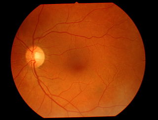
<span class="mw-page-title-main">Eye examination</span> Series of tests assessing vision and pertaining to the eyes

An eye examination is a series of tests performed to assess vision and ability to focus on and discern objects. It also includes other tests and examinations pertaining to the eyes. Eye examinations are primarily performed by an optometrist, ophthalmologist, or an orthoptist. Health care professionals often recommend that all people should have periodic and thorough eye examinations as part of routine primary care, especially since many eye diseases are asymptomatic.

<span class="mw-page-title-main">Slit lamp</span> Device for examining the eye

In ophthalmology and optometry, a slit lamp is an instrument consisting of a high-intensity light source that can be focused to shine a thin sheet of light into the eye. It is used in conjunction with a biomicroscope. The lamp facilitates an examination of the anterior segment and posterior segment of the human eye, which includes the eyelid, sclera, conjunctiva, iris, natural crystalline lens, and cornea. The binocular slit-lamp examination provides a stereoscopic magnified view of the eye structures in detail, enabling anatomical diagnoses to be made for a variety of eye conditions. A second, hand-held lens is used to examine the retina.

<span class="mw-page-title-main">Posterior segment of eyeball</span> Back two-thirds of the eye

The posterior segment or posterior cavity is the back two-thirds of the eye that includes the anterior hyaloid membrane and all of the optical structures behind it: the vitreous humor, retina, choroid, and optic nerve. The portion of the posterior segment visible during ophthalmoscopy is sometimes referred to as the posterior pole, or fundus. Some ophthalmologists specialize in the treatment and management of posterior segment disorders and diseases.

<span class="mw-page-title-main">Fundus (eye)</span> Concave interior of the eye

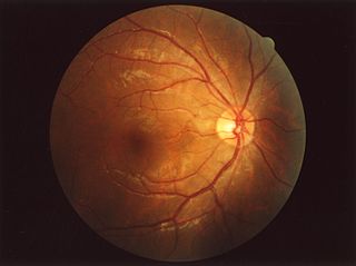
The fundus of the eye is the interior surface of the eye opposite the lens and includes the retina, optic disc, macula, fovea, and posterior pole. The fundus can be examined by ophthalmoscopy and/or fundus photography.
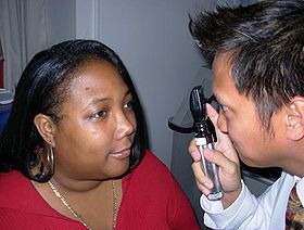
<span class="mw-page-title-main">Dilated fundus examination</span> Enlarging the pupil of the eye

Dilated fundus examination or dilated-pupil fundus examination (DFE) is a diagnostic procedure that employs the use of mydriatic eye drops to dilate or enlarge the pupil in order to obtain a better view of the fundus of the eye. Once the pupil is dilated, examiners use ophthalmoscopy (funduscopy) to view the eye's interior, allowing assessment of the retina, optic nerve head, blood vessels, and other features. They also often use specialized equipment such as a fundus camera. DFE has been found to be a more effective method for evaluation of internal ocular health than non-dilated examination. It is frequently performed by ophthalmologists and optometrists as part of an eye examination.

The red reflex refers to the reddish-orange reflection of light from the back of the eye, or fundus, observed when using an ophthalmoscope or retinoscope. The reflex relies on the transparency of optical media and reflects off the fundus back through media into the aperture of the ophthalmoscope. The red reflex is considered abnormal if there is any asymmetry between the eyes, dark spots, or white reflex (Leukocoria).

<span class="mw-page-title-main">Andreas Anagnostakis</span> Cretan ophthalmologist

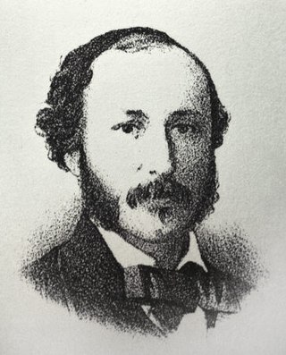
Andreas Anagnostakis was a Greek ophthalmologist, physician, and educator. He is best known for inventing the ophthalmoscope, a handheld tool used in diagnostics and still relevant today. He is credited as the first ophthalmologist in Greece.

<span class="mw-page-title-main">Fundus photography</span> Medical imaging of the eyes

Fundus photography involves photographing the rear of an eye, also known as the fundus. Specialized fundus cameras consisting of an intricate microscope attached to a flash enabled camera are used in fundus photography. The main structures that can be visualized on a fundus photo are the central and peripheral retina, optic disc and macula. Fundus photography can be performed with colored filters, or with specialized dyes including fluorescein and indocyanine green.

<span class="mw-page-title-main">Vitreous hemorrhage</span> Medical condition

Vitreous hemorrhage is the extravasation, or leakage, of blood into the areas in and around the vitreous humor of the eye. The vitreous humor is the clear gel that fills the space between the lens and the retina of the eye. A variety of conditions can result in blood leaking into the vitreous humor, which can cause impaired vision, floaters, and photopsia.

The eye is made up of the sclera, the iris, and the pupil, a black hole located at the center of the eye with the main function of allowing light to pass to the retina. Due to certain muscle spasms in the eye, the pupil can resemble a tadpole, which consists of a circular body, no arms or legs, and a tail.

<span class="mw-page-title-main">Eagle eye</span>

The eagle eye is among the sharpest in the animal kingdom, with an eyesight estimated at 4 to 8 times stronger than that of the average human. Although an eagle may only weigh 10 pounds (4.5 kg), its eyes are roughly the same size as those of a human. Eagle weight varies: a small eagle could weigh 700 grams (1.5 lb), while a larger one could weigh 6.5 kilograms (14 lb); an eagle of about 10 kilograms (22 lb) weight could have eyes as big as that of a human who weighs 200 pounds (91 kg). Although the size of the eagle eye is about the same as that of a human being, the back side shape of the eagle eye is flatter. Their eyes are stated to be larger in size than their brain, by weight. Color vision with resolution and clarity are the most prominent features of eagles' eyes, hence sharp-sighted people are sometimes referred to as "eagle-eyed". Eagles can identify five distinctly colored squirrels and locate their prey even if hidden.

<span class="mw-page-title-main">Teleophthalmology</span>

Teleophthalmology is a branch of telemedicine that delivers eye care through digital medical equipment and telecommunications technology. Today, applications of teleophthalmology encompass access to eye specialists for patients in remote areas, ophthalmic disease screening, diagnosis and monitoring; as well as distant learning.

Sickle cell retinopathy can be defined as retinal changes due to blood vessel damage in the eye of a person with a background of sickle cell disease. It can likely progress to loss of vision in late stages due to vitreous hemorrhage or retinal detachment. Sickle cell disease is a structural red blood cell disorder leading to consequences in multiple systems. It is characterized by chronic red blood cell destruction, vascular injury, and tissue ischemia causing damage to the brain, eyes, heart, lungs, kidneys, spleen, and musculoskeletal system.

<span class="mw-page-title-main">Heidelberg Retinal Tomography</span>

The Heidelberg Retinal Tomography is a diagnostic procedure used in ophthalmology. The Heidelberg Retina Tomograph (HRT) is an ophthalmological confocal point scanning laser ophthalmoscope for examining the cornea and certain areas of the retina using different diagnostic modules. However, the most widely used area of application for HRT is the inspection of the optic nerve head (papilla) for early detection and follow-up of glaucoma. The procedure has established itself as an integral part of routine glaucoma diagnostics alongside the visual field examination (perimetry), the chamber angle examination (gonioscopy) and the measurement of intraocular pressure (tonometry). The HRT is the most widely used application of confocal scanning laser ophthalmoscopy.

References

  1. 1 2 "Ophthalmoscopy | Michigan Medicine". Healthwise. Retrieved 10 July 2019.
  2. "Ophthalmoscopes Part 1" . Retrieved 2013-01-25.
  3. "Principles of Ophthalmoscopy". lifehugger.com. Retrieved 2009-05-24.
  4. Hoovers Citation
  5. Allyn, Welch. "PanOptic Ophthalmoscope". www.welchallyn.com. Retrieved 2019-01-29.
  6. Merriam-Webster, Merriam-Webster's Medical Dictionary, Merriam-Webster.
  7. Merriam-Webster, Merriam-Webster's Unabridged Dictionary, Merriam-Webster.
  8. Elsevier, Dorland's Illustrated Medical Dictionary, Elsevier.
  9. Houghton Mifflin Harcourt, The American Heritage Dictionary of the English Language, Houghton Mifflin Harcourt, archived from the original on 2015-09-25, retrieved 2015-10-21.
  10. Wolters Kluwer, Stedman's Medical Dictionary, Wolters Kluwer.