
A cataract is a cloudy area in the lens of the eye that leads to a decrease in vision of the eye. Cataracts often develop slowly and can affect one or both eyes. Symptoms may include faded colours, blurry or double vision, halos around light, trouble with bright lights, and difficulty seeing at night. This may result in trouble driving, reading, or recognizing faces. Poor vision caused by cataracts may also result in an increased risk of falling and depression. Cataracts cause 51% of all cases of blindness and 33% of visual impairment worldwide.

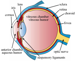
The vitreous body is the clear gel that fills the space between the lens and the retina of the eyeball in humans and other vertebrates. It is often referred to as the vitreous humor or simply "the vitreous". Vitreous fluid or "liquid vitreous" is the liquid component of the vitreous gel, found after a vitreous detachment. It is not to be confused with the aqueous humor, the other fluid in the eye that is found between the cornea and lens.

Vitrectomy is a surgery to remove some or all of the vitreous humor from the eye.

Eye surgery, also known as ophthalmic surgery or ocular surgery, is surgery performed on the eye or its adnexa. Eye surgery is part of ophthalmology and is performed by an ophthalmologist or eye surgeon. The eye is a fragile organ, and requires due care before, during, and after a surgical procedure to minimize or prevent further damage. An eye surgeon is responsible for selecting the appropriate surgical procedure for the patient, and for taking the necessary safety precautions. Mentions of eye surgery can be found in several ancient texts dating back as early as 1800 BC, with cataract treatment starting in the fifth century BC. It continues to be a widely practiced class of surgery, with various techniques having been developed for treating eye problems.

Phacoemulsification is a cataract surgery method in which the internal lens of the eye which has developed a cataract is emulsified with the tip of an ultrasonic handpiece and aspirated from the eye. Aspirated fluids are replaced with irrigation of balanced salt solution to maintain the volume of the anterior chamber during the procedure. This procedure minimises the incision size and reduces the recovery time and risk of surgery induced astigmatism.

An Intraocular lens (IOL) is a lens implanted in the eye usually as part of a treatment for cataracts or for correcting other vision problems such as short sightedness and long sightedness, a form of refractive surgery. If the natural lens is left in the eye, the IOL is known as phakic, otherwise it is a pseudophakic lens. Both kinds of IOLs are designed to provide the same light-focusing function as the natural crystalline lens. This can be an alternative to LASIK, but LASIK is not an alternative to an IOL for treatment of cataracts.
Cataract surgery, also called lens replacement surgery, is the removal of the natural lens of the eye that has developed a cataract, an opaque or cloudy area. The eye's natural lens is usually replaced with an artificial intraocular lens (IOL) implant.
A posterior vitreous detachment (PVD) is a condition of the eye in which the vitreous membrane separates from the retina. It refers to the separation of the posterior hyaloid membrane from the retina anywhere posterior to the vitreous base.
Trypan blue is an azo dye. It is a direct dye for cotton textiles. In biosciences, it is used as a vital stain to selectively colour dead tissues or cells blue.
Aphakia is the absence of the lens of the eye, due to surgical removal, such as in cataract surgery, a perforating wound or ulcer, or congenital anomaly. It causes a loss of ability to maintain focus (accommodation), high degree of farsightedness (hyperopia), and a deep anterior chamber. Complications include detachment of the vitreous or retina, and glaucoma.

An iridectomy, also known as a surgical iridectomy or corectomy, is the surgical removal of part of the iris. These procedures are most frequently performed in the treatment of closed-angle glaucoma and iris melanoma.
Howard V. Gimbel FRCSC, AOE, FACS, CABES, is a Canadian ophthalmologist, university professor, senior editor, and amateur musician. He is better known for his invention, along with Thomas Neuhann, of the continuous curvilinear capsulorhexis (CCC), a technique employed in modern cataract surgery.

Capsulotomy is a type of eye surgery in which an incision is made into the capsule of the crystalline lens of the eye. In modern cataract operations, the lens capsule is usually not removed. The most common forms of cataract surgery remove nearly all of the crystalline lens but do not remove the crystalline lens capsule. The crystalline lens capsule is retained and used to contain and position the intraocular lens implant (IOL).

In ophthalmology, glued intraocular lens or glued IOL is a surgical technique for implantation, with the use of biological glue, of a posterior chamber IOL in eyes with deficient or absent posterior capsules. A quick-acting surgical fibrin sealant derived from human blood plasma, with both hemostatic and adhesive properties, is used.
Intraocular lens scaffold, or IOL scaffold technique, is a surgical procedure in ophthalmology. In cases where the posterior lens capsule is ruptured and the cataract is present, an intraocular lens (IOL) can be inserted under the cataract. The IOL acts as a scaffold, and prevents the cataract pieces from falling to the back of the eye. The cataract can then be safely removed by emulsifying it with ultrasound and aspiration. This technique is called IOL scaffold, and was initiated by Amar Agarwal at Dr. Agarwal's Eye Hospital in Chennai, India.
Intravitreal injection is the method of administration of drugs into the eye by injection with a fine needle. The medication will be directly applied into the vitreous humor. It is used to treat various eye diseases, such as age-related macular degeneration (AMD), diabetic retinopathy, and infections inside the eye such as endophthalmitis. As compared to topical administration, this method is beneficial for a more localized delivery of medications to the targeted site, as the needle can directly pass through the anatomical eye barrier and dynamic barrier. It could also minimize adverse drug effects on other body tissues via the systemic circulation, which could be a possible risk for intravenous injection of medications. Although there are risks of infections or other complications, with suitable precautions throughout the injection process, chances for these complications could be lowered.
Uveitis–glaucoma–hyphaema (UGH) syndrome, also known as Ellingson syndrome, is a complication of cataract surgery, caused by intraocular lens subluxation or dislocation. The chafing of mispositioned intraocular lens over iris, ciliary body or iridocorneal angle cause elevated intraocular pressure (IOP) anterior uveitis and hyphema. It is most commonly caused by anterior chamber IOLs and sulcus IOLs but, the condition can be seen with any type of IOL, including posterior chamber lenses and cosmetic iris implants.
Ophthalmic viscosurgical devices (OVDs) are a class of clear gel-like material used in eye surgery to maintain the volume and shape of the anterior chamber of the eye, and protect the intraocular tissues during the procedure. They were originally called viscoelastic substances, or just viscoelastics. Their consistency allows the surgical instruments to move through them, but when there is low shear stress they do not flow, and retain their shape, preventing collapse of the anterior chamber. OVDs are available in several formulations which may be combined or used individually as best suits the procedure, and are introduced into the anterior chamber at the start of the procedure, and removed at the end. Their tendency to remain coherent helps with removal, as the cohesive variants tend to be drawn into the aspiration orifice without breaking up.
Manual small incision cataract surgery (MSICS) is an evolution of extracapsular cataract extraction (ECCE); the lens is removed from the eye through a self-sealing scleral tunnel wound. A well-constructed scleral tunnel is held closed by internal pressure, is watertight, and does not require suturing. The wound is relatively smaller than that in ECCE but is still markedly larger than a phacoemulsification wound. Comparative trials of MSICS against phaco in dense cataracts have found no difference in outcomes but MSICS had shorter operating times and significantly lower costs. MSICS has become the method of choice in the developing world because it provides high-quality outcomes with less surgically induced astigmatism than ECCE, no suture-related problems, quick rehabilitation, and fewer post-operative visits. MSICS is easy and fast to learn for the surgeon, cost effective, simple, and applicable to almost all types of cataract.
Cataract surgery has a long history in Europe, Asia, and Africa. It is one of the most common and successful surgical procedures in worldwide use, thanks to improvements in techniques for cataract removal and developments in intraocular lens (IOL) replacement technology, in implantation techniques, and in IOL design, construction, and selection. Surgical techniques that have contributed to this success include microsurgery, viscoelastics, and phacoemulsification.
