LASIK

Last updated
LASIK
US Navy 070501-N-5319A-007 Capt. Joseph Pasternak, an ophthalmology surgeon at National Naval Medical Center Bethesda, lines up the laser on Marine Corps Lt. Col. Lawrence Ryder's eye before beginning LASIK IntraLase surgery.jpg
Specialty Ophthalmology, optometry
ICD-9-CM 11.71
MeSH D020731
MedlinePlus 007018

LASIK or Lasik ( laser-assisted in situ keratomileusis ), commonly referred to as laser eye surgery or laser vision correction, is a type of refractive surgery for the correction of myopia, hyperopia, and an actual cure for astigmatism, since it is in the cornea. [1] LASIK surgery is performed by an ophthalmologist who uses a laser or microkeratome to reshape the eye's cornea in order to improve visual acuity. [2]

Contents

LASIK is very similar to another surgical corrective procedure, photorefractive keratectomy (PRK), and LASEK. All represent advances over radial keratotomy in the surgical treatment of refractive errors of vision. For patients with moderate to high myopia or thin corneas which cannot be treated with LASIK and PRK, the phakic intraocular lens is an alternative. [3] [4]

As of 2018, roughly 9.5 million Americans have had LASIK [1] [5] and, globally, between 1991 and 2016, more than 40 million procedures were performed. [6] [7] However, the procedure seemed to be a declining option as of 2015. [8]

Process

The planning and analysis of corneal reshaping techniques such as LASIK have been standardized by the American National Standards Institute, an approach based on the Alpins method of astigmatism analysis. The FDA website on LASIK states,

"Before undergoing a refractive procedure, you should carefully weigh the risks and benefits based on your own personal value system, and try to avoid being influenced by friends that have had the procedure or doctors encouraging you to do so." [9]

The procedure involves creating a thin flap on the eye, folding it to enable remodeling of the tissue beneath with a laser and repositioning the flap.

Preoperative procedures

Contact lenses

Patients wearing soft contact lenses are instructed to stop wearing them 5 to 21 days before surgery. One industry body recommends that patients wearing hard contact lenses should stop wearing them for a minimum of six weeks plus another six weeks for every three years the hard contacts have been worn. The cornea is avascular because it must be transparent to function normally. Its cells absorb oxygen from the tear film. Thus, low-oxygen-permeable contact lenses reduce the cornea's oxygen absorption, sometimes resulting in corneal neovascularization—the growth of blood vessels into the cornea. This causes a slight lengthening of inflammation duration and healing time and some pain during surgery, because of greater bleeding. Although some contact lenses (notably modern RGP and soft silicone hydrogel lenses) are made of materials with greater oxygen permeability that help reduce the risk of corneal neovascularization, patients considering LASIK are warned to avoid over-wearing their contact lenses.[ citation needed ]

Pre-operative examination and education

In the United States, the FDA has approved LASIK for age 18 or 22 and over because the vision has to stabilize. More importantly the patient's eye prescription should be stable for at least one year prior to surgery. The patient may be examined with pupillary dilation and education given prior to the procedure. Before the surgery, the patient's corneas are examined with a pachymeter to determine their thickness, and with a topographer, or corneal topography machine, [2] to measure their surface contour. Using low-power lasers, a topographer creates a topographic map of the cornea. The procedure is contraindicated if the topographer finds difficulties such as keratoconus [2] The preparatory process also detects astigmatism and other irregularities in the shape of the cornea. Using this information, the surgeon calculates the amount and the location of corneal tissue to be removed. The patient is prescribed and self-administers an antibiotic beforehand to minimize the risk of infection after the procedure and is sometimes offered a short acting oral sedative medication as a pre-medication. Prior to the procedure, anaesthetic eye drops are instilled. Factors that may rule out LASIK for some patients include large pupils, thin corneas and extremely dry eyes. [10]

Operative procedure

Flap creation

Flap creation with femtosecond laser Lasik Femtosegundo.gif
Flap creation with femtosecond laser
Flaporhexis as an alternative method to lift a femtosecond laser flap

A soft corneal suction ring is applied to the eye, holding the eye in place. This step in the procedure can sometimes cause small blood vessels to burst, resulting in bleeding or subconjunctival hemorrhage into the white (sclera) of the eye, a harmless side effect that resolves within several weeks. Increased suction causes a transient dimming of vision in the treated eye. Once the eye is immobilized, a flap is created by cutting through the corneal epithelium and Bowman's layer. This process is achieved with a mechanical microkeratome using a metal blade, or a femtosecond laser that creates a series of tiny closely arranged bubbles within the cornea. A hinge is left at one end of this flap. The flap is folded back, revealing the stroma, the middle section of the cornea. The process of lifting and folding back the flap can sometimes be uncomfortable.[ citation needed ]

Laser remodeling

The second step of the procedure uses an excimer laser (193 nm) to remodel the corneal stroma. The laser vaporizes the tissue in a finely controlled manner without damaging the adjacent stroma. No burning with heat or actual cutting is required to ablate the tissue. The layers of tissue removed are tens of micrometers thick.[ citation needed ]

Performing the laser ablation in the deeper corneal stroma provides for more rapid visual recovery and less pain than the earlier technique, photorefractive keratectomy (PRK). [11]

During the second step, the patient's vision becomes blurry, once the flap is lifted. They will be able to see only white light surrounding the orange light of the laser, which can lead to mild disorientation. The excimer laser uses an eye tracking system that follows the patient's eye position up to 4,000 times per second, redirecting laser pulses for precise placement within the treatment zone. Typical pulses are around 1 millijoule (mJ) of pulse energy in 10 to 20 nanoseconds. [12]

Repositioning of the flap

After the laser has reshaped the stromal layer, the LASIK flap is carefully repositioned over the treatment area by the surgeon and checked for the presence of air bubbles, debris, and proper fit on the eye. The flap remains in position by natural adhesion until healing is completed.

Postoperative care

Patients are usually given a course of antibiotic and anti-inflammatory eye drops. These are continued in the weeks following surgery. Patients are told to rest and are given dark eyeglasses to protect their eyes from bright lights and occasionally protective goggles to prevent rubbing of the eyes when asleep and to reduce dry eyes. They also are required to moisturize the eyes with preservative-free tears and follow directions for prescription drops. Occasionally after the procedure a bandage contact lens is placed to aid the healing, and typically removed after 3–4 days. Patients should be adequately informed by their surgeons of the importance of proper post-operative care to minimize the risk of complications. [13]

Wavefront-guided

Wavefront-guided LASIK is a variation of LASIK surgery in which, rather than applying a simple correction of only long/short-sightedness and astigmatism (only lower order aberrations as in traditional LASIK), an ophthalmologist applies a spatially varying correction, guiding the computer-controlled excimer laser with measurements from a wavefront sensor. The goal is to achieve a more optically perfect eye, though the result still depends on the physician's success at predicting changes that occur during healing and other factors that may have to do with the regularity/irregularity of the cornea and the axis of any residual astigmatism. Another important factor is whether the excimer laser can correctly register eye position in 3 dimensions, and to track the eye in all the possible directions of eye movement. If a wavefront guided treatment is performed with less than perfect registration and tracking, pre-existing aberrations can be worsened. In older patients, scattering from microscopic particles (cataract or incipient cataract) may play a role that outweighs any benefit from wavefront correction. [14] [15] [16] [17]

When treating a patient with preexisting astigmatism, most wavefront-guided LASIK lasers are designed to treat regular astigmatism as determined externally by corneal topography. In patients who have an element of internally induced astigmatism, therefore, the wavefront-guided astigmatism correction may leave regular astigmatism behind (a cross-cylinder effect). If the patient has preexisting irregular astigmatism, wavefront-guided approaches may leave both regular and irregular astigmatism behind. This can result in less-than-optimal visual acuity compared with a wavefront-guided approach combined with vector planning, as shown in a 2008 study. [18] Thus, vector-planning offers a better alignment between corneal astigmatism and laser treatment, and leaves less regular astigmatism behind on the cornea, which is advantageous whether irregular astigmatism coexists or not.[ citation needed ]

The "leftover" astigmatism after a purely surface-guided laser correction can be calculated beforehand, and is called ocular residual astigmatism (ORA). ORA is a calculation of astigmatism due to the noncorneal surface (internal) optics. The purely refraction-based approach represented by wavefront analysis actually conflicts with corneal surgical experience developed over many years. [17]

The pathway to "super vision" thus may require a more customized approach to corneal astigmatism than is usually attempted, and any remaining astigmatism ought to be regular (as opposed to irregular), which are both fundamental principles of vector planning overlooked by a purely wavefront-guided treatment plan. [17] This was confirmed by the 2008 study mentioned above, which found a greater reduction in corneal astigmatism and better visual outcomes under mesopic conditions using wavefront technology combined with vector analysis than using wavefront technology alone, and also found equivalent higher-order aberrations (see below). [18] Vector planning also proved advantageous in patients with keratoconus. [19]

No good data can be found that compare the percentage of LASIK procedures that employ wavefront guidance versus the percentage that do not, nor the percentage of refractive surgeons who have a preference one way or the other. Wavefront technology continues to be positioned as an "advance" in LASIK with putative advantages; [20] [21] [22] [23] however, it is clear that not all LASIK procedures are performed with wavefront guidance. [24]

Still, surgeons claim patients are generally more satisfied with this technique than with previous methods, particularly regarding lowered incidence of "halos," the visual artifact caused by spherical aberration induced in the eye by earlier methods. A meta-analysis of eight trials showed a lower incidence of these higher order aberrations in patients who had wavefront-guided LASIK compared to non-wavefront-guided LASIK. [25] Based on their experience, the United States Air Force has described WFG-Lasik as giving "superior vision results". [26]

Topography-assisted

Topography-assisted LASIK is intended to be an advancement in precision and reduce night-vision side effects. The first topography-assisted device received FDA approval September 13, 2013. [27] [28]

History

Barraquer's early work

In the 1950s, the microkeratome and keratomileusis technique were developed in Bogotá, Colombia, by the Spanish ophthalmologist José Barraquer. In his clinic, he would cut thin (one hundredth of a mm thick) flaps in the cornea to alter its shape. Barraquer also investigated how much of the cornea had to be left unaltered in order to provide stable long-term results. [29] This work was followed by that of the Russian scientist, Svyatoslav Fyodorov, who developed radial keratotomy (RK) in the 1970s and designed the first posterior chamber implantable contact lenses (phakic intraocular lens) in the 1980s.[ citation needed ]

Laser refractive surgery

In 1980, Rangaswamy Srinivasan, Samuel E. Blum and James J. Wynne at the IBM Research laboratory, discovered that an ultraviolet excimer laser could etch living tissue, with precision and with no thermal damage to the surrounding area. The phenomenon was termed "ablative photo-decomposition" (APD). [30] [31] Five years later, in 1985, Steven Trokel at the Edward S. Harkness Eye Institute, Columbia University in New York City, published his work using the excimer laser in radial keratotomy. He wrote,

"The central corneal flattening obtained by radial diamond knife incisions has been duplicated by radial laser incisions in 18 enucleated human eyes. The incisions, made by 193 nm far-ultraviolet light radiation emitted by the excimer laser, produced corneal flattening ranging from 0.12 to 5.35 diopters. Both the depth of the corneal incisions and the degree of central corneal flattening correlated with the laser energy applied. Histopathology revealed the remarkably smooth edges of the laser incisions." [32]

Together with his colleagues, Charles Munnerlyn and Terry Clapham, Trokel founded VISX USA inc. [33] Marguerite B. MacDonald MD performed the first human VISX refractive laser eye surgery in 1989. [34]

Patent

A number of patents have been issued for several techniques related to LASIK. Rangaswamy Srinivasan and James Wynne filed a patent application on the ultraviolet excimer laser, in 1986, issued in 1988. [35] In 1989, Gholam A. Peyman was granted a US patent for using an excimer laser to modify corneal curvature. [36] It was,

"A method and apparatus for modifying the curvature of a live cornea via use of an excimer laser. The live cornea has a thin layer removed therefrom, leaving an exposed internal surface thereon. Then, either the surface or thin layer is exposed to the laser beam along a predetermined pattern to ablate desired portions. The thin layer is then replaced onto the surface. Ablating a central area of the surface or thin layer makes the cornea less curved, while ablating an annular area spaced from the center of the surface or layer makes the cornea more curved. The desired predetermined pattern is formed by use of a variable diaphragm, a rotating orifice of variable size, a movable mirror or a movable fiber optic cable through which the laser beam is directed towards the exposed internal surface or removed thin layer." [35]

The patents related to so-called broad-beam LASIK and PRK technologies were granted to US companies including Visx and Summit during 1990–1995 based on the fundamental US patent issued to IBM (1988) which claimed the use of UV laser for the ablation of organic tissues. [35]

Implementation in the U.S.

The LASIK technique was implemented in the U.S. after its successful application elsewhere. The Food and Drug Administration (FDA) commenced a trial of the excimer laser in 1989. The first enterprise to receive FDA approval to use an excimer laser for photo-refractive keratectomy was Summit Technology (founder and CEO, Dr. David Muller). [37] In 1992, under the direction of the FDA, Greek ophthalmologist Ioannis Pallikaris introduced LASIK to ten VISX centers. In 1998, the "Kremer Excimer Laser", serial number KEA 940202, received FDA approval for its singular use for performing LASIK. [38] Subsequently, Summit Technology was the first company to receive FDA approval to mass manufacture and distribute excimer lasers. VISX and other companies followed. [38]

The excimer laser that was used for the first LASIK surgeries by I. Pallikaris MEL60-UOC.jpg
The excimer laser that was used for the first LASIK surgeries by I. Pallikaris

Pallikaris suggested a flap of cornea could be raised by microkeratome prior to the performing of PRK with the excimer laser. The addition of a flap to PRK became known as LASIK.

Recent years

The procedure seems to be a declining option for many in the United States, dropping more than 50 percent, from about 1.5 million surgeries in 2007 to 604,000 in 2015, according to the eye-care data source Market Scope. [39] A study in the journal Cornea determined the frequency with which LASIK was searched on Google from 2007 to 2011. [40] Within this time frame, LASIK searches declined by 40% in the United States. [40] Countries such as the U.K. and India also showed a decline, 22% and 24% respectively. [40] Canada, however, showed an increase in LASIK searches by 8%. [40] This decrease in interest can be attributed to several factors: the emergence of refractive cataract surgery, the economic recession in 2008, and unfavorable media coverage from the FDA's 2008 press release on LASIK. [8]

Effectiveness

In 2006, the British National Health Service's National Institute for Health and Clinical Excellence (NICE) considered evidence of the effectiveness and the potential risks of the laser surgery stating "current evidence suggests that photorefractive (laser) surgery for the correction of refractive errors is safe and effective for use in appropriately selected patients. Clinicians undertaking photorefractive (laser) surgery for the correction of refractive errors should ensure that patients understand the benefits and potential risks of the procedure. Risks include failure to achieve the expected improvement in unaided vision, development of new visual disturbances, corneal infection and flap complications. These risks should be weighed against those of wearing spectacles or contact lenses." [41] The FDA reports "The safety and effectiveness of refractive procedures has not been determined in patients with some diseases." [42]

Satisfaction

Surveys of LASIK surgery find rates of patient satisfaction between 92 and 98 percent. [43] [44] [45] In March 2008, the American Society of Cataract and Refractive Surgery published a patient satisfaction meta-analysis of over 3,000 peer-reviewed articles from international clinical journals. Data from a systematic literature review conducted from 1988 to 2008, consisting of 309 peer-reviewed articles about "properly conducted, well-designed, randomized clinical trials" found a 95.4 percent patient satisfaction rate among LASIK patients. [46]

A 2017 JAMA study claims that overall, preoperative symptoms decreased significantly, and visual acuity excelled. A meta-analysis discovered that 97% of patients achieved uncorrected visual acuity (UCVA) of 20/40, while 62% achieved 20/20. [47] The increase in visual acuity allows individuals to enter occupations that were previously not an option due to their vision.[ citation needed ]

Dissatisfaction

Some people with poor outcomes from LASIK surgical procedures report a significantly reduced quality of life because of vision problems or pain associated with the surgery. [1] A small percentage of patients may need further surgery because their condition is over- or under-corrected. Some patients need to wear contact lenses or glasses even after treatment. [48]

The most common reason for dissatisfaction in LASIK patients is chronic severe dry eye. Independent research indicates 95% of patients experience dry eye in the initial post-operative period. This number has been reported to up to 60% after one month. Symptoms begin to improve in the vast majority of patients in the 6 to 12 months following the surgery. [49] However, 30% of post-LASIK referrals to tertiary ophthalmology care centers have been shown to be due to chronic dry eye. [50] [51]

Morris Waxler, a former FDA official who was involved in the approval of LASIK, subsequently criticized its widespread use. In 2010, Waxler made media appearances and claimed that the procedure had a failure rate greater than 50%. The FDA responded that Waxler's information was "filled with false statements, incorrect citations" and "mischaracterization of results". [52]

A 2016 JAMA study indicates that the prevalence of complications from LASIK are higher than indicated, with the study indicating many patients experience glare, halos or other visual symptoms. [53] Forty-three percent of participants in a JAMA study (published in 2017) reported new visual symptoms they had not experienced before.

Presbyopia

A type of LASIK, known as presbyLasik, may be used in presbyopia. Results are, however, more variable and some people have a decrease in visual acuity. [54]

Risks

Higher-order aberrations

Higher-order aberrations are visual problems that require special testing for diagnosis and are not corrected with normal spectacles (eyeglasses). These aberrations include 'starbursts', 'ghosting', 'halos' and others. [1] [55] Some patients describe these symptoms post-operatively and associate them with the LASIK technique including the formation of the flap and the tissue ablation. [56]

There is a correlation between pupil size and aberrations. This correlation may be the result of irregularity in the corneal tissue between the untouched part of the cornea and the reshaped part. Daytime post-LASIK vision is optimal, since the pupil size is smaller than the LASIK flap.[ citation needed ]

Others propose that higher-order aberrations are present preoperatively. [57] They can be measured in micrometers (µm) whereas the smallest laser-beam size approved by the FDA is about 1000 times larger, at 0.65 mm. In situ keratomileusis effected at a later age increases the incidence of corneal higher-order wavefront aberrations. [58] [59] These factors demonstrate the importance of careful patient selection for LASIK treatment.

A subconjunctival hemorrhage is a common and minor post-LASIK complication. Eye hemorrhage.jpg
A subconjunctival hemorrhage is a common and minor post-LASIK complication.

Dry eyes

95% of patients report dry-eye symptoms after LASIK. [1] [60] Although it is usually temporary, it can develop into chronic and severe dry eye syndrome. Quality of life can be severely affected by dry-eye syndrome. [61]

Underlying conditions with dry eye such as Sjögren's syndrome are considered contraindications to Lasik. [62]

Treatments include artificial tears, prescription tears, and punctal occlusion. Punctal occlusion is accomplished by placing a collagen or silicone plug in the tear duct, which normally drains fluid from the eye. Some patients complain of ongoing dry-eye symptoms despite such treatments and the symptoms may be permanent. [63]

Halos

Some post-LASIK patients see halos and starbursts around bright lights at night. [1] At night, the pupil may dilate to be larger than the flap leading to the edge of the flap or stromal changes causing visual distortion of light that does not occur during the day when the pupil is smaller. The eyes can be examined for large pupils pre-operatively and the risk of this symptom assessed.[ citation needed ]

Complications due to LASIK have been classified as those that occur due to preoperative, intraoperative, early postoperative, or late postoperative sources: [64] According to the UK National Health Service, complications occur in fewer than 5% of cases. [60]

Other complications

FDA's position

In October 2009, the FDA, the National Eye Institute (NEI), and the Department of Defense (DoD) launched the LASIK Quality of Life Collaboration Project (LQOLCP) to help better understand the potential risk of severe problems that can result from LASIK [82] in response to widespread reports of problems experienced by patients after LASIK laser eye surgery. [83] This project examined patient-reported outcomes with LASIK (PROWL). The project consisted of three phases: pilot phase, phase I, phase II (PROWL-1) and phase III (PROWL-2). [84] The last two phases were completed in 2014.

The results of the LASIK Quality of Life Study were published in October 2014. [82]

Based on our initial analyses of our studies:
  • Up to 46 percent of participants, who had no visual symptoms before surgery, reported at least one visual symptom at three months after surgery.
  • Participants who developed new visual symptoms after surgery, most often developed halos. Up to 40 percent of participants with no halos before LASIK had halos three months following surgery.
  • Up to 28 percent of participants with no symptoms of dry eyes before LASIK, reported dry eye symptoms at three months after their surgery.
  • Less than 1 percent of study participants experienced "a lot of" difficulty with or inability to do usual activities without corrective lenses because of their visual symptoms (halos, glare, et al.) after LASIK surgery.
  • Participants who were not satisfied with the LASIK surgery reported all types of visual symptoms the questionnaire measured (double vision/ghosting, starbursts, glare, and halos).

The FDA's director of the Division of Ophthalmic Devices, said about the LASIK study "Given the large number of patients undergoing LASIK annually, dissatisfaction and disabling symptoms may occur in a significant number of patients". [85] Also in 2014, FDA published an article highlighting the risks and a list of factors and conditions individuals should consider when choosing a doctor for their refractive surgery. [86]

Contraindications

Not everyone is eligible to receive LASIK. Severe keratoconus or thin corneas may disqualify patients from LASIK, though other procedures may be viable options. Those with Fuchs' corneal endothelial dystrophy, corneal epithelial basement membrane dystrophy, retinal tears, autoimmune diseases, severe dry eyes, and significant blepharitis should be treated before consideration for LASIK. Women who are pregnant or nursing are generally not eligible to undergo LASIK. [87]

People with large pupils (e.g. due to taking medications or in the younger age group) may be particularly prone to symptoms such as glare, halos, starbursts, and ghost images (double vision) in dim light after surgery. Because the laser can only work on the inner section of the cornea, the outer rim is left unaffected. In dim lighting, a patient's pupils dilate and may be predisposed to optic aberrations due to refractive asynchrony of the two regions with regards to the incoming light. [88]

Further research

Since 1991, there have been further developments such as faster lasers; larger spot areas; bladeless flap incisions; intraoperative corneal pachymetry; and "wavefront-optimized" and "wavefront-guided" techniques which were introduced by the University of Michigan's Center for Ultrafast Optical Science. The goal of replacing standard LASIK in refractive surgery is to avoid permanently weakening the cornea with incisions and to deliver less energy to the surrounding tissues. More recently, techniques like Epi-Bowman Keratectomy have been developed that avoid touching the epithelial basement membrane or Bowman's layer. [89]

Experimental techniques

Comparison to photorefractive keratectomy

A systematic review that compared PRK and LASIK concluded that LASIK has shorter recovery time and less pain. [95] The two techniques after a period of one year have similar results. [95]

A 2017 systematic review found uncertainty in visual acuity, but found that in one study, those receiving PRK were less likely to achieve a refractive error, and were less likely to have an over-correction than compared to LASIK. [96]

Related Research Articles

<span class="mw-page-title-main">Farsightedness</span> Eye condition in which light is focused behind instead of on the retina

Far-sightedness, also known as long-sightedness, hypermetropia, and hyperopia, is a condition of the eye where distant objects are seen clearly but near objects appear blurred. This blur is due to incoming light being focused behind, instead of on, the retina due to insufficient accommodation by the lens. Minor hypermetropia in young patients is usually corrected by their accommodation, without any defects in vision. But, due to this accommodative effort for distant vision, people may complain of eye strain during prolonged reading. If the hypermetropia is high, there will be defective vision for both distance and near. People may also experience accommodative dysfunction, binocular dysfunction, amblyopia, and strabismus. Newborns are almost invariably hypermetropic, but it gradually decreases as the newborn gets older.

<span class="mw-page-title-main">Photorefractive keratectomy</span> Refractive eye surgery procrdure

Photorefractive keratectomy (PRK) and laser-assisted sub-epithelial keratectomy (LASEK) are laser eye surgery procedures intended to correct a person's vision, reducing dependency on glasses or contact lenses. LASEK and PRK permanently change the shape of the anterior central cornea using an excimer laser to ablate a small amount of tissue from the corneal stroma at the front of the eye, just under the corneal epithelium. The outer layer of the cornea is removed prior to the ablation.

A microkeratome is a precision surgical instrument with an oscillating blade designed for creating the corneal flap in LASIK or ALK surgery. The normal human cornea varies from around 500 to 600 μm in thickness; and in the LASIK procedure, the microkeratome creates an 83 to 200 μm thick flap. The microkeratome uses an oscillating blade system, which has a blade that oscillates horizontally as the blade travels vertically for a precise cut. This piece of equipment is used all around the world to cut the cornea flap. The microkeratome is also used in Descemet's stripping automated endothelial keratoplasty (DSAEK), where it is used to slice a thin layer from the back of the donor cornea, which is then transplanted into the posterior cornea of the recipient. It was invented by Jose Barraquer and Cesar Carlos Carriazo in the 1950s in Colombia.

<span class="mw-page-title-main">Radial keratotomy</span> Refractive surgical procedure to correct myopia (nearsightedness

Radial keratotomy (RK) is a refractive surgical procedure to correct myopia (nearsightedness). It was developed in 1974 by Svyatoslav Fyodorov, a Russian ophthalmologist. It has been largely supplanted by newer, more accurate operations, such as photorefractive keratectomy, LASIK, Epi-LASIK and the phakic intraocular lens.

<span class="mw-page-title-main">Refractive surgery</span> Surgery to treat common vision disorders

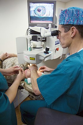
Refractive surgery is optional eye surgery used to improve the refractive state of the eye and decrease or eliminate dependency on glasses or contact lenses. This can include various methods of surgical remodeling of the cornea (keratomileusis), lens implantation or lens replacement. The most common methods today use excimer lasers to reshape the curvature of the cornea. Refractive eye surgeries are used to treat common vision disorders such as myopia, hyperopia, presbyopia and astigmatism.

<span class="mw-page-title-main">Intraocular lens</span> Lens implanted in the eye to treat cataracts or myopia

An Intraocular lens (IOL) is a lens implanted in the eye usually as part of a treatment for cataracts or for correcting other vision problems such as short sightedness and long sightedness, a form of refractive surgery. If the natural lens is left in the eye, the IOL is known as phakic, otherwise it is a pseudophakic lens. Both kinds of IOLs are designed to provide the same light-focusing function as the natural crystalline lens. This can be an alternative to LASIK, but LASIK is not an alternative to an IOL for treatment of cataracts.

<span class="mw-page-title-main">Refractive error</span> Problem with focusing light accurately on the retina due to the shape of the eye

Refractive error is a problem with focusing light accurately on the retina due to the shape of the eye and/or cornea. The most common types of refractive error are near-sightedness, far-sightedness, astigmatism, and presbyopia. Near-sightedness results in far away objects being blurry, far-sightedness and presbyopia result in close objects being blurry, and astigmatism causes objects to appear stretched out or blurry. Other symptoms may include double vision, headaches, and eye strain.

<span class="mw-page-title-main">Phakic intraocular lens</span> Lens implanted in eye in addition to the natural lens

A phakic intraocular lens (PIOL) is an intraocular lens that is implanted surgically into the eye to correct refractive errors without removing the natural lens. Intraocular lenses that are implanted into eyes after the eye's natural lens has been removed during cataract surgery are known as pseudophakic.

Automated lamellar keratoplasty (ALK), also known as keratomileusis in situ, is a non-laser lamellar refractive procedure used to correct high degree refractive errors. This procedure can correct large amounts of myopia and hyperopia. However, the resultant change is not as predictable as with other procedures.

ReLExSmall incision lenticule extraction (SMILE), second generation of ReLEx Femtosecond lenticule extraction (FLEx), is a form of laser based refractive eye surgery developed by Carl Zeiss Meditec used to correct myopia, and cure astigmatism. Although similar to LASIK laser surgery, the intrastromal procedure uses a single femtosecond laser referenced to the corneal surface to cleave a thin lenticule from the corneal stroma for manual extraction.

Stephen Updegraff, M.D., FACS is an American refractive surgeon best known for his early involvement in, and contributions to, LASIK. He is a Fellow of the American College of Surgeons, a board-certified member of the American Board of Ophthalmology, a founding member of the American College of Ophthalmic Surgeons, and a member of the International Society of Refractive Surgery, the American Academy of Ophthalmology, the American Society of Cataract and Refractive Surgery, and the Pine Ridge Eye Study Society. Updegraff currently serves as the medical director of Updegraff Vision in St. Petersburg, Florida.

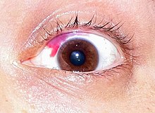
Diffuse lamellar keratitis (DLK) is a sterile inflammation of the cornea which may occur after refractive surgery, such as LASIK. Its incidence has been estimated to be 1 in 500 patients, though this may be as high as 32% in some cases.

<span class="mw-page-title-main">Gholam A. Peyman</span> Iranian-American ophthalmologist and retina surgeon known for inventing LASIK eye surgery

Gholam A. Peyman is an Iranian American ophthalmologist, retina surgeon, and inventor. He is best known for his invention of LASIK eye surgery, a vision correction procedure designed to allow people to see clearly without glasses. He was awarded the first US patent for the procedure in 1989.

Laser blended vision is a laser eye treatment which is used to treat presbyopia or other age-related eye conditions. It can be used to help people that simply need reading glasses, and also those who have started to need bifocal or varifocal spectacle correction due to ageing changes in the eye. It can be used for people who are also short-sighted (myopia) or long-sighted (hyperopia) and who also may have astigmatism.

Jeff Machat MD, FRCSC, DABO is an ophthalmologist in the United States and Canada specializing in surgical vision correction better known as refractive eye surgery.

The Alpins Method is a system to plan and analyze the results of refractive surgical procedures, such as laser in-situ keratomileus (LASIK). The Alpins Method is also used to plan cataract/toric intraocular lens (IOL) surgical procedures.

Peter S. Hersh is an American ophthalmologist and specialist in LASIK eye surgery, keratoconus, and diseases of the cornea. He co-authored the article in the journal Ophthalmology that presented the results of the study that led to the first approval by the U.S. Food and Drug Administration (FDA) of the excimer laser for the correction of nearsightedness in the United States. Hersh was also medical monitor of the study that led to approval of corneal collagen crosslinking for the treatment of keratoconus.

The eye, like any other optical system, suffers from a number of specific optical aberrations. The optical quality of the eye is limited by optical aberrations, diffraction and scatter. Correction of spherocylindrical refractive errors has been possible for nearly two centuries following Airy's development of methods to measure and correct ocular astigmatism. It has only recently become possible to measure the aberrations of the eye and with the advent of refractive surgery it might be possible to correct certain types of irregular astigmatism.

Post-LASIK ectasia is a condition similar to keratoconus where the cornea starts to bulge forwards at a variable time after LASIK, PRK, or SMILE corneal laser eye surgery. However, the physiological processes of post-LASIK ectasia seem to be different from keratoconus. The visible changes in the basal epithelial cell and anterior and posterior keratocytes linked with keratoconus were not observed in post-LASIK ectasia.

<span class="mw-page-title-main">Noel Alpins</span> Australian ophthalmologist

Noel Alpins is an Australian ophthalmologist who developed the Alpins method of astigmatism analysis used in refractive, corneal, and cataract surgery, used in the research of LASIK.

References

  1. 1 2 3 4 5 6 Rabin, Roni Caryn (June 11, 2018). "Lasik's Risks Are Coming Into Sharper Focus – Some patients who undergo the eye surgery report a variety of side effects. They may persist for years, studies show". The New York Times . Retrieved June 11, 2018.
  2. 1 2 3 Finn, Peter (20 December 2012). "Medical Mystery: Preparation for surgery revealed cause of deteriorating eyesight". The Washington Post.
  3. Lovisolo CF, Reinstein DZ (Nov–Dec 2005). "Phakic intraocular lenses". Survey of Ophthalmology. 50 (6): 549–87. doi:10.1016/j.survophthal.2005.08.011. PMID   16263370.
  4. Sanders DR, Vukich JA (May 2003). "Comparison of implantable contact lens and laser assisted in situ keratomileusis for moderate to high myopia". Cornea. 22 (4): 324–31. doi:10.1097/00003226-200305000-00009. PMID   12792475. S2CID   21142105.
  5. Lindfield D, Poole T. "Nd:YAG Treatment of Epithelial Ingrowth". Cataract and Refractive Surgery Today. Retrieved 12 September 2013.
  6. Stodola, Ellen (April 1, 2016). "LASIK worldwide". EyeWorld.org. Archived from the original on June 12, 2018. Retrieved June 12, 2018.
  7. "A Look at LASIK Past, Present and Future". EyeNet Magazine. Archived from the original on 31 July 2013. Retrieved 12 September 2013.
  8. 1 2 Corcoran KJ (July 2015). "Macroeconomic landscape of refractive surgery in the United States". Current Opinion in Ophthalmology. 26 (4): 249–54. doi:10.1097/ICU.0000000000000159. PMID   26058020. S2CID   11842503.
  9. "US FDA/CDRH: LASIK – What are the risks and how can I find the right doctor for me?". Fda.gov. June 9, 2014. Retrieved December 23, 2016.
  10. "Am I a Candidate For LASIK Surgery? TLC Laser Eye Centers". Tlcvision.com. Retrieved 20 December 2018.
  11. Shortt AJ, Allan BD, Evans JR (January 2013). "Laser-assisted in-situ keratomileusis (LASIK) versus photorefractive keratectomy (PRK) for myopia". The Cochrane Database of Systematic Reviews. 1 (1): CD005135. doi:10.1002/14651858.CD005135.pub3. PMID   23440799. There was evidence that LASIK gives a faster visual recovery than PRK and is a less painful technique. Results at one year after surgery were comparable: most analyses favoured LASIK but they were not statistically significant.
  12. "Patent: ultraviolet solid state laser". Freepatentsonline.com. Retrieved 2011-12-10.
  13. Azar DT, Gatinel D (2007). Refractive surgery (2nd ed.). Philadelphia: Mosby Elsevier. ISBN   978-0-323-03599-6.
  14. Walsh MJ. Is the future of refractive surgery based on corneal topography or wavefront? "Ocular Surgery News". August 1, 2000, page 26.
  15. Walsh MJ. Wavefront is showing signs of success, but can it do it alone? Ocular Surgery News. September 1, 2000, page 41.
  16. EW Dialogue: the future of wavefront refraction as a diagnostic tool. "EyeWorld". May 2000, pages 64 and 65.
  17. 1 2 3 Alpins NA (2002). "Wavefront technology: a new advance that fails to answer old questions on corneal vs. refractive astigmatism correction". Journal of Refractive Surgery. 18 (6): 737–9. doi:10.3928/1081-597X-20021101-12. PMID   12458868.
  18. 1 2 Alpins N, Stamatelatos G (August 2008). "Clinical outcomes of laser in situ keratomileusis using combined topography and refractive wavefront treatments for myopic astigmatism". Journal of Cataract and Refractive Surgery. 34 (8): 1250–9. doi:10.1016/j.jcrs.2008.03.028. PMID   18655973. S2CID   29819060.
  19. Alpins N, Stamatelatos G (April 2007). "Customized photoastigmatic refractive keratectomy using combined topographic and refractive data for myopia and astigmatism in eyes with forme fruste and mild keratoconus". Journal of Cataract and Refractive Surgery. 33 (4): 591–602. doi:10.1016/j.jcrs.2006.12.014. PMID   17397730. S2CID   14881153.
  20. American Academy of Ophthalmology. "Refractive Laser Surgery: An In-Depth Look at LASIK and Brief Overview of PRK, Epi-LASIK, and LASEK: A Science Writer's Guide" Archived 2012-06-16 at the Wayback Machine . Accessed January 29, 2012.
  21. Abbott Medical Optics website. "WaveScan WaveFront System". Accessed August 15, 2012.
  22. Emory Healthcare website. "Wavefront technology". Accessed August 15, 2012.
  23. Croes K. AllAboutVision website. "Custom LASIK or wavefront LASIK: individualized vision correction". Accessed August 15, 2012.
  24. Segre L. "Cost of LASIK eye surgery and other corrective procedures". Allaboutvision.com. Retrieved 2012-08-15.
  25. Fares U, Suleman H, Al-Aqaba MA, Otri AM, Said DG, Dua HS (August 2011). "Efficacy, predictability, and safety of wavefront-guided refractive laser treatment: metaanalysis". Journal of Cataract and Refractive Surgery. 37 (8): 1465–75. doi:10.1016/j.jcrs.2011.02.029. PMID   21782089. S2CID   26968756.
  26. Campbell S. "Air Force aims for 'weapons-grade' vision". Af.mil. Archived from the original on 2012-07-28. Retrieved 2011-12-10.
  27. "Nidek EC-5000 Excimer Laser System – P970053/S011". Food and Drug Administration. 2013-10-13. Retrieved 2016-05-01.
  28. Stulting D (2014-04-28). "Topography-guided LASIK: A paradigm shift in refractive laser treatment" (PDF). EyeWorld Daily News. Retrieved 2016-05-01.
  29. Troutman RC, Swinger C (1978). "Refractive keratoplasty: keratophakia and keratomileusis". Transactions of the American Ophthalmological Society. 76: 329–39. PMC   1311630 . PMID   382579.
  30. "Prize for the Industrial Application of Physics Winner – American Institute of Physics". Aip.org. Archived from the original on 2011-09-28. Retrieved 2011-12-10.
  31. "James Wynne". laserfest.org. Retrieved 30 December 2021.
  32. Cotliar AM, Schubert HD, Mandel ER, Trokel SL (February 1985). "Excimer laser radial keratotomy". Ophthalmology. 92 (2): 206–8. doi:10.1016/s0161-6420(85)34052-6. PMID   3982798.
  33. "VISX - World Leader in Laser Vision Correction Technology used for Conventional LASIK, Wavefront Guided LASIK, PRK, LASEK, and PTK Procedures". Archived from the original on 2015-10-19. Retrieved 2019-01-25.
  34. McDonald MB, Kaufman HE, Frantz JM, Shofner S, Salmeron B, Klyce SD (May 1989). "Excimer laser ablation in a human eye. Case report". Archives of Ophthalmology. 107 (5): 641–2. doi:10.1001/archopht.1989.01070010659013. PMID   2719572.
  35. 1 2 3 US4784135,Samuel E. Blum, Rangaswamy Srinivasan, James J. Wynne,"Far ultraviolet surgical and dental procedures",issued 1988-11-15
  36. US4840175,Gholam A. Peyman,"Method for modifying corneal curvature",issued 1988-6-20
  37. "FDA-Approved Lasers for PRK and Other Refractive Surgeries". Fda.gov. Retrieved 2011-12-10.
  38. 1 2 "List of FDA-Approved Lasers for LASIK". Fda.gov. Retrieved 2011-12-10.
  39. Schoenberg, Nara (May 23, 2016). "Lasik surgery falling out of favor with patients". Chicago Tribune.
  40. 1 2 3 4 Stein JD, Childers DM, Nan B, Mian SI (July 2013). "Gauging interest of the general public in laser-assisted in situ keratomileusis eye surgery". Cornea. 32 (7): 1015–8. doi:10.1097/ICO.0b013e318283c85a. PMC   3679260 . PMID   23538615.
  41. "Photorefractive (laser) surgery for the correction of refractive errors" (pdf). National Health Service. March 2006.
  42. "LASIK – When is LASIK not for me?". FDA. 2018-11-03. Retrieved 20 December 2018.
  43. Saragoussi D, Saragoussi JJ (September 2004). "[Lasik, PRK and quality of vision: a study of prognostic factors and a satisfaction survey]". Journal Français d'Ophtalmologie (in French). 27 (7): 755–64. doi:10.1016/S0181-5512(04)96210-9. PMID   15499272.
  44. Bailey MD, Mitchell GL, Dhaliwal DK, Boxer Wachler BS, Zadnik K (July 2003). "Patient satisfaction and visual symptoms after laser in situ keratomileusis". Ophthalmology. 110 (7): 1371–8. doi:10.1016/S0161-6420(03)00455-X. PMID   12867394.
  45. McGhee CN, Craig JP, Sachdev N, Weed KH, Brown AD (April 2000). "Functional, psychological, and satisfaction outcomes of laser in situ keratomileusis for high myopia". Journal of Cataract and Refractive Surgery. 26 (4): 497–509. doi:10.1016/S0886-3350(00)00312-6. PMID   10771222. S2CID   13304987.
  46. Solomon KD, Fernández de Castro LE, Sandoval HP, Biber JM, Groat B, Neff KD, et al. (April 2009). "LASIK world literature review: quality of life and patient satisfaction". Ophthalmology. 116 (4): 691–701. doi:10.1016/j.ophtha.2008.12.037. PMID   19344821.
  47. Sugar A, Hood CT, Mian SI (January 2017). "Patient-Reported Outcomes Following LASIK: Quality of Life in the PROWL Studies". JAMA. 317 (2): 204–205. doi:10.1001/jama.2016.19323. PMID   28097345.
  48. 1 2 "LASIK Eye Surgery". The New York Times. Retrieved 10 September 2013.
  49. Shtein RM (October 2011). "Post-LASIK dry eye". Expert Review of Ophthalmology. 6 (5): 575–582. doi:10.1586/eop.11.56. PMC   3235707 . PMID   22174730.
  50. Levinson BA, Rapuano CJ, Cohen EJ, Hammersmith KM, Ayres BD, Laibson PR (January 2008). "Referrals to the Wills Eye Institute Cornea Service after laser in situ keratomileusis: reasons for patient dissatisfaction". Journal of Cataract and Refractive Surgery. 34 (1): 32–9. doi:10.1016/j.jcrs.2007.08.028. PMID   18165078. S2CID   11133295.
  51. Jabbur NS, Sakatani K, O'Brien TP (September 2004). "Survey of complications and recommendations for management in dissatisfied patients seeking a consultation after refractive surgery". Journal of Cataract and Refractive Surgery. 30 (9): 1867–74. doi:10.1016/j.jcrs.2004.01.020. PMID   15342048. S2CID   25054973.
  52. Rodemich, Karen (2010). "Former FDA official warns of LASIK risks: the man who OK'd LASIK now warns of an "epidemic" of eye problems". Review of Optometry. 147 (10): 4.
  53. Cha, Ariana Eunjung (2016-11-23). "Many LASIK patients may wind up with glare, halos or other visual symptoms, study suggests". Washington Post. ISSN   0190-8286 . Retrieved 2018-04-04.
  54. Pallikaris IG, Panagopoulou SI (July 2015). "PresbyLASIK approach for the correction of presbyopia". Current Opinion in Ophthalmology. 26 (4): 265–72. doi:10.1097/icu.0000000000000162. PMID   26058023. S2CID   35434343.
  55. Pop M, Payette Y (January 2004). "Risk factors for night vision complaints after LASIK for myopia". Ophthalmology. 111 (1): 3–10. doi: 10.1016/j.ophtha.2003.09.022 . PMID   14711706.
  56. Padmanabhan P, Basuthkar SS, Joseph R (Jul–Aug 2010). "Ocular aberrations after wavefront optimized LASIK for myopia". Indian Journal of Ophthalmology. 58 (4): 307–12. doi: 10.4103/0301-4738.64139 . PMC   2907032 . PMID   20534921.
  57. "Individual Risk Factors of Halos, Loss of Contrast Sensitivity, Glare and Starbursts after LASIK." operationauge.com
  58. Yamane N, Miyata K, Samejima T, Hiraoka T, Kiuchi T, Okamoto F, et al. (November 2004). "Ocular higher-order aberrations and contrast sensitivity after conventional laser in situ keratomileusis". Investigative Ophthalmology & Visual Science. 45 (11): 3986–90. doi: 10.1167/iovs.04-0629 . PMID   15505046.
  59. Oshika T, Miyata K, Tokunaga T, Samejima T, Amano S, Tanaka S, et al. (June 2002). "Higher order wavefront aberrations of cornea and magnitude of refractive correction in laser in situ keratomileusis". Ophthalmology. 109 (6): 1154–8. doi:10.1016/S0161-6420(02)01028-X. PMID   12045059.
  60. 1 2 "Laser eye surgery". NHS Choices. 5 March 2012. Retrieved 26 October 2013.
  61. "LASIK – What are the risks and how can I find the right doctor for me?". Food and Drug Administration. 12 September 2011. Retrieved 26 October 2013.
  62. Simpson RG, Moshirfar M, Edmonds JN, Christiansen SM, Behunin N (2012). "Laser in situ keratomileusis in patients with collagen vascular disease: a review of the literature". Clinical Ophthalmology. 6: 1827–37. doi: 10.2147/OPTH.S36690 . PMC   3497460 . PMID   23152662.
  63. "LASIK". Fda.gov. 2018-08-08. Retrieved 2022-07-17.
  64. Majmudar, PA. "LASIK Complications." Focal Points: Clinical Modules for Ophthalmologists. American Academy of Ophthalmology. September, 2004. Archived March 11, 2006, at the Wayback Machine
  65. Carrillo C, Chayet AS, Dougherty PJ, Montes M, Magallanes R, Najman J, et al. (2005). "Incidence of complications during flap creation in LASIK using the NIDEK MK-2000 microkeratome in 26,600 cases". Journal of Refractive Surgery. 21 (5 Suppl): S655-7. doi:10.3928/1081-597X-20050902-20. PMID   16212299.
  66. "Eye Surgery Education Council". Lasikinstitute.org. Archived from the original on 2011-09-28. Retrieved 2011-12-10.
  67. Tham VM, Maloney RK (May 2000). "Microkeratome complications of laser in situ keratomileusis". Ophthalmology. 107 (5): 920–4. doi:10.1016/S0161-6420(00)00004-X. PMID   10811084.
  68. 1 2 Vesaluoma M, Pérez-Santonja J, Petroll WM, Linna T, Alió J, Tervo T (February 2000). "Corneal stromal changes induced by myopic LASIK". Investigative Ophthalmology & Visual Science. 41 (2): 369–76. PMID   10670464.
  69. 1 2 3 4 Sun L, Liu G, Ren Y, Li J, Hao J, Liu X, Zhang Y (2005). "Efficacy and safety of LASIK in 10,052 eyes of 5081 myopic Chinese patients". Journal of Refractive Surgery. 21 (5 Suppl): S633-5. doi:10.3928/1081-597X-20050902-15. PMID   16212294.
  70. "Ectasia After LASIK". American Academy of Ophthalmology.
  71. Galvis V, Tello A, Ortiz AI, Quintero MP, Parra MM, Blanco NA (April–June 2019). "Traumatic corneal flap avulsion and loss 13 years after LASIK". Saudi Journal of Ophthalmology. 33 (2): 172–176. doi: 10.1016/j.sjopt.2018.08.001 . PMC   6664271 . PMID   31384163.
  72. 1 2 Ruiz-Moreno JM, Alió JL (2003). "Incidence of retinal disease following refractive surgery in 9,239 eyes". Journal of Refractive Surgery. 19 (5): 534–47. doi:10.3928/1081-597X-20030901-08. PMID   14518742.
  73. Suarez E, Torres F, Vieira JC, Ramirez E, Arevalo JF (October 2002). "Anterior uveitis after laser in situ keratomileusis". Journal of Cataract and Refractive Surgery. 28 (10): 1793–8. doi:10.1016/S0886-3350(02)01364-0. PMID   12388030. S2CID   11880947.
  74. Boes DA, Omura AK, Hennessy MJ (December 2001). "Effect of high-altitude exposure on myopic laser in situ keratomileusis". Journal of Cataract and Refractive Surgery. 27 (12): 1937–41. doi:10.1016/S0886-3350(01)01074-4. PMID   11738908. S2CID   45468164.
  75. Dimmig JW, Tabin G (2003). "The ascent of Mount Everest following laser in situ keratomileusis". Journal of Refractive Surgery. 19 (1): 48–51. doi:10.3928/1081-597X-20030101-10. PMID   12553606.
  76. Hammer T, Heynemann M, Naumann I, Duncker GI (March 2006). "[Correction and induction of high-order aberrations after standard and wavefront-guided LASIK and their influence on the postoperative contrast sensitivity]". Klinische Monatsblätter für Augenheilkunde (in German). 223 (3): 217–24. doi:10.1055/s-2005-858864. PMID   16552654. S2CID   71611964.
  77. Alió JL, Montés-Mico R (February 2006). "Wavefront-guided versus standard LASIK enhancement for residual refractive errors". Ophthalmology. 113 (2): 191–7. doi:10.1016/j.ophtha.2005.10.004. PMID   16378639.
  78. Caster AI, Hoff JL, Ruiz R (2005). "Conventional vs wavefront-guided LASIK using the LADARVision4000 excimer laser". Journal of Refractive Surgery. 21 (6): S786-91. doi:10.3928/1081-597X-20051101-28. PMID   16329381.
  79. Health, Center for Devices and Radiological (2018-11-03). "LASIK – What are the risks and how can I find the right doctor for me?". FDA. Retrieved 20 December 2018.
  80. "LASIK laser eye surgery". Webmd.boots.com. Archived from the original on 2016-05-15. Retrieved 2016-05-06.
  81. St. Philip, Elizabeth; Favaro, Avis (April 7, 2019). "Families deal with repercussions after rare but severe complications from laser eye surgery". CTV. Retrieved 26 November 2019.
  82. 1 2 "LASIK Quality of Life Collaboration Project". U.S. Food and Drug Administration. Retrieved 28 November 2014.
  83. "Latest on FDA's LASIK Program". U.S. Food and Drug Administration. 2019-04-26.
  84. Eydelman MB, LASIK Quality of Life Collaboration Project (LQOLCP) (PDF), U.S. Food and Drug Administration
  85. LASIK Quality of Life Collaboration Project: Study Results Presented at the Refractive Surgery Subspecialty Day of the American Academy of Ophthalmology (AAO) on October 17, 2014 (PDF – 1.8MB)
  86. "What are the risks and how can I find the right doctor for me?". U.S. Food and Drug Administration. Retrieved 2015-12-03.
  87. "LASIK for Myopia and Astigmatism: Safety and Efficacy – EyeWiki". eyewiki.aao.org. Retrieved 2019-08-06.
  88. Health, Center for Devices and Radiological (2018-11-03). "When is LASIK not for me?". FDA.
  89. Editor, Sean McKinney, Senior. "Time to Revisit Surface Ablation?". www.reviewofophthalmology.com.{{cite web}}: |last= has generic name (help)CS1 maint: multiple names: authors list (link)
  90. "You are being redirected..." (PDF). Avedro.com. Archived from the original (PDF) on 4 November 2013. Retrieved 20 December 2018.
  91. "CRSTodayEurope.com > May 2010 > Industry interview: Aiming to change the face of refractive surgery—again". Bmctoday.net. 2010-04-16. Retrieved 2011-12-10.
  92. "IntraCOR for presbyopia" (PDF). 2010pv.com. Archived from the original (PDF) on 2011-09-02. Retrieved 20 December 2018.
  93. "IntraCOR for myopia" (PDF). 2010pv.com. Archived from the original (PDF) on 2011-09-02. Retrieved 20 December 2018.
  94. Chen S, Feng Y, Stojanovic A, Jankov MR, Wang Q (January 2012). "IntraLase femtosecond laser vs mechanical microkeratomes in LASIK for myopia: a systematic review and meta-analysis" (PDF). Journal of Refractive Surgery. 28 (1): 15–24. doi:10.3928/1081597x-20111228-02. PMID   22233436.
  95. 1 2 Shortt AJ, Allan BD, Evans JR (January 2013). "Laser-assisted in-situ keratomileusis (LASIK) versus photorefractive keratectomy (PRK) for myopia". The Cochrane Database of Systematic Reviews. 1 (1): CD005135. doi:10.1002/14651858.CD005135.pub3. PMID   23440799.
  96. Kuryan J, Cheema A, Chuck RS (February 2017). "Laser-assisted subepithelial keratectomy (LASEK) versus laser-assisted in-situ keratomileusis (LASIK) for correcting myopia". The Cochrane Database of Systematic Reviews. 2017 (2): CD011080. doi:10.1002/14651858.CD011080.pub2. PMC   5408355 . PMID   28197998.