Corneal topography

Last updated
Corneal
Corneal topography right ax.jpg
A corneal topogram of an eye affected by keratoconus. Blue shows the flattest areas, and red the steepest.
MeSH D019781

Corneal topography, also known as photokeratoscopy or videokeratography, is a non-invasive medical imaging technique for mapping the anterior curvature of the cornea, the outer structure of the eye. Since the cornea is normally responsible for some 70% of the eye's refractive power, [1] its topography is of critical importance in determining the quality of vision and corneal health.

Contents

The three-dimensional map is therefore a valuable aid to the examining ophthalmologist or optometrist and can assist in the diagnosis and treatment of a number of conditions; in planning cataract surgery and intraocular lens implantation; in planning refractive surgery such as LASIK, and evaluating its results; or in assessing the fit of contact lenses. A development of keratoscopy, corneal topography extends the measurement range from the four points a few millimeters apart that is offered by keratometry to a grid of thousands of points covering the entire cornea. The procedure is carried out in seconds and is painless.

Operation

Oculus Pentacam(tm) Corneal Eye Scan Oculus Pentacam Corneal Eye Scan.jpg
Oculus Pentacam™ Corneal Eye Scan

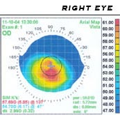
The patient is seated facing the device, which is raised to eye level. One design consists of a bowl containing an illuminated pattern, such as a series of concentric rings. Another type uses a mechanically rotated arm bearing a light source. In either type, light is focused on the anterior surface of the patient's cornea and reflected back to a digital camera at the device. The topology of the cornea is revealed by the shape taken by the reflected pattern. A computer provides the necessary analysis, typically determining the position and height of several thousand points across the cornea. The topographical map can be represented in a number of graphical formats, such as a sagittal map, which color-codes the steepness of curvature according to its dioptric value.

Development

A Medmont E300 topographer Computerised Corneal Topography.jpg
A Medmont E300 topographer

The corneal topograph owes its heritage to the Portuguese ophthalmologist Antonio Placido, who, in 1880, viewed a painted disk (Placido's disk) of alternating black and white rings reflected in the cornea. [2] The rings showed as contour lines projected on the corneal tear film. The French ophthalmologist Louis Émile Javal incorporated the rings in his ophthalmometer and mounted an eyepiece which magnified the image of the eye. He proposed that the image should be photographed or diagrammatically represented to allow analysis of the image. [3]

In 1896, Allvar Gullstrand incorporated the disk in his ophthalmoscope, examining photographs of the cornea via a microscope and was able to manually calculate the curvature by means of a numerical algorithm. Gullstrand recognized the potential of the technique and commented that despite its laboriousness it could "give a resultant accuracy that previously could not be obtained in any other way". [4] The flat field of Placido's disk reduced the accuracy close to the corneal periphery and in the 1950s the Wesley-Jessen company made use of a curved bowl to reduce the field defects. [2] The curvature of the cornea could be determined from comparison of photographs of the rings against standardized images.

In the 1980s, photographs of the projected images became hand-digitized and then analysed by computer. Automation of the process soon followed with the image captured by a digital camera and passed directly to a computer. [5] In the 1990s, systems became commercially available from a number of suppliers. The first completely automatic system was the Corneal Modeling System (CMS-1) developed by Computed Anatomy, Inc. in New York City, under the direction of Martin Gersten and a group of surgeons at the New York Eye and Ear Infirmary. The price of the early instruments was initially very high ($75,000), largely confining their use to research establishments. However, prices have fallen substantially over time, bringing corneal topographs into the budget of smaller clinics and increasing the number of patients that can be examined.

Use

Corneal topography showing stage II keratoconus. Corneal topography, stage II keratoconus (Elise A. Slim et al.).svg
Corneal topography showing stage II keratoconus.

Computerized corneal topography can be employed for diagnostics. It is, in fact, one of the exams the patients have to undergo prior to the Cross-linking and the Mini Asymmetric Radial Keratotomy (M.A.R.K.). For example, the KISA% index (keratometry, I-S, skew percentage, astigmatism) is used to arrive at a diagnosis of keratoconus, to screen the suspect keratoconic patients and analyse the degree of corneal steepness changes in healthy relatives. [6]

Nevertheless, topography in itself is a measurement of the first reflective surface of the eye (tear film) and is not giving any additional information beside the shape of this layer expressed in curvature. Keratoconus in itself is a pattern of the entire cornea, therefore every measurement just focusing on one layer, might not be enough for a state of the art diagnosis. Especially early cases of keratoconus might be missed by a plain topographic measurement, which is critical if refractive surgery is being considered. [7] The measurement is also sensitive to unstable tear films. Also, the alignment of the measurement can be difficult, especially with eyes that have Keratoconus, a significant astigmatism, or sometimes after refractive surgery.

Corneal topography instruments generate a measurement called simulated keratometry (SimK), which approximates the classic measurement of the widely used keratometer. Another novel use of corneal topographic data is called CorT, which has been shown to quantify refractive astigmatism more accurately than SimK and other approaches. [8] CorT utilizes data from all Placido rings across the cornea compared with SimK, which is based on only one ring. [9] [10]

While corneal topography relies on reflected light from the front (anterior) of the cornea, a technique called corneal tomography also provides a measure of the back (posterior) shape of the cornea. [11] [12] [13] A measure called CorT total includes this posterior corneal data and more accurately reflects refraction compared with regular CorT, SimK, and other techniques. [12] [13]

Related Research Articles

<span class="mw-page-title-main">Keratoconus</span> Medical condition involving the eye

Keratoconus (KC) is a disorder of the eye that results in progressive thinning of the cornea. This may result in blurry vision, double vision, nearsightedness, irregular astigmatism, and light sensitivity leading to poor quality-of-life. Usually both eyes are affected. In more severe cases a scarring or a circle may be seen within the cornea.

<span class="mw-page-title-main">Farsightedness</span> Eye condition in which light is focused behind instead of on the retina

Far-sightedness, also known as long-sightedness, hypermetropia, and hyperopia, is a condition of the eye where distant objects are seen clearly but near objects appear blurred. This blur is due to incoming light being focused behind, instead of on, the retina due to insufficient accommodation by the lens. Minor hypermetropia in young patients is usually corrected by their accommodation, without any defects in vision. But, due to this accommodative effort for distant vision, people may complain of eye strain during prolonged reading. If the hypermetropia is high, there will be defective vision for both distance and near. People may also experience accommodative dysfunction, binocular dysfunction, amblyopia, and strabismus. Newborns are almost invariably hypermetropic, but it gradually decreases as the newborn gets older.

<span class="mw-page-title-main">LASIK</span> Corrective ophthalmological surgery

LASIK or Lasik, commonly referred to as laser eye surgery or laser vision correction, is a type of refractive surgery for the correction of myopia, hyperopia, and an actual cure for astigmatism, since it is in the cornea. LASIK surgery is performed by an ophthalmologist who uses a laser or microkeratome to reshape the eye's cornea in order to improve visual acuity.

<span class="mw-page-title-main">Astigmatism (optical systems)</span> Optical aberration

An optical system with astigmatism is one where rays that propagate in two perpendicular planes have different foci. If an optical system with astigmatism is used to form an image of a cross, the vertical and horizontal lines will be in sharp focus at two different distances. The term comes from the Greek α- (a-) meaning "without" and στίγμα (stigma), "a mark, spot, puncture".

<span class="mw-page-title-main">Radial keratotomy</span> Refractive surgical procedure to correct myopia (nearsightedness

Radial keratotomy (RK) is a refractive surgical procedure to correct myopia (nearsightedness). It was developed in 1974 by Svyatoslav Fyodorov, a Russian ophthalmologist. It has been largely supplanted by newer, more accurate operations, such as photorefractive keratectomy, LASIK, Epi-LASIK and the phakic intraocular lens.

<span class="mw-page-title-main">Refractive surgery</span> Surgery to treat common vision disorders

Refractive surgery is optional eye surgery used to improve the refractive state of the eye and decrease or eliminate dependency on glasses or contact lenses. This can include various methods of surgical remodeling of the cornea (keratomileusis), lens implantation or lens replacement. The most common methods today use excimer lasers to reshape the curvature of the cornea. Refractive eye surgeries are used to treat common vision disorders such as myopia, hyperopia, presbyopia and astigmatism.

<span class="mw-page-title-main">Intraocular lens</span> Lens implanted in the eye to treat cataracts or myopia

An Intraocular lens (IOL) is a lens implanted in the eye usually as part of a treatment for cataracts or for correcting other vision problems such as short sightedness and long sightedness, a form of refractive surgery. If the natural lens is left in the eye, the IOL is known as phakic, otherwise it is a pseudophakic lens. Both kinds of IOLs are designed to provide the same light-focusing function as the natural crystalline lens. This can be an alternative to LASIK, but LASIK is not an alternative to an IOL for treatment of cataracts.

<span class="mw-page-title-main">Corneal cross-linking</span> Surgical procedure

Corneal cross-linking (CXL) with riboflavin (vitamin B2) and UV-A light is a surgical treatment for corneal ectasia such as keratoconus, PMD, and post-LASIK ectasia.

<span class="mw-page-title-main">Keratoscope</span>

A keratoscope, sometimes known as Placido's disk, is an ophthalmic instrument used to assess the shape of the anterior surface of the cornea. A series of concentric rings is projected onto the cornea and their reflection viewed by the examiner through a small hole in the centre of the disk. A regular-shaped cornea should show equally spaced symmetric reflections. If the patient is suffering from astigmatism or from a corneal dystrophy, the rings will be distorted.

<span class="mw-page-title-main">Astigmatism</span> Type of eye defect

Astigmatism is a type of refractive error due to rotational asymmetry in the eye's refractive power. This results in distorted or blurred vision at any distance. Other symptoms can include eyestrain, headaches, and trouble driving at night. Astigmatism often occurs at birth and can change or develop later in life. If it occurs in early life and is left untreated, it may result in amblyopia.

<span class="mw-page-title-main">Pellucid marginal degeneration</span> Degenerative corneal condition

Pellucid marginal degeneration (PMD) is a degenerative corneal condition, often confused with keratoconus. It typically presents with painless vision loss affecting both eyes. Rarely, it may cause acute vision loss with severe pain due to perforation of the cornea. It is typically characterized by a clear, bilateral thinning (ectasia) in the inferior and peripheral region of the cornea, although some cases affect only one eye. The cause of the disease remains unclear.

<span class="mw-page-title-main">Corneal pachymetry</span>

Corneal pachymetry is the process of measuring the thickness of the cornea. A pachymeter is a medical device used to measure the thickness of the eye's cornea. It is used to perform corneal pachymetry prior to refractive surgery, for Keratoconus screening, LRI surgery and is useful in screening for patients suspected of developing glaucoma among other uses.

<span class="mw-page-title-main">Avi Wallerstein</span> Canadian ophthalmologist and laser eye surgeon

Avi Wallerstein is a Canadian ophthalmologist and laser eye surgeon who specializes in surgical vision correction, also termed refractive eye surgery. He practises in Montreal and Toronto. In 2001, he co-founded LASIK MD with Mark Cohen. LASIK MD is Canada's largest provider of laser refractive surgery, performing over 60,000 procedures a year. He is one of only 14 certified CLasik instructors in North America.

<span class="mw-page-title-main">Keratoprosthesis</span> Surgical procedure where a diseased cornea is replaced with an artificial one

Keratoprosthesis is a surgical procedure where a diseased cornea is replaced with an artificial cornea. Traditionally, keratoprosthesis is recommended after a person has had a failure of one or more donor corneal transplants. More recently, a less invasive, non-penetrating artificial cornea has been developed which can be used in more routine cases of corneal blindness. While conventional cornea transplant uses donor tissue for transplant, an artificial cornea is used in the keratoprosthesis procedure. The surgery is performed to restore vision in patients with severely damaged cornea due to congenital birth defects, infections, injuries and burns.

The Alpins Method is a system to plan and analyze the results of refractive surgical procedures, such as laser in-situ keratomileus (LASIK). The Alpins Method is also used to plan cataract/toric intraocular lens (IOL) surgical procedures.

Peter S. Hersh is an American ophthalmologist and specialist in LASIK eye surgery, keratoconus, and diseases of the cornea. He co-authored the article in the journal Ophthalmology that presented the results of the study that led to the first approval by the U.S. Food and Drug Administration (FDA) of the excimer laser for the correction of nearsightedness in the United States. Hersh was also medical monitor of the study that led to approval of corneal collagen crosslinking for the treatment of keratoconus.

The aim of an accurate intraocular lens power calculation is to provide an intraocular lens (IOL) that fits the specific needs and desires of the individual patient. The development of better instrumentation for measuring the eye's axial length (AL) and the use of more precise mathematical formulas to perform the appropriate calculations have significantly improved the accuracy with which the surgeon determines the IOL power.

Post-LASIK ectasia is a condition similar to keratoconus where the cornea starts to bulge forwards at a variable time after LASIK, PRK, or SMILE corneal laser eye surgery. However, the physiological processes of post-LASIK ectasia seem to be different from keratoconus. The visible changes in the basal epithelial cell and anterior and posterior keratocytes linked with keratoconus were not observed in post-LASIK ectasia.

Corneal ectatic disorders or corneal ectasia are a group of uncommon, noninflammatory, eye disorders characterised by bilateral thinning of the central, paracentral, or peripheral cornea.

Anastasios John Kanellopoulos is a Greek-American eye surgeon specializing in corneal transplantation, cornea crosslinking for keratoconus, complicated cataract surgery and complicated glaucoma. Widely known for research and clinical contributions in micro-incision cataract, customized laser refractive surgery and corneal cross-linking propagation and most innovations, reducing corneal transplants for advanced keratoconus.

References

  1. Pavan-Langston, Deborah (2007). Manual of Ocular Diagnosis and Therapy. Hagerstown, MD: Lippincott Williams & Wilkins. p. 405. ISBN   978-0-7817-6512-1.
  2. 1 2 Goss D, Gerstman D (2000). "The Optical Science Underlying the Quantification of Corneal Contour" (PDF). Indiana Journal of Optometry. 3 (1). Archived from the original (PDF) on 2006-05-06. Retrieved 2006-04-01.
  3. Corneal Topography measuring and modifying the cornea - Schanzlin 1992, page 4-5
  4. Gullstrand A (1909). "Procedure of rays in the eye". Helmholtz's Treatise on Physiological Optics.
  5. Busin M, Wilmanns I, Spitznas M (1989). "Automated corneal topography: computerized analysis of photokeratoscope images". Graefes Arch. Clin. Exp. Ophthalmol. 227 (3): 230–6. doi:10.1007/bf02172754. PMID   2737484. S2CID   33893859.{{cite journal}}: CS1 maint: multiple names: authors list (link)
  6. Rabinowitz YS, Rasheed K (October 1999). "KISA% index: a quantitative videokeratography algorithm embodying minimal topographic criteria for diagnosing keratoconus". Journal of Cataract and Refractive Surgery. 25 (10): 1327–35. doi:10.1016/S0886-3350(99)00195-9. PMID   10511930.
  7. "Placido-based corneal topography outdated, limited in scope | Ophthalmology". Osnsupersite.com. Retrieved 2013-06-18.
  8. Alpins, Noel; JK Ong; G Stamatelatos (2012). "New method of quantifying corneal topographic astigmatism that corresponds with manifest refractive cylinder". Journal of Cataract and Refractive Surgery. 38 (11): 1978–1988. doi:10.1016/j.jcrs.2012.07.026. PMID   23010252. S2CID   27999083.
  9. Biro, A (25 November 2012). "New measurement method quantifies corneal astigmatism". Ocular Surgery News. US edition. Retrieved 2017-05-06.
  10. Ngoei, Enette (February 2013). "Refractive editor's corner of the world: CorT'ing accuracy". EyeWorld. Archived from the original on 2016-03-03. Retrieved 2017-05-06.
  11. Fung, MW, et al (2015). "Corneal Topography and Imaging". Medscape website. Updated 17 March 2016. Accessed 6 May 2017.
  12. 1 2 Guttman Krader, C (2015). "Astigmatism measurement based on total corneal power". EuroTimes website. Posted 3 November 2015. Accessed 6 May 2017.
  13. 1 2 Alpins, Noel; JK Ong; G Stamatelatos (2015). "Corneal topographic astigmatism (CorT) to quantify total corneal astigmatism". Journal of Refractive Surgery. 31 (3): 182–186. doi:10.3928/1081597X-20150224-02. PMID   25751835.

Further reading