Intravitreal injection

Last updated
An anatomical diagram of the human eye, showing vitreous humor in the vitreous chamber. Three Main Layers of the Eye.png
An anatomical diagram of the human eye, showing vitreous humor in the vitreous chamber.

Intravitreal injection is the method of administration of drugs into the eye by injection with a fine needle. The medication will be directly applied into the vitreous humor. [1] It is used to treat various eye diseases, such as age-related macular degeneration (AMD), diabetic retinopathy, and infections inside the eye such as endophthalmitis. [1] As compared to topical administration, this method is beneficial for a more localized delivery of medications to the targeted site, as the needle can directly pass through the anatomical eye barrier (e.g. cornea, conjunctiva and lens) and dynamic barrier (e.g. tears and aqueous humor). [2] [3] It could also minimize adverse drug effects on other body tissues via the systemic circulation, which could be a possible risk for intravenous injection of medications. [2] [4] Although there are risks of infections or other complications, with suitable precautions throughout the injection process, chances for these complications could be lowered. [5]

Contents

History

Intravitreal injection was first mentioned in a study in 1911, in which the injection of air was used to repair a detached retina. [6] [7] [8] There were also investigations evaluating intravitreal antibiotics injection using sulfanilamide and penicillin to treat endophthalmitis in the 1940s, yet due to the inconsistency of results and safety concerns, this form of drug delivery was only for experimental use and not applied in patients. [8] It was until 1998, that fomivirsen (Vitravene™), the first intravitreal administered medication, was approved by the U.S. Food and Drug Administration (FDA). [8]

In 2004, when Aiello et al. published the first guidelines for intravitreal injection in the journal 'Retina', fomivirsen was still the only medication licensed by the FDA for intravitreal injection. [8] At the end of the year, on December 17, the first intravitreal anti-VEGF drug pegaptanib (Macugen) was also licensed by FDA for treatment of wet age-related macular degeneration (wet AMD). [2] [9]

Intravitreal injection has then become more common and a surge in the number of injections performed could be seen. [10] Six extra medications, namely triamcinolone acetonide, ranibizumab (Lucentis), aflibercept (Eylea/Zaltrap), dexamethasone, ocriplasmin and fluocinolone acetonide were approved for this injection by the end of 2014. [2] There are also increasing off-label use of bevacizumab (Avastin) for the management of various ophthalmologic diseases, like AMD, retinal vein occlusion and diabetic macular edema. [2] [9] On top of that, the number of intravitreal injections has escalated from less than 3000 per year in 1999, to an estimation of near 6 million in 2016. [2] [7]

Applications

Intravitreal injection is used to inject a drug into the eye to reduce inflammation (anti-inflammatory), inhibit the growth and development of new blood vessels (angiostatic), or lower the permeability of blood vessels (anti-permeability), in turn curing various eye diseases. [11]

Disorders/diseases that can be treated with intravitreal injection include:

Sometimes, an intravitreal injection of antibiotics and steroids is given as part of routine cataract surgery. This avoids having to use drops after surgery. [1]

Common medications used in injection

Antimicrobials

Antimicrobials are intravitreally injected to treat eye infections, such as endophthalmitis and retinitis. [10] The medication used depends on the pathogen responsible for the disease.

Antibiotics

This type of drug targets on bacterial infection. The first use of intravitreal antibiotics was dated back to experiments in the 1940s, in which penicillin and sulfonamides were used to treat the rabbit endophthalmitis models. [10] [14] Later, more studies proved the beneficial effects of intravitreal antibiotics on acute postoperative endophthalmitis. [10] [14] In the 1970s, Peyman's research on the suggested doses for the medications was published. [14] Intravitreal antibiotics then has gradually become the major treatment to manage bacterial endophthalmitis. [14] Some common antibiotics administered nowadays are vancomycin (for Gram-positive bacteria) and ceftazidime (for Gram-negative bacteria). [15]

The dosage of antibiotics injected intravitreally is usually low to avoid possible retina toxicity. [10] [14] Some alternative antibiotics have also been tested to replace those that have a higher risk of causing macular toxicity (e.g. aminoglycosides). [10] [14] In light of the raised occurrence of antibiotics resistance, the medications should be chosen and evaluated with the support of bacterial culture and antibiotics sensitivity test results. [14] Sometimes, combinations of different antibiotics may be needed to treat polymicrobial infections (infections that are caused by more than one type of microorganisms), or as an empirical treatment. [14]

Antibiotics, such as moxifloxacin, vancomycin, etc., are used perioperatively and postoperatively as a common method of endophthalmitis prevention in cataract surgery. Researches show such injection of antibiotics is more useful to prevent infection as compared to chemoprophylaxis(chemoprevention) given topically. [16] However, it has recently been controversial whether it has sufficient efficacy for endophthalmitis prophylaxis, and whether it improves the effectiveness in preventing endophthalmitis by perioperative povidone-iodine when used in combination with the antiseptic. [16]

Antifungals

If the endophthalmitis is suspected to be a fungal infection, antifungals, such as amphotericin B and voriconazole, could be intravitreally injected to treat the disease. [10] [17] Although amphotericin B has a broad spectrum, voriconazole is more commonly used now as it has a higher efficacy and lower toxicity. [10]

Antivirals

Since the 1990s, intravitreal antivirals have been used to treat cytomegalovirus retinitis (CMV retinitis) in immunodeficient patients, such as AIDS patients. [10] [18] Some medications that could be used include ganciclovir, foscarnet, and cidofovir. [10] [19] The amount and frequency of the intravitreal agent injected varies among the drug chosen: for example, foscarnet has to be given more frequently than ganciclovir as it has a shorter intravitreal half-life. [10] If the traditional antiviral therapy fails, a combination of these two medications may be injected. [10] On the other hand, antiviral drugs could also be administered for patients with acute retinal necrosis due to varicella-zoster virus retinitis. [10]

Anti-VEGF drugs

Vascular endothelial growth factor (VEGF) is a type of protein the body cells produce to stimulate the growth of new blood vessels. [20] Anti-VEGF agents are chemicals that could inhibit these growth factors to reduce or prevent the abnormal growth of blood vessels, which could lead to damage to the eye and vision. [21]

Anti-VEGF drugs are often injected to reduce the swelling or bleeding of the retina, which can be used to treat wet age-related macular degeneration (AMD), macular oedema (which could be diabetic), diabetic retinopathy, retinal vein occlusion, etc. [22]

Some common anti-VEGF drugs are bevacizumab (Avastin), ranibizumab (Lucentis) and aflibercept (Eylea/Zaltrap). [23]

Corticosteroids

The primary use of the corticosteroids is to reduce the inflammation by inhibiting the inflammatory cytokines. [11] It could be used to treat numerous eye disorders, such as diabetic retinopathy and retinal vein occlusion. [10] [11]

Below are some examples of this type of medication:

Triamcinolone acetonide

Triamcinolone acetonide is one of the most commonly used steroid agents for the treatment of several retinal conditions. The drug is often seen as an ester in commercial drugs and appears as a white- to cream-colored crystalline powder. [11] It is much more soluble in alcohol than in water, which could be the reason for its longer duration of action (around 3 months after 4 mg intravitreal injection of the drug). [11] [24] The drug is also 5 times more potent than hydrocortisone while only has a tenth of its sodium-retaining potency. [11]

It has proven to be effective for the management of abnormal endothelial cell proliferation-associated disorders, and the accumulation of intraretinal and subretinal fluid. [11]

Dexamethasone

Dexamethasone is a potent cytokine inhibitor that is naturally released from human pericytes. [11] It is shown to be able to significantly decrease intercellular adhesion molecule-1 mRNA and protein levels and therefore reduce leukostasis and help maintain the blood-retinal-barrier. [11] Its potency is 5 times greater than triamcinolone acetonide. [11] Due to its relatively short half-life, the medication is often given as an intravitreal implant for a continuous and stable release to the target site. [11] [25] Some newly developed dexamethasone implants, such as Ozurdex, are made from biodegradable materials that could be intravitreally injected rather than surgically implanted. [11] [26]

This corticosteroid is usually used to treat disorders and diseases including macular edema secondary to retinal vein occlusion, pseudophakic cystoid macular edema, macular edema secondary to uveitis, diabetic macular edema, and age-related macular degeneration. [11]

Fluocinolone acetonide

Fluocinolone acetonide is a synthetic corticosteroid as potent as dexamethasone, but with a much lower water solubility, which could be accounted for the extended period of release from the intravitreal implant injected. [11] It was also proven to have a localized effect in the posterior segment of the eye and is not absorbed into the systemic circulation, thus less likely to give rise to systemic adverse effect. [11]

The medication could be used in treatment for noninfectious posterior uveitis and diabetic macular edema, while applications in the management of other ophthalmic diseases are still under research. [11] [27] [28]

Injection site

A photo of a right eye divided into 4 quadrants: superotemporal, superonasal, inferotemporal, and inferonasal. Right eye- 4 quadrants-white margins removed.jpg
A photo of a right eye divided into 4 quadrants: superotemporal, superonasal, inferotemporal, and inferonasal.

The injection is usually done at the inferotemporal quadrant (i.e. the lower quadrant away from the nose) of the eye undergoing the procedure, as it is usually more accessible. [6] [12] However, depending on the eye's condition, patient's and the ophthalmologist's preference, other regions could also be used. [6] [22]

Patient with aphakic (without lens due to cataract surgery), or pseudophakic eye (with implanted lens after removal of natural lens) would have the injection 3.0-3.5 mm posterior to the limbus, while injection to the phakic eye (with natural lens) is done 3.5-4.0 mm posterior to the limbus. [6]

An anatomical diagram showing the limbus of the eye. Limbus.png
An anatomical diagram showing the limbus of the eye.

Procedures

Location

Like many injections, intravitreal injection is commonly performed in the office setting. [29] [30] An operation room may be a better option to provide a more sterile environment for the procedure to lower the chance of infections, yet it will also increase the costs. [29] Since the occurrence of serious post-injection infection (e.g. endophthalmitis) is low, in-office intravitreal injection is preferred. [29] [31]

Steps

The exact procedures and techniques of the intravitreal injection varies among different guidelines, and may depend on the practices of the person performing the injection. Below is an example of the steps for the injection:

The patient usually leans back on the chair (in supine position), in which the headrest is stable and the patient is comfortable. [12] [30] Sterile drape is sometimes used to cover the face of the patient and only show the eye for the injection. [12] [32]

The specialist first applies anesthetics to the eye and eyelid to numb the area, so that the patient will not feel the pain during the procedure. [12] [22] [30] The type of anesthetic used depends on the practitioner practices and the patient's history. Some common forms of anesthetic used are eye drops (e.g. tetracaine/proparacaine) or gel (e.g. lidocaine 2% or 4% jelly), which is applied topically. [12] [30] Other choices of anesthesia include the use of lidocaine soaked pledget (a small cotton or wool pad) and subconjunctival injection (injection under the conjunctiva) of anesthetic agents. [23] However, the latter may cause a raised chance of subconjunctival hemorrhage. [12] Sometimes, for an eye with inflammation, a retrobulbar block may be given, but usually the topical or subconjunctival anesthesia is sufficient. [12] The anesthetic takes time to show the numbing effect, ranging from 1–5 minutes, depending on the chemical chosen. [12]

The specialist then sterilizes the eye and the surrounding area, often with povidone-iodine (PVP-I) solution, to prevent any infection in the injected site. [22] [30] Aqueous chlorhexidine is used instead in case of adverse effects to povidone-iodine. [23]

Next, an eyelid speculum is placed to retract the eyelids and thus hold the eye open. [22] [30] It helps to prevent contamination of the needle and the injection site by the eyelid or eyelashes. [33] Povidone-iodine solution is applied to the conjunctiva at the site of injection. [22] Another dose of local anesthetic may be given to the conjunctival surface again (for example, by placing a cotton swab soaked with the anesthetic drug solution over the targeted region), which is followed by the reapplication of PVP-I solution. [22]

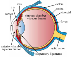
The injection site is measured and marked with a measuring caliper or other devices. [6] [32] The patient is then told to look away from the injection site to show the quadrant to be injected, and the doctor inserts the needle at the target site in a single motion into the mid-vitreous cavity. [12] Once the needle is in the vitreous cavity, the doctor pushes the plunger to release the drug into the cavity. [12] After that, the needle is removed, and the injection site is immediately covered with a cotton swab to avoid vitreous reflux (reflux of fluid from the vitreous cavity). [6] [12] The excess PVP-I solution is rinsed away. [12]

Finally, the doctor checks the patient's vision and intraocular pressure (IOP) of the eye. [12] The injection of certain medications, such as triamcinolone acetonide (Kenalog or Triesence), may cause a sudden increase in the IOP, [34] [35] and the patient should be monitored until the pressure returns to a normal level. If a large volume of drug is injected, paracentesis may be required. [12]

Possible risks and complications

Side effects of intravitreal injection can be classified into two categories: drug-related side effects and injection-related side effects. [11] For example, in an intravitreal steroid injection, complications could be divided into steroid-related adverse effects and injection-related adverse effects, in which the former most commonly include cataract formation and increase in intraocular pressure (IOP). [11]

Other examples of potential adverse effects are listed as follows:

A surgery may be required to treat certain severe complications. Some of the above complications could also lead to blindness, or even loss of the eye (in the case of a severe infection). [12]

Precautions

Precautions should be taken before, during, and after the injection to lower the chances of complications:

Pre-treatment

During the injection

Post-treatment

See also

Related Research Articles

<span class="mw-page-title-main">Diabetic retinopathy</span> Medical condition

Diabetic retinopathy, is a medical condition in which damage occurs to the retina due to diabetes mellitus. It is a leading cause of blindness in developed countries.

<span class="mw-page-title-main">Vitrectomy</span> Surgery to remove vitreous humor from the eye

Vitrectomy is a surgery to remove some or all of the vitreous humor from the eye.

The National Eye Institute (NEI) is part of the U.S. National Institutes of Health (NIH), an agency of the U.S. Department of Health and Human Services. The mission of NEI is "to eliminate vision loss and improve quality of life through vision research." NEI consists of two major branches for research: an extramural branch that funds studies outside NIH and an intramural branch that funds research on the NIH campus in Bethesda, Maryland. Most of the NEI budget funds extramural research.

<span class="mw-page-title-main">Macular edema</span> Medical condition

Macular edema occurs when fluid and protein deposits collect on or under the macula of the eye and causes it to thicken and swell (edema). The swelling may distort a person's central vision, because the macula holds tightly packed cones that provide sharp, clear, central vision to enable a person to see detail, form, and color that is directly in the centre of the field of view.

<span class="mw-page-title-main">Triamcinolone acetonide</span> Medicinal chemical compound, steroid

Triamcinolone acetonide, sold under the brand name Kenalog among others, is a synthetic corticosteroid medication used topically to treat various skin conditions, to relieve the discomfort of mouth sores, and by injection into joints to treat various joint conditions. It is also injected into lesions to treat inflammation in some parts of the body, particularly the skin. In nasal spray form, it is used to treat allergic rhinitis. It is used for the treatment of macular edema associated with uveitis. It is a more potent derivative of triamcinolone, and is about eight times as potent as prednisone.

<span class="mw-page-title-main">Intravitreal administration</span>

Intravitreal is a route of administration of a drug, or other substance, in which the substance is delivered into the vitreous humor of the eye. "Intravitreal" literally means "inside an eye". Intravitreal injections were first introduced in 1911 when Ohm gave an injection of air into the vitreous humor to repair a detached retina. In the mid-1940s, intravitreal injections became a standard way to administer drugs to treat endophthalmitis and cytomegalovirus retinitis.

Ranibizumab, sold under the brand name Lucentis among others, is a monoclonal antibody fragment (Fab) created from the same parent mouse antibody as bevacizumab. It is an anti-angiogenic that is approved to treat the "wet" type of age-related macular degeneration, diabetic retinopathy, and macular edema due to branch retinal vein occlusion or central retinal vein occlusion.

<span class="mw-page-title-main">Endophthalmitis</span> Medical condition

Endophthalmitis, or endophthalmia, is inflammation of the interior cavity of the eye, usually caused by an infection. It is a possible complication of all intraocular surgeries, particularly cataract surgery, and can result in loss of vision or loss of the eye itself. Infection can be caused by bacteria or fungi, and is classified as exogenous, or endogenous. Other non-infectious causes include toxins, allergic reactions, and retained intraocular foreign bodies. Intravitreal injections are a rare cause, with an incidence rate usually less than 0.05%.

<span class="mw-page-title-main">Pegaptanib</span> Drug to treat macular degeneration

Pegaptanib sodium injection is an anti-angiogenic medicine for the treatment of neovascular (wet) age-related macular degeneration (AMD). It was discovered by NeXstar Pharmaceuticals and licensed in 2000 to EyeTech Pharmaceuticals, now OSI Pharmaceuticals, for late stage development and marketing in the United States. Gilead Sciences continues to receive royalties from the drugs licensing. Outside the US pegaptanib is marketed by Pfizer. Approval was granted by the U.S. Food and Drug Administration (FDA) in December 2004.

<span class="mw-page-title-main">Intermediate uveitis</span> Medical condition

Intermediate uveitis is a form of uveitis localized to the vitreous and peripheral retina. Primary sites of inflammation include the vitreous of which other such entities as pars planitis, posterior cyclitis, and hyalitis are encompassed. Intermediate uveitis may either be an isolated eye disease or associated with the development of a systemic disease such as multiple sclerosis or sarcoidosis. As such, intermediate uveitis may be the first expression of a systemic condition. Infectious causes of intermediate uveitis include Epstein–Barr virus infection, Lyme disease, HTLV-1 virus infection, cat scratch disease, and hepatitis C.

Aflibercept, sold under the brand names Eylea among others, is a medication used to treat wet macular degeneration and metastatic colorectal cancer. It was developed by Regeneron Pharmaceuticals and is approved in the United States and the European Union.

<span class="mw-page-title-main">Macular telangiectasia</span> Disease of the retina affecting central vision

Macular telangiectasia is a condition of the retina, the light-sensing tissue at the back of the eye that causes gradual deterioration of central vision, interfering with tasks such as reading and driving.

<span class="mw-page-title-main">Laser coagulation</span> Procedure widely used in eye surgery

Laser coagulation or laser photocoagulation surgery is used to treat a number of eye diseases and has become widely used in recent decades. During the procedure, a laser is used to finely cauterize ocular blood vessels to attempt to bring about various therapeutic benefits.

<span class="mw-page-title-main">Branch retinal vein occlusion</span> Medical condition

Branch retinal vein occlusion is a common retinal vascular disease of the elderly. It is caused by the occlusion of one of the branches of central retinal vein.

<span class="mw-page-title-main">Vitreomacular adhesion</span> Human medical condition

Vitreomacular adhesion (VMA) is a human medical condition where the vitreous gel of the human eye adheres to the retina in an abnormally strong manner. As the eye ages, it is common for the vitreous to separate from the retina. But if this separation is not complete, i.e. there is still an adhesion, this can create pulling forces on the retina that may result in subsequent loss or distortion of vision. The adhesion in of itself is not dangerous, but the resulting pathological vitreomacular traction (VMT) can cause severe ocular damage.

Anti–vascular endothelial growth factor therapy, also known as anti-VEGF therapy or medication, is the use of medications that block vascular endothelial growth factor. This is done in the treatment of certain cancers and in age-related macular degeneration. They can involve monoclonal antibodies such as bevacizumab, antibody derivatives such as ranibizumab (Lucentis), or orally-available small molecules that inhibit the tyrosine kinases stimulated by VEGF: sunitinib, sorafenib, axitinib, and pazopanib.

Brolucizumab sold under trade name Beovu among others, is a humanized single-chain antibody fragment for the treatment of neovascular (wet) age-related macular degeneration (AMD).

<span class="mw-page-title-main">Faricimab</span> Medication for macular degeneration

Faricimab, sold under the brand name Vabysmo, is a monoclonal antibody used for the treatment of neovascular age-related macular degeneration (nAMD) and diabetic macular edema (DME). Faricimab is the first bispecific monoclonal antibody to target both vascular endothelial growth factor (VEGF) and angiopoietin 2 (Ang-2). By targeting these pathways, faricimab stabilizes blood vessels in the retina. It is given by intravitreal injection by an ophthalmologist.

Sickle cell retinopathy can be defined as retinal changes due to blood vessel damage in the eye of a person with a background of sickle cell disease. It can likely progress to loss of vision in late stages due to vitreous hemorrhage or retinal detachment. Sickle cell disease is a structural red blood cell disorder leading to consequences in multiple systems. It is characterized by chronic red blood cell destruction, vascular injury, and tissue ischemia causing damage to the brain, eyes, heart, lungs, kidneys, spleen, and musculoskeletal system.

Conbercept, sold under the commercial name Lumitin, is a novel vascular endothelial growth factor (VEGF) inhibitor used to treat neovascular age-related macular degeneration (AMD) and diabetic macular edema (DME). The anti-VEGF was approved for the treatment of neovascular AMD by the China State FDA (CFDA) in December 2013. As of December 2020, conbercept is undergoing phase III clinical trials through the U.S. Food and Drug Administration’s PANDA-1 and PANDA-2 development programs.

References

  1. 1 2 3 4 5 6 7 8 9 10 11 "Intravitreal injection: MedlinePlus Medical Encyclopedia". medlineplus.gov. Retrieved 2020-03-26.
  2. 1 2 3 4 5 6 "Intravitreal Injections and Endophthalmitis". Medscape. Retrieved 2020-04-21.
  3. Cholkar, Kishore; Dasari, Supriya Reddy; Pal, Dhananjay; Mitra, Ashim K. (2013), "Eye: anatomy, physiology and barriers to drug delivery", Ocular Transporters and Receptors, Elsevier, pp. 1–36, doi:10.1533/9781908818317.1, ISBN   978-1-907568-86-2
  4. "INTRAVITREAL INJECTIONS INFORMATION | Eye Associates" . Retrieved 2020-04-23.
  5. Artunay, O.; Yuzbasioglu, E.; Rasier, R.; Sengül, A.; Bahcecioglu, H. (2009-02-13). "Incidence and management of acute endophthalmitis after intravitreal bevacizumab (Avastin) injection". Eye. 23 (12): 2187–2193. doi: 10.1038/eye.2009.7 . ISSN   1476-5454.
  6. 1 2 3 4 5 6 7 8 "How to Give Intravitreal Injections". American Academy of Ophthalmology. 2013-04-01. Retrieved 2020-04-07.
  7. 1 2 Grzybowski, Andrzej; Told, Reinhard; Sacu, Stefan; Bandello, Francesco; Moisseiev, Elad; Loewenstein, Anat; Schmidt-Erfurth, Ursula; on behalf of the Euretina Board (2018). "2018 Update on Intravitreal Injections: Euretina Expert Consensus Recommendations". Ophthalmologica. 239 (4): 181–193. doi: 10.1159/000486145 . ISSN   0030-3755. PMID   29393226. S2CID   12528415.
  8. 1 2 3 4 AIELLO, LLOYD P.; BRUCKER, ALEXANDER J.; CHANG, STANLEY; CUNNINGHAM, EMMETT T.; D'AMICO, DONALD J.; FLYNN, HARRY W.; GRILLONE, LISA R.; HUTCHERSON, STEVE; LIEBMANN, JEFFREY M.; O'BRIEN, TERRENCE P.; SCOTT, INGRID U. (October 2004). "Evolving Guidelines for Intravitreous Injections". Retina. 24 (Supplement): S3–S19. doi:10.1097/00006982-200410001-00002. ISSN   0275-004X. PMID   15483476. S2CID   23532836.
  9. 1 2 Colin A. McCannel; Harry W. Flynn Jr.; Emmett T. Cunningham Jr. "Updated Guidelines for Intravitreal Injection". Review of Ophthalmology. Retrieved 2020-04-21.
  10. 1 2 3 4 5 6 7 8 9 10 11 12 13 14 15 Shikari, Hasanain; Samant, PreetamM (2016). "Intravitreal injections: A review of pharmacological agents and techniques". Journal of Clinical Ophthalmology and Research. 4 (1): 51. doi: 10.4103/2320-3897.174429 . ISSN   2320-3897. S2CID   53481704.
  11. 1 2 3 4 5 6 7 8 9 10 11 12 13 14 15 16 17 18 19 20 21 22 23 Sarao, Valentina; Veritti, Daniele; Boscia, Francesco; Lanzetta, Paolo (8 January 2014). "Intravitreal Steroids for the Treatment of Retinal Diseases". The Scientific World Journal. 2014: 989501. doi: 10.1155/2014/989501 . PMC   3910383 . PMID   24526927. S2CID   184726.
  12. 1 2 3 4 5 6 7 8 9 10 11 12 13 14 15 16 17 18 19 "Intravitreal Injections - EyeWiki". eyewiki.aao.org. Retrieved 2020-04-07.
  13. "Macular Edema | National Eye Institute". www.nei.nih.gov. Retrieved 2020-04-06.
  14. 1 2 3 4 5 6 7 8 Radhika, Medikonda; Mithal, Kopal; Bawdekar, Abhishek; Dave, Vivek; Jindal, Animesh; Relhan, Nidhi; Albini, Thomas; Pathengay, Avinash; Flynn, Harry W (December 2014). "Pharmacokinetics of intravitreal antibiotics in endophthalmitis". Journal of Ophthalmic Inflammation and Infection. 4 (1): 22. doi: 10.1186/s12348-014-0022-z . ISSN   1869-5760. PMC   4306439 . PMID   25667683.
  15. Cornut, P.-L.; Chiquet, C. (October 2008). "Injections intravitréennes d'antibiotiques et endophtalmies". Journal Français d'Ophtalmologie (in French). 31 (8): 815–823. doi:10.1016/S0181-5512(08)74405-X. PMID   19107050.
  16. 1 2 Kindle, Trevor; Ferguson, Tanner; Ibach, Mitch; Greenwood, Michael; Schweitzer, Justin; Swan, Russell; Sudhagoni, Ramu G.; Berdahl, John P. (January 2018). "Safety and efficacy of intravitreal injection of steroid and antibiotics in the setting of cataract surgery and trabecular microbypass stent". Journal of Cataract & Refractive Surgery. 44 (1): 56–62. doi:10.1016/j.jcrs.2017.10.040. ISSN   0886-3350. PMID   29502618.
  17. Maryann Scholl; Justin D. Scholl. "Treatment of Endophthalmitis Resulting From Traumatic Eye Injury". www.uspharmacist.com. Retrieved 2020-04-25.
  18. "Inflammatory Retinal Diseases. Medical information". patient.info. Retrieved 2020-04-25.
  19. "Cytomegalovirus Adult and Adolescent Opportunistic Infection". AIDSinfo. Archived from the original on 2020-08-31. Retrieved 2020-04-25.
  20. "Vascular Endothelial Growth Factor - Health Encyclopedia - University of Rochester Medical Center". www.urmc.rochester.edu. Retrieved 2020-04-25.
  21. "Anti-VEGF Treatments". American Academy of Ophthalmology. 2019-03-02. Retrieved 2020-04-25.
  22. 1 2 3 4 5 6 7 8 9 Ingrid U. Scott; Harry W. Flynn Jr. "An Update on the Intravitreal Injection Procedure". www.retina-specialist.com. Retrieved 2020-04-05.
  23. 1 2 3 Fagan, Xavier J; Al-Qureshi, Salmaan (2013). "Intravitreal injections: a review of the evidence for best practice: Intravitreal injections: evidence for best practice". Clinical & Experimental Ophthalmology. 41 (5): 500–507. doi: 10.1111/ceo.12026 . PMID   23078366.
  24. Sides Media, www sidesmedia com. "Retina Today - Dexamethasone Intravitreal Implant: Pharmacology and Clinical Update". Retina Today. Retrieved 2020-04-26.
  25. Lambiase, A.; Abdolrahimzadeh, S.; Solmaz, S.M. (2014). "An update on intravitreal implants in use for eye disorders". Drugs of Today. 50 (3): 239–49. doi:10.1358/dot.2014.050.03.2103755. ISSN   1699-3993. PMID   24696869.
  26. Christoforidis, John B.; Chang, Susie; Jiang, Angela; Wang, Jillian; Cebulla, Colleen M. (2012). "Intravitreal Devices for the Treatment of Vitreous Inflammation". Mediators of Inflammation. 2012: 126463. doi: 10.1155/2012/126463 . ISSN   0962-9351. PMC   3441042 . PMID   22988344.
  27. Syed, Yahiya Y. (April 2017). "Fluocinolone Acetonide Intravitreal Implant 0.19 mg (ILUVIEN®): A Review in Diabetic Macular Edema". Drugs. 77 (5): 575–583. doi:10.1007/s40265-017-0722-4. ISSN   0012-6667. PMID   28283896. S2CID   42529291.
  28. "Retisert - FDA prescribing information, side effects and uses". Drugs.com. Retrieved 2020-04-26.
  29. 1 2 3 "Intravitreal Injection Technique: a primer". www.eyerounds.org. Retrieved 2020-04-07.
  30. 1 2 3 4 5 6 7 "Intravitreal Injections - The American Society of Retina Specialists". www.asrs.org. Retrieved 2020-04-07.
  31. Tabandeh, Homayoun; Boscia, Francesco; Sborgia, Alessandra; Ciracì, Lorenza; Dayani, Pouya; Mariotti, Cesare; Furino, Claudio; Flynn, Harry W. (January 2014). "ENDOPHTHALMITIS ASSOCIATED WITH INTRAVITREAL INJECTIONS: Office-Based Setting and Operating Room Setting". Retina. 34 (1): 18–23. doi:10.1097/IAE.0000000000000008. ISSN   0275-004X. PMID   24362413. S2CID   34637729.
  32. 1 2 Yorston, David (2014). "Intravitreal injection technique". Community Eye Health. 27 (87): 47. ISSN   0953-6833. PMC   4322739 . PMID   25918462.
  33. 1 2 3 4 5 6 7 8 9 10 11 Avery, Robert L.; Bakri, Sophie J.; Blumenkranz, Mark S.; Brucker, Alexander J.; Cunningham, Emmett T.; DʼAmico, Donald J.; Dugel, Pravin U.; Flynn, Harry W.; Freund, K. Bailey; Haller, Julia A.; Jumper, J. Michael (2014). "Intravitreal Injection Technique and Monitoring". Retina. 34: S1–S18. doi:10.1097/iae.0000000000000399. ISSN   0275-004X. PMID   25489719. S2CID   205645429.
  34. "Intravitreal Kenalog Injections". American Academy of Ophthalmology. 2004-10-01. Retrieved 2020-04-06.
  35. "Trivaris Intravitreal, Triesence (triamcinolone intravitreal) dosing, indications, interactions, adverse effects, and more". reference.medscape.com. Retrieved 2020-04-07.
  36. Brodie, Frank L.; Ruggiero, Jason; Ghodasra, Devon H.; Hui, James Z.; VanderBeek, Brian L.; Brucker, Alexander J. (July 2014). "Volume and Composition of Reflux After Intravitreal Injection". Retina. 34 (7): 1473–1476. doi:10.1097/IAE.0000000000000098. ISSN   0275-004X. PMC   4065616 . PMID   24451925.
  37. "Eye floaters - Symptoms and causes". Mayo Clinic. Retrieved 2020-04-07.