Indiana University Health Proton Therapy Center

Last updated
Indiana University Health Proton Therapy Center
MPRI Front.jpg
IU Health Proton Therapy Center
USA Indiana location map.svg
Red pog.svg
Located in Bloomington, Indiana
Geography
Location Bloomington, Indiana, United States
Coordinates 39°11′24″N86°31′22″W / 39.19000°N 86.52278°W / 39.19000; -86.52278
Organization
Care system Public
Type Specialist
Affiliated university Indiana University
Specialty Oncology
History
Opened2004
Closed2014
Links
Website http://iuhealthproton.org [ dead link ]
Lists Hospitals in Indiana

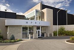
The Indiana University Health Proton Therapy Center, formerly known as the Midwest Proton Radiotherapy Institute (MPRI), [1] was the first proton facility in the Midwest. The center was located on the Indiana University campus in Bloomington, Indiana, United States. The IU Health Proton Therapy Center was the only proton therapy center in the U.S. to use a uniform-scanning beam for dose delivery, [2] which decreases undesirable neutron dose to patients. [3] The Center opened in 2004, and ceased operations in 2014.

Contents

Affiliations

The center was affiliated with the Melvin and Bren Simon Cancer Center and Indiana University Health and was the only U.S. proton therapy center associated with a university-based proton therapy technology research group, IU Cyclotron Operations. The center's pediatric program was affiliated with Riley Hospital for Children.

History

Proton therapy is not experimental and has been used in the United States since 1946. [2] In 2014 there were only 12 centers in the U.S that provided proton therapy. The scarcity of proton centers was due to the cost of the cyclotron that produces a proton beam. [4] IU Health Proton Therapy Center was able to repurpose the cyclotron located at the Indiana University Cyclotron Facility (IUCF), adapting the purpose from a research facility into a proton therapy center.

The Indiana University Cyclotron Facility (IUCF). [5] was a cyclotron located on the Indiana University campus in Bloomington, Indiana, United States. It accelerated protons to an energy of 200 MeV, as well as light ions: deuterium, 3He 4He, 6Li and 7Li. [6] The beam could be polarized and was delivered to experimental halls. The facility was operated between 1976 and 2010. in 1985 the IUCF was upgraded to operate a cooled beam (Cooler storage ring) able to accelerate protons to 500 MeV. [7] In 2004, the IUCF was repurposed for medical use and became the Indiana University Health Proton Therapy Center [8]

The proton therapy center and the cyclotron closed operations on December 5, 2014. [9] The decision was made due to a lack of revenue and debt incurred by the center, as well as advances in proton therapy around the country that "now make the equipment and methods at the proton therapy center out of date." [10] The proton center was able to produce spot scanning beams in 2008 and gate to both lung and heart.

Proton therapy

Radiation oncologists have been using proton therapy to treat cancer since the 1950s. Long recognized for their targeting capability, proton beams achieve greater precision than traditional X-rays, while exposing healthy tissue to less radiation. This allows physicians to deliver high doses of radiation even when tumors are close to sensitive organs and tissue. A proton therapy beam’s powerful energy is focused directly on a patient’s tumor. Once released, the energy stops – there is no exit dose and no additional radiation unlike X-ray beams and gamma knife rays. [11]

Related Research Articles

<span class="mw-page-title-main">Radiation therapy</span> Therapy using ionizing radiation, usually to treat cancer

Radiation therapy or radiotherapy is a treatment using ionizing radiation, generally provided as part of cancer therapy to either kill or control the growth of malignant cells. It is normally delivered by a linear particle accelerator. Radiation therapy may be curative in a number of types of cancer if they are localized to one area of the body, and have not spread to other parts. It may also be used as part of adjuvant therapy, to prevent tumor recurrence after surgery to remove a primary malignant tumor. Radiation therapy is synergistic with chemotherapy, and has been used before, during, and after chemotherapy in susceptible cancers. The subspecialty of oncology concerned with radiotherapy is called radiation oncology. A physician who practices in this subspecialty is a radiation oncologist.

<span class="mw-page-title-main">External beam radiotherapy</span> Treatment of cancer with ionized radiation

External beam radiation therapy (EBRT) is a form of radiotherapy that utilizes a high-energy collimated beam of ionizing radiation, from a source outside the body, to target and kill cancer cells. A radiotherapy beam is composed of particles which travel in a consistent direction; each radiotherapy beam consists of one type of particle intended for use in treatment, though most beams contain some contamination by other particle types.

A radiation therapist, therapeutic radiographer or radiotherapist is an allied health professional who works in the field of radiation oncology. Radiation therapists plan and administer radiation treatments to cancer patients in most Western countries including the United Kingdom, Australia, most European countries, and Canada, where the minimum education requirement is often a baccalaureate degree or postgraduate degrees in radiation therapy. Radiation therapists can also prescribe medications and radiation, interpret tests results, perform follow ups, reviews, and provide consultations to cancer patients in the United Kingdom and Ontario, Canada . In the United States, radiation therapists have a lower educational requirement and often require postgraduate education and certification in order to plan treatments.

<span class="mw-page-title-main">Paul Scherrer Institute</span> Swiss federal research institute

The Paul Scherrer Institute (PSI) is a multi-disciplinary research institute for natural and engineering sciences in Switzerland. It is located in the Canton of Aargau in the municipalities Villigen and Würenlingen on either side of the River Aare, and covers an area over 35 hectares in size. Like ETH Zurich and EPFL, PSI belongs to the Swiss Federal Institutes of Technology Domain of the Swiss Confederation. The PSI employs around 2,100 people. It conducts basic and applied research in the fields of matter and materials, human health, and energy and the environment. About 37% of PSI's research activities focus on material sciences, 24% on life sciences, 19% on general energy, 11% on nuclear energy and safety, and 9% on particle physics.

<span class="mw-page-title-main">Proton therapy</span> Medical Procedure

In medicine, proton therapy, or proton radiotherapy, is a type of particle therapy that uses a beam of protons to irradiate diseased tissue, most often to treat cancer. The chief advantage of proton therapy over other types of external beam radiotherapy is that the dose of protons is deposited over a narrow range of depth; hence in minimal entry, exit, or scattered radiation dose to healthy nearby tissues.

<span class="mw-page-title-main">Radiosurgery</span> Surgical Specialty

Radiosurgery is surgery using radiation, that is, the destruction of precisely selected areas of tissue using ionizing radiation rather than excision with a blade. Like other forms of radiation therapy, it is usually used to treat cancer. Radiosurgery was originally defined by the Swedish neurosurgeon Lars Leksell as "a single high dose fraction of radiation, stereotactically directed to an intracranial region of interest".

<span class="mw-page-title-main">Bragg peak</span> Path length of maximum energy loss of ionizing radiation

The Bragg peak is a pronounced peak on the Bragg curve which plots the energy loss of ionizing radiation during its travel through matter. For protons, α-rays, and other ion rays, the peak occurs immediately before the particles come to rest. It is named after William Henry Bragg, who discovered it in 1903.

<span class="mw-page-title-main">Fast neutron therapy</span>

Fast neutron therapy utilizes high energy neutrons typically between 50 and 70 MeV to treat cancer. Most fast neutron therapy beams are produced by reactors, cyclotrons (d+Be) and linear accelerators. Neutron therapy is currently available in Germany, Russia, South Africa and the United States. In the United States, one treatment center is operational, in Seattle, Washington. The Seattle center uses a cyclotron which produces a proton beam impinging upon a beryllium target.

<span class="mw-page-title-main">Radiation treatment planning</span>

In radiotherapy, radiation treatment planning (RTP) is the process in which a team consisting of radiation oncologists, radiation therapist, medical physicists and medical dosimetrists plan the appropriate external beam radiotherapy or internal brachytherapy treatment technique for a patient with cancer.

Electron therapy or electron beam therapy (EBT) is a kind of external beam radiotherapy where electrons are directed to a tumor site for medical treatment of cancer.

<span class="mw-page-title-main">Tomotherapy</span> Type of radiation therapy

Tomotherapy is a type of radiation therapy treatment machine. In tomotherapy a thin radiation beam is modulated as it rotates around the patient, while they are moved through the bore of the machine. The name comes from the use of a strip-shaped beam, so that only one “slice” of the target is exposed at any one time by the radiation. The external appearance of the system and movement of the radiation source and patient can be considered analogous to a CT scanner, which uses lower doses of radiation for imaging. Like a conventional machine used for X-ray external beam radiotherapy, a linear accelerator generates the radiation beam, but the external appearance of the machine, the patient positioning, and treatment delivery is different. Conventional linacs do not work on a slice-by-slice basis but typically have a large area beam which can also be resized and modulated.

FLUKA is a fully integrated Monte Carlo simulation package for the interaction and transport of particles and nuclei in matter. FLUKA has many applications in particle physics, high energy experimental physics and engineering, shielding, detector and telescope design, cosmic ray studies, dosimetry, medical physics, radiobiology. A recent line of development concerns hadron therapy.

<span class="mw-page-title-main">Cobalt therapy</span> Medical use of gamma rays

Cobalt therapy is the medical use of gamma rays from the radioisotope cobalt-60 to treat conditions such as cancer. Beginning in the 1950s, cobalt-60 was widely used in external beam radiotherapy (teletherapy) machines, which produced a beam of gamma rays which was directed into the patient's body to kill tumor tissue. Because these "cobalt machines" were expensive and required specialist support, they were often housed in cobalt units. Cobalt therapy was a revolutionary advance in radiotherapy in the post-World War II period but is now being replaced by other technologies such as linear accelerators.

Particle therapy is a form of external beam radiotherapy using beams of energetic neutrons, protons, or other heavier positive ions for cancer treatment. The most common type of particle therapy as of August 2021 is proton therapy.

Radiobiology is a field of clinical and basic medical sciences that involves the study of the effects of ionizing radiation on living things, in particular health effects of radiation. Ionizing radiation is generally harmful and potentially lethal to living things but can have health benefits in radiation therapy for the treatment of cancer and thyrotoxicosis. Its most common impact is the induction of cancer with a latent period of years or decades after exposure. High doses can cause visually dramatic radiation burns, and/or rapid fatality through acute radiation syndrome. Controlled doses are used for medical imaging and radiotherapy.

Intraoperative electron radiation therapy is the application of electron radiation directly to the residual tumor or tumor bed during cancer surgery. Electron beams are useful for intraoperative radiation treatment because, depending on the electron energy, the dose falls off rapidly behind the target site, therefore sparing underlying healthy tissue.

<span class="mw-page-title-main">Harvard Cyclotron Laboratory</span> From 1961 to 2002, a proton accelerator used for research and development

The Harvard Cyclotron Laboratory operated from 1949 to 2002. It was most notable for its contributions to the development of proton therapy.

Pencil beam scanning is the practice of steering a beam of radiation or charged particles across an object. It is often used in proton therapy, to reduce unnecessary radiation exposure to surrounding non-cancerous cells.

<span class="mw-page-title-main">Neutron capture therapy of cancer</span> Nonsurgical therapeutic modality for treating locally invasive malignant tumors

Neutron capture therapy (NCT) is a type of radiotherapy for treating locally invasive malignant tumors such as primary brain tumors, recurrent cancers of the head and neck region, and cutaneous and extracutaneous melanomas. It is a two-step process: first, the patient is injected with a tumor-localizing drug containing the stable isotope boron-10 (10B), which has a high propensity to capture low energy "thermal" neutrons. The neutron cross section of 10B is 1,000 times more than that of other elements, such as nitrogen, hydrogen, or oxygen, that occur in tissue. In the second step, the patient is radiated with epithermal neutrons, the sources of which in the past have been nuclear reactors and now are accelerators that produce higher energy epithermal neutrons. After losing energy as they penetrate tissue, the resultant low energy "thermal" neutrons are captured by the 10B atoms. The resulting decay reaction yields high-energy alpha particles that kill the cancer cells that have taken up enough 10B.

<span class="mw-page-title-main">The Svedberg Laboratory</span>

The The Svedberg Laboratory (TSL) is a university facility, based in Uppsala, Sweden. The activities at TSL are based around the particle accelerator Gustaf Werner cyclotron.

References

  1. "Midwest Proton Radiotherapy Institute to be named IU Health Proton Therapy Center" (PDF). IU Health Proton Therapy Center. Indiana University Health. October 26, 2010. Archived from the original (PDF) on March 26, 2012. Retrieved June 20, 2016.
  2. 1 2 "About the Proton Therapy Center Team". IU Health Proton Therapy Center. Indiana University Health. Archived from the original on March 9, 2014. Retrieved June 20, 2016.
  3. Hecksel, D.; Anferov, V.; Fitzek, M.; Shahnazi, K. (June 2010). "Influence of beam efficiency through the patient-specific collimator on secondary neutron dose equivalent in double scattering and uniform scanning modes of proton therapy". Medical Physics. 37 (6): 2910–7. Bibcode:2010MedPh..37.2910H. doi:10.1118/1.3431575. PMID   20632602.
  4. "History". IU Health Proton Therapy Center. Indiana University Health. Archived from the original on May 2, 2012. Retrieved June 20, 2016.
  5. IUCF page at Indiana University
  6. Indiana University Cyclotron Facility (1976-2010)
  7. H.-O. Meyer (2007) “The Indiana Cooler: A Retrospective” Annu. Rev. Nucl. Part. Sci. 57, 1
  8. B. von Przewoski, R. Tayloe and J. Whitmore (2003) “A new life for Indiana’s cyclotron“, CERN Courier
  9. "IU's proton therapy center closes after 2,079 patients". Indianapolis Business Journal. Associated Press. December 5, 2014. Retrieved June 20, 2016.
  10. "IU closing proton therapy center, cyclotron". Indianapolis Business Journal. August 21, 2014. Retrieved June 20, 2016.
  11. "How proton therapy works". IU Health Proton Therapy Center. Indiana University Health. Archived from the original on February 5, 2012.