Interocular transfer

Last updated

Interocular transfer (IOT) is a phenomenon of visual perception in which information available to one eye will produce an effect in the other eye. For example, the state of adaptation of one eye can have a small effect on the state of light adaptation of the other. Aftereffects induced through one eye can be measured through the other. [1] [2]

Contents

IOT can occur in various tasks involving motion aftereffects (MAE), depth perception (stereopsis) and visual learning. [3] Some of the pioneering research in IOT also hypothesizes its process and location of occurrence, though there is no actual evidence that localizes the process of IOT.

Most of the early research in interocular transfer introduces the role of binocular neurons in the process of interocular transfer, [4] the role of adaptation in the IOT process occurring in motion aftereffects (MAE), [5] and use of IOT in determining the stereoscopic vision in those with visual disorders like amblyopia (lazy eye syndrome) and strabismus. [6]

Research

The groundwork for interocular transfer research began after Hubel and Wiesel's (1962) study on understanding the binocular interaction in visual cortex. Their research laid the foundation for further interocular research by studying the neurons in the cat's visual cortex when stimuli is presented to both eyes (binocular neurons). Their study provided a crucial step in understanding how brain integrates visual information from both eyes. [7]  One of the earliest research on interocular transfer was conducted by Wolfgang Kohler in 1917 where one of the chickens' eyes used in the experiment were patched shut. Interocular transfer was observed between the eyes of the chicken who were made to discriminate gray colored sheets of varying brightness. [8]
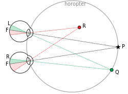
Binocular Vision Assisted by Binocular Neurons Binocular vision.svg
Binocular Vision Assisted by Binocular Neurons

Two hypothesis to explain the occurrence of interocular transfer have been advanced - the retinal locus hypothesis and sensorimotor integration hypothesis. The retinal locus hypothesis attempted to explain the occurrence of interocular transfer when the stimuli are presented in the dorsotemporal part of the retina. The sensorimotor integration hypothesis to their study proposed that pigeons can transfer information depending on whether the response key and the visual stimulus are presented in the same location. [9]

Motion aftereffects

Motion aftereffect is a phenomena that causes a visual stimuli to undergo apparent motion. A prolonged exposure to motion of a stimulus in a particular direction causes a perception of motion in the opposite direction. The middle temporal area (known as MT or V5), is associated with motion aftereffects. [10]

Interocular transfer occurs in the relational-motion detectors, which are of the binocular or monocular class. The study confirms the presence on interocular transfer of MAE, and localizes the adaptation for MAE in either an individual's eye, or the brain areas which combine the information from both set of eyes. [11]

A neural model for interocular transfer to explain its occurrence in motion aftereffects has been developed. In a pool of many neurons, some are adapted to a specific visual stimulus (like a certain orientation of lines) and some are not. This model suggests that all these neurons somehow combine their information, allowing transfer of information between the two eyes. This means that all the adapted and unadapted neurons contribute to a process where information is pooled together and transferred. [12]

The visual aftereffects in the unstimulated eye occurred due to the merging of the monocular visual fields of the two eyes, and not due to one central origin of vision. The study supplies two experiments. In the first experimental study, one eye was stimulated with a colored patch on a white background while the other eye remained closed. After sometime, when the covered eye was uncovered, a negative after-image was perceived by the unstimulated eye. This inferenced the merging of the monocular visual fields, rather than transfer from the stimulated to unstimulated eye. The second experimental study asked participants to fix the uncovered right eye to the left of a moving stimulus on a homogenous background, while the left eye remained closed. Then, the participants were asked to uncover their left eye and focus attention slightly to the right of the moving stimulus, while keeping the stimulated right eye covered. The after-effect was seldom observed. Here, the authors again argued that the effect is observed due to a merging of the monocular visual cues, and not due to the transfer of information between the two hemispheres of the brain. [13]

In adaptation

The Human Visual Cortex Human visual cortex V1.png
The Human Visual Cortex

Adaptation is caused by the prolonged viewing of unchanging patterns. IOT in adaptation within the primary visual cortex has been explored. IOT as the ability to experience aftereffects in the eye that did not view the adapting pattern occurring in the primary visual cortex (V1) of cats. IOT may be mediated by callosal connections between the two hemispheres, and is not dependent on the conventional binocularity of neurons. The study attempted to provide the physiological evidence to the existence of IOT. [14]  There is also FMRI evidence that observed binocular visual interactions in the visual cortex in humans. [15]

Stereopsis

The visual experience on the development of binocularity in the visual cortex. Stereoscopic vision is absent in people with amblyopia and strabismus. When IOT of the tilt aftereffect was investigated for binocularity, it was found that normal subjects have a high degree of interocular transfer, while strabismic subjects have very little. [16]

Applications

Interocular transfer is shown to be a catalyst in the process of rehabilitation in patients suffering from amblyopia (lazy eye). The use of virtual reality games strengthens the interocular transfer between the two eyes of an amblyopic patient, leading to an enhanced visual acuity, contrast sensitivity and stereopsis in the amblyopic patient. This occurs because the amblyopic eye and fellow eye share the same neural pathways. Thus, when the fellow eye is stimulated through exposure to the VR game, it creates new connections, and strengthens the connects in this pathways. This leads to eventual improvements in vision in the amblyopic eye. [17]

Related Research Articles

<span class="mw-page-title-main">Visual cortex</span> Region of the brain that processes visual information

The visual cortex of the brain is the area of the cerebral cortex that processes visual information. It is located in the occipital lobe. Sensory input originating from the eyes travels through the lateral geniculate nucleus in the thalamus and then reaches the visual cortex. The area of the visual cortex that receives the sensory input from the lateral geniculate nucleus is the primary visual cortex, also known as visual area 1 (V1), Brodmann area 17, or the striate cortex. The extrastriate areas consist of visual areas 2, 3, 4, and 5.

<span class="mw-page-title-main">Binocular vision</span> Type of vision with two eyes facing the same direction

In biology, binocular vision is a type of vision in which an animal has two eyes capable of facing the same direction to perceive a single three-dimensional image of its surroundings. Binocular vision does not typically refer to vision where an animal has eyes on opposite sides of its head and shares no field of view between them, like in some animals.

<span class="mw-page-title-main">Depth perception</span> Visual ability to perceive the world in 3D

Depth perception is the ability to perceive distance to objects in the world using the visual system and visual perception. It is a major factor in perceiving the world in three dimensions. Depth perception happens primarily due to stereopsis and accommodation of the eye.

<span class="mw-page-title-main">Visual system</span> Body parts responsible for vision

The visual system is the physiological basis of visual perception. The system detects, transduces and interprets information concerning light within the visible range to construct an image and build a mental model of the surrounding environment. The visual system is associated with the eye and functionally divided into the optical system and the neural system.

<span class="mw-page-title-main">Amblyopia</span> Failure of the brain to process input from one eye

Amblyopia, also called lazy eye, is a disorder of sight in which the brain fails to fully process input from one eye and over time favors the other eye. It results in decreased vision in an eye that typically appears normal in other aspects. Amblyopia is the most common cause of decreased vision in a single eye among children and younger adults.

<span class="mw-page-title-main">Torsten Wiesel</span> Swedish neuroscientist

Torsten Nils Wiesel is a Swedish neurophysiologist. With David H. Hubel, he received the 1981 Nobel Prize in Physiology or Medicine, for their discoveries concerning information processing in the visual system; the prize was shared with Roger W. Sperry for his independent research on the cerebral hemispheres.

<span class="mw-page-title-main">David H. Hubel</span> Canadian neurophysiologist

David Hunter Hubel was an American Canadian neurophysiologist noted for his studies of the structure and function of the visual cortex. He was co-recipient with Torsten Wiesel of the 1981 Nobel Prize in Physiology or Medicine, for their discoveries concerning information processing in the visual system. For much of his career, Hubel worked as the Professor of Neurobiology at Johns Hopkins University and Harvard Medical School. In 1978, Hubel and Wiesel were awarded the Louisa Gross Horwitz Prize from Columbia University. In 1983, Hubel received the Golden Plate Award of the American Academy of Achievement.

The receptive field, or sensory space, is a delimited medium where some physiological stimuli can evoke a sensory neuronal response in specific organisms.

The McCollough effect is a phenomenon of human visual perception in which colorless gratings appear colored contingent on the orientation of the gratings. It is an aftereffect requiring a period of induction to produce it. For example, if someone alternately looks at a red horizontal grating and a green vertical grating for a few minutes, a black-and-white horizontal grating will then look greenish and a black-and-white vertical grating will then look pinkish. The effect is remarkable because, although it diminishes rapidly with repeated testing, it has been reported to last up to 2.8 months when exposure to testing is limited.

<span class="mw-page-title-main">Motion perception</span> Inferring the speed and direction of objects

Motion perception is the process of inferring the speed and direction of elements in a scene based on visual, vestibular and proprioceptive inputs. Although this process appears straightforward to most observers, it has proven to be a difficult problem from a computational perspective, and difficult to explain in terms of neural processing.

Stereopsis is the component of depth perception retrieved by means of binocular disparity through binocular vision. It is not the only contributor to depth perception, but it is a major one. Binocular vision occurs because each eye receives a different image due to their slightly different positions in one's head. These positional differences are referred to as "horizontal disparities" or, more generally, "binocular disparities". Disparities are processed in the visual cortex of the brain to yield depth perception. While binocular disparities are naturally present when viewing a real three-dimensional scene with two eyes, they can also be simulated by artificially presenting two different images separately to each eye using a method called stereoscopy. The perception of depth in such cases is also referred to as "stereoscopic depth".

Complex cells can be found in the primary visual cortex (V1), the secondary visual cortex (V2), and Brodmann area 19 (V3).

Flash suppression is a phenomenon of visual perception in which an image presented to one eye is suppressed by a flash of another image presented to the other eye.

Dichoptic is viewing a separate and independent field by each eye. In dichoptic presentation, stimulus A is presented to the left eye and a different stimulus B is presented to the right eye.

<span class="mw-page-title-main">Visual tilt effects</span>

Due to the effect of a spatial context or temporal context, the perceived orientation of a test line or grating pattern can appear tilted away from its physical orientation. The tilt illusion (TI) is the phenomenon that the perceived orientation of a test line or grating is altered by the presence of surrounding lines or grating with a different orientation. And the tilt aftereffect (TAE) is the phenomenon that the perceived orientation is changed after prolonged inspection of another oriented line or grating.

<span class="mw-page-title-main">Stereopsis recovery</span> Medical phenomenon

Stereopsis recovery, also recovery from stereoblindness, is the phenomenon of a stereoblind person gaining partial or full ability of stereo vision (stereopsis).

Alternating occlusion training, also referred to as electronic rapid alternate occlusion, is an approach to amblyopia and to intermittent central suppression in vision therapy, in which electronic devices such as programmable shutter glasses or goggles are used to block the field of view of one eye in rapid alternation.

Binocular neurons are neurons in the visual system that assist in the creation of stereopsis from binocular disparity. They have been found in the primary visual cortex where the initial stage of binocular convergence begins. Binocular neurons receive inputs from both the right and left eyes and integrate the signals together to create a perception of depth.

Stereoscopic motion, as introduced by Béla Julesz in his book Foundations of Cyclopean Perception of 1971, is a translational motion of figure boundaries defined by changes in binocular disparity over time in a real-life 3D scene, a 3D film or other stereoscopic scene. This translational motion gives rise to a mental representation of three dimensional motion created in the brain on the basis of the binocular motion stimuli. Whereas the motion stimuli as presented to the eyes have a different direction for each eye, the stereoscopic motion is perceived as yet another direction on the basis of the views of both eyes taken together. Stereoscopic motion, as it is perceived by the brain, is also referred to as cyclopean motion, and the processing of visual input that takes place in the visual system relating to stereoscopic motion is called stereoscopic motion processing.

Binocular switch suppression (BSS) is a technique to suppress usually salient images from an individual's awareness, a type of experimental manipulation used in visual perception and cognitive neuroscience. In BSS, two images of differing signal strengths are repetitively switched between the left and right eye at a constant rate of 1 Hertz. During this process of switching, the image of lower contrast and signal strength is perceptually suppressed for a period of time.

References

  1. Howarth, C.M., Vorobyov, V., & Sengpiel, F., (2008). "Interocular transfer of adaptation in the primary visual cortex." Cerebral Cortex, 19(8), 1835–1843.
  2. Wade NJ, Swanston MT, de Weert CM (1993). "On interocular transfer of motion aftereffects". Perception. 22 (11): 1365–1380. doi:10.1068/p221365. PMID   8047420.
  3. Millodot M (2018). Dictionary of optometry and vision science (8th ed.). Amsterdam: Elsevier. ISBN   978-0-7020-7222-2.
  4. Hubel DH, Wiesel TN (January 1962). "Receptive fields, binocular interaction and functional architecture in the cat's visual cortex". The Journal of Physiology. 160 (1): 106–154. doi:10.1113/jphysiol.1962.sp006837. PMC   1359523 . PMID   14449617.
  5. Wade NJ, Swanston MT, de Weert CM (November 1993). "On interocular transfer of motion aftereffects". Perception. 22 (11): 1365–1380. doi:10.1068/p221365. PMID   8047420.
  6. Mitchell DE, Ware C (February 1974). "Interocular transfer of a visual after-effect in normal and stereoblind humans". The Journal of Physiology. 236 (3): 707–721. doi:10.1113/jphysiol.1974.sp010461. PMC   1350858 . PMID   4822580.
  7. Hubel DH, Wiesel TN (January 1962). "Receptive fields, binocular interaction and functional architecture in the cat's visual cortex". The Journal of Physiology. 160 (1): 106–154. doi:10.1113/jphysiol.1962.sp006837. PMC   1359523 . PMID   14449617.
  8. Ortega LJ, Stoppa K, Güntürkün O, Troje NF (November 2008). "Limits of intraocular and interocular transfer in pigeons". Behavioural Brain Research. 193 (1): 69–78. doi:10.1016/j.bbr.2008.04.022. PMID   18547658.
  9. Ortega LJ, Stoppa K, Güntürkün O, Troje NF (November 2008). "Limits of intraocular and interocular transfer in pigeons". Behavioural Brain Research. 193 (1): 69–78. doi:10.1016/j.bbr.2008.04.022. PMID   18547658.
  10. Biber U, Ilg UJ (January 2011). "Visual stability and the motion aftereffect: a psychophysical study revealing spatial updating". PLOS ONE. 6 (1): e16265. Bibcode:2011PLoSO...616265B. doi: 10.1371/journal.pone.0016265 . PMC   3027650 . PMID   21298104.
  11. Wade NJ, Swanston MT, de Weert CM (November 1993). "On interocular transfer of motion aftereffects". Perception. 22 (11): 1365–1380. doi:10.1068/p221365. PMID   8047420.
  12. Blake R, Overton R, Lema-Stern S (April 1981). "Interocular transfer of visual aftereffects". Journal of Experimental Psychology. Human Perception and Performance. 7 (2): 367–381. doi:10.1037/0096-1523.7.2.367. PMID   6453930.
  13. Day RH (1958). "On Interocular Transfer and the Central Origin of Visual After-Effects". The American Journal of Psychology. 71 (4): 784–790. doi:10.2307/1420346. ISSN   0002-9556. JSTOR   1420346.
  14. Howarth CM, Vorobyov V, Sengpiel F (August 2009). "Interocular transfer of adaptation in the primary visual cortex". Cerebral Cortex. 19 (8): 1835–1843. doi:10.1093/cercor/bhn211. PMID   19015372.
  15. Büchert M, Greenlee MW, Rutschmann RM, Kraemer FM, Luo F, Hennig J (August 2002). "Functional magnetic resonance imaging evidence for binocular interactions in human visual cortex" (PDF). Experimental Brain Research. 145 (3): 334–339. doi:10.1007/s00221-002-1121-x. PMID   12136383.
  16. Mitchell DE, Ware C (February 1974). "Interocular transfer of a visual after-effect in normal and stereoblind humans". The Journal of Physiology. 236 (3): 707–721. doi:10.1113/jphysiol.1974.sp010461. PMC   1350858 . PMID   4822580.
  17. Jiménez-Rodríguez C, Yélamos-Capel L, Salvestrini P, Pérez-Fernández C, Sánchez-Santed F, Nieto-Escámez F (March 2023). "Rehabilitation of visual functions in adult amblyopic patients with a virtual reality videogame: a case series". Virtual Reality. 27 (1): 385–396. doi:10.1007/s10055-021-00605-3. ISSN   1434-9957.