Binocular neurons

Last updated

Binocular neurons are neurons in the visual system that assist in the creation of stereopsis from binocular disparity. They have been found in the primary visual cortex where the initial stage of binocular convergence begins. [1] [2] Binocular neurons receive inputs from both the right and left eyes and integrate the signals together to create a perception of depth.

Contents

History

In the 19th century Charles Wheatstone determined that retinal disparity was a large contributor to depth perception. [1] Using a stereoscope, he showed that horizontal disparity is used by the brain to calculate the relative depths of different objects in 3-dimensional space in reference to a fixed point. This process is called stereopsis. Two main classes of cells in visual cortex were identified by David H. Hubel and Torsten Wiesel in 1962 through their investigation of the cat's primary visual cortex. [3] These classes were called simple and complex cells, which differ in how their receptive fields respond to light and dark stimuli. Béla Julesz in 1971 used random dot stereograms to find that monocular depth cues, such as shading, are not required for stereoscopic vision. [1] Disparity selective cells were first recorded in the striate cortex (V1) of the cat by Peter Orlebar Bishop and John Douglas Pettigrew in the late 1960s, [1] however this discovery was unexpected and was not published until 1986. [4] These disparity selective cells, also known as binocular neurons, were again found in the awake behaving macaque monkey in 1985. [5] Additionally, population responses of binocular neurons have been found in human ventral and dorsal pathways using fMRI. [6]

Neuroanatomy

The dorsal pathway (green) and ventral pathway (purple) are shown. They originate from the primary visual cortex. Binocular neurons are found throughout both pathways. Ventral-dorsal streams.svg
The dorsal pathway (green) and ventral pathway (purple) are shown. They originate from the primary visual cortex. Binocular neurons are found throughout both pathways.

Both the dorsal and ventral pathways contribute to the perception of depth. [7] Binocular neurons, in the sense of being activated by stimuli in either eye, are first found in the visual cortex in layer 4. [7] [8] Binocular neurons appear in the striate cortex (V1), the prestriate cortex (V2), the ventral extrastriate area (V4), the dorsal extrastriate area (V5/MT), medial superior temporal area, caudal intraparietal area, and a collection of areas in the anterior inferior temporal cortex. [7] Neurons in the prestriate cortex (V2) are more sensitive to different disparities than those in the striate cortex (V1). [7] Binocular neurons in the striate cortex (V1) are only sensitive to absolute disparity, where in other visual cortical areas they are sensitive to relative disparity. [7] [9]

In the prestriate cortex (V2) and ventral extrastriate area (V4), binocular neurons respond most readily to a centre-surround stimulus. [7] A centre-surround stimulus consists of a fixed object with another object rotating in a circle around the fixed object. Areas in the anterior inferior temporal cortex respond to surface curvature. [7] Binocular neurons in both the caudal intraparietal area and the dorsal extrastriate area (V5/MT) respond to surface slants. [7] Binocular neurons in both the medial superior temporal area and dorsal extrastriate area (V5/MT) respond to surface depth sparation.[ clarification needed ] [7] On one hand, the anticorrelated response of the binocular neurons in the striate cortex (V1), the prestriate cortex (V2), dorsal extrastriate area (V5/MT), and medial superior temporal area, all show similar responses. [7] On the other hand, binocular neurons in the ventral extrastriate area (V4) show weaker anticorrelated responses in comparison to the other areas. Finally, areas in the anterior inferior temporal cortex do not show any anticorrelated response. [7]

Function

Binocular neurons create depth perception through computation of relative and absolute disparity created by differences in the distance between the left and right eyes. Binocular neurons in the dorsal and ventral pathways combine to create depth perception, however, the two pathways perform differ in the type of stereo computation they perform. [7] The dorsal pathway generally performs a cross-correlation based upon the region of the different retinal images, while the ventral pathway fixes the multiple matching problem. In combination, the two pathways allow for judgments about stereo depth. [7] In general the ventral pathway is more sensitive to relative disparity. The cells in this pathway are sensitive to the relative depth between different objects or features close to one another in the physical world which is called fine stereopsis. The dorsal pathway contains cells that are more sensitive to coarse stereopsis. This allows for simple computations of depth based upon the different images in both the left and right eyes, but this computation only occurs when the surfaces analyzed contain a gradient of different depths. [1]

Receptive Fields

Disparity from planes of different depths. Far cells would respond to disparities on planes 1 and 2. Near cells would respond to disparities on planes -1 and -2. Tuned zero cells would respond to disparities on plane 0, or the plane of fixation. Binocular disparity 2D.png
Disparity from planes of different depths. Far cells would respond to disparities on planes 1 and 2. Near cells would respond to disparities on planes -1 and -2. Tuned zero cells would respond to disparities on plane 0, or the plane of fixation.

Simple cells have separate regions in their receptive field that respond to light and dark stimuli. Unlike simple cells, the receptive field of complex cells have a mix of regions that respond to light and dark stimuli. The prevailing theory of how simple and complex cells interact is that cells in the lateral geniculate nucleus stimulate simple cells, and simple cells in turn stimulate complex cells where then a combination of complex cells create depth perception. [1] [7] [10] Three different cell types exist: far cells, near cells, and tuned zero cells. Far cells respond to disparities in planes further away from the plane of fixation, near cells are stimulated by disparities in planes closer than the plane of fixation, and tuned zero cells respond to disparities on the plane of fixation. [8] [11] The plane of fixation is the plane in 3-dimensional space on which the two eyes are focused and is parallel to the coronal plane of the head.

Correspondence Problem

The correspondence problem questions how the visual system determines what features or objects contained within the two retinal images come from the same real world objects. [1] For example, when looking at a picture of a tree, the visual system must determine that the two retinal images of the tree come from the same actual object in space. If the correspondence problem is not overcome in this case, the organism would perceive two trees when there is only one. In order to solve this problem, the visual system must have a way of avoiding false-matches of the two retinal images. [12] A possible way the visual system avoids false-matches is that binocular complex cells have cross-matching patches between their receptive fields, meaning that multiple complex cells would be stimulated by same feature. [1] [13] Simulation of real binocular complex cells involves a hierarchical squared summation of multiple simple cell receptive fields where the simple cells sum the contribution from both the right and left retinal images. [1]

Energy Models

An energy model, a kind of stimulus-response model, of binocular neurons allows for investigation behind the computational function these disparity tuned cells play in the creation of depth perception. [1] [13] [14] [15] Energy models of binocular neurons involve the combination of monocular receptive fields that are either shifted in position or phase. [1] [13] These shifts in either position or phase allow for the simulated binocular neurons to be sensitive to disparity. The relative contributions of phase and position shifts in simple and complex cells combine together in order to create depth perception of an object in 3-dimensional space. [13] [14] Binocular simple cells are modeled as linear neurons. Due to the linear nature of these neurons, positive and negative values are encoded by two neurons where one neuron encodes the positive part and the other the negative part. This results in the neurons being complements of each other where the excitatory region of one binocular simple cell overlaps with the inhibitory region of another. [13] [14] Each neuron's response is limited such that only one may have a non-zero response for any time. This kind of limitation is called halfwave-rectifing. Binocular complex cells are modeled as energy neurons since they do not have discrete on and off regions in their receptive fields. [1] [3] [13] [14] Energy neurons sum the squared responses of two pairs of linear neurons which must be 90 degrees out of phase. [13] Alternatively, they can also be the sum the squared responses of four halfwave-rectified linear neurons. [14]

Stereo Model

The stereo model is an energy model that integrates both the position-shift model and the phase-difference model. [13] [14] The position-shift model suggests that the receptive fields of left and right simple cells are identical in shape but are shifted horizontally relative to each other. This model was proposed by Bishop and Pettigrew in 1986. [1] According to the phase-difference model the excitatory and inhibitory sub-regions of the left and right receptive fields of simple cells are shifted in phase such that their boundaries overlap. This model was developed by Ohzawa in 1990. [1] The stereo model uses Fourier phase dependence of simple cell responses, and it suggests that the use of the response of only simple cells is not enough to accurately depict the physiological observations found in cat, monkey, and human visual pathways. [1] In order to make the model more representative of physiological observations, the stereo model combines the responses of both simple and complex cells into a single signal. [1] How this combination is done depends on the incoming stimulus. As one example, the model uses independent Fourier phases for some types of stimuli, and finds the preferred disparity of the complex cells equal to the left-right receptive field shift. [1] [14] For other stimuli, the complex cell becomes less phase sensitive than the simple cells alone, and when the complex cells larger receptive field is included in the model, the phase sensitivity is returns to results similar to normal physiological observations. [1] In order to include the larger receptive fields of complex cells, the model averages several pairs of simple cells nearby and overlaps their receptive fields to construct the complex cell model. This allows the complex cell to be phase independent for all stimuli presented while still maintaining an equal receptive field shift to the simple cells it is composed of in the model. [14]

The stereo model is then made from a multitude of complex cell models that have differing disparities covering a testable range of disparities. [14] Any individual stimulus is then distinguishable through finding the complex cell in the population with the strongest response to the stimuli. [1] [14] The stereo model accounts for most non-temporal physiological observations of binocular neurons as well as the correspondence problem. [1] [14] [16] An important aspect of the stereo model is it accounts for disparity attraction and repulsion. [1] An example of disparity attraction and repulsion is that at a close distance two objects appear closer in depth than in actuality, and at further distances from each other they appear further in depth than in actuality. [1] Disparity attraction and repulsion is believed to be directly related to the physiological properties of binocular neurons in the visual cortex. [1] Use of the stereo model has allowed for interpretation of the source of differing peak locations found in disparity tuning curves of some cells in visual cortex. These differing peak locations of the disparity tuning curves are called characteristic disparity. Due to the lack of defined disparity tuning curves for simple cells, they cannot have characteristic disparities., [1] but the characteristic disparities can be attributed to complex cells instead. [1] [17] Two limitations of the stereo model is that it does not account for the response of binocular neurons in time, and that it does not give much insight into connectivity of binocular neurons. [16] [18]

See also

Related Research Articles

<span class="mw-page-title-main">Visual cortex</span> Region of the brain that processes visual information

The visual cortex of the brain is the area of the cerebral cortex that processes visual information. It is located in the occipital lobe. Sensory input originating from the eyes travels through the lateral geniculate nucleus in the thalamus and then reaches the visual cortex. The area of the visual cortex that receives the sensory input from the lateral geniculate nucleus is the primary visual cortex, also known as visual area 1 (V1), Brodmann area 17, or the striate cortex. The extrastriate areas consist of visual areas 2, 3, 4, and 5.

<span class="mw-page-title-main">Visual system</span> Body parts responsible for vision

The visual system is the physiological basis of visual perception. The system detects, transduces and interprets information concerning light within the visible range to construct an image and build a mental model of the surrounding environment. The visual system is associated with the eye and functionally divided into the optical system and the neural system.

<span class="mw-page-title-main">Lateral geniculate nucleus</span> Component of the visual system in the brains thalamus

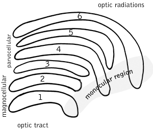
In neuroanatomy, the lateral geniculate nucleus is a structure in the thalamus and a key component of the mammalian visual pathway. It is a small, ovoid, ventral projection of the thalamus where the thalamus connects with the optic nerve. There are two LGNs, one on the left and another on the right side of the thalamus. In humans, both LGNs have six layers of neurons alternating with optic fibers.

<span class="mw-page-title-main">Occipital lobe</span> Part of the brain at the back of the head

The occipital lobe is one of the four major lobes of the cerebral cortex in the brain of mammals. The name derives from its position at the back of the head, from the Latin ob, 'behind', and caput, 'head'.

The receptive field, or sensory space, is a delimited medium where some physiological stimuli can evoke a sensory neuronal response in specific organisms.

<span class="mw-page-title-main">Motion perception</span> Inferring the speed and direction of objects

Motion perception is the process of inferring the speed and direction of elements in a scene based on visual, vestibular and proprioceptive inputs. Although this process appears straightforward to most observers, it has proven to be a difficult problem from a computational perspective, and difficult to explain in terms of neural processing.

Stereopsis is the component of depth perception retrieved by means of binocular disparity through binocular vision. It is not the only contributor to depth perception, but it is a major one. Binocular vision occurs because each eye receives a different image due to their slightly different positions in one's head. These positional differences are referred to as "horizontal disparities" or, more generally, "binocular disparities". Disparities are processed in the visual cortex of the brain to yield depth perception. While binocular disparities are naturally present when viewing a real three-dimensional scene with two eyes, they can also be simulated by artificially presenting two different images separately to each eye using a method called stereoscopy. The perception of depth in such cases is also referred to as "stereoscopic depth".

<span class="mw-page-title-main">Koniocellular cell</span> Type of neuron found in the thalamus of primates

In neuroscience, a koniocellular cell is a neuron with a small cell body that is located in the koniocellular layer of the lateral geniculate nucleus (LGN) of the thalamus of primates, including humans.

In neuroscience, neuronal tuning refers to the hypothesized property of brain cells by which they selectively represent a particular type of sensory, association, motor, or cognitive information. Some neuronal responses have been hypothesized to be optimally tuned to specific patterns through experience. Neuronal tuning can be strong and sharp, as observed in primary visual cortex, or weak and broad, as observed in neural ensembles. Single neurons are hypothesized to be simultaneously tuned to several modalities, such as visual, auditory, and olfactory. Neurons hypothesized to be tuned to different signals are often hypothesized to integrate information from the different sources. In computational models called neural networks, such integration is the major principle of operation. The best examples of neuronal tuning can be seen in the visual, auditory, olfactory, somatosensory, and memory systems, although due to the small number of stimuli tested the generality of neuronal tuning claims is still an open question.

The two-streams hypothesis is a model of the neural processing of vision as well as hearing. The hypothesis, given its initial characterisation in a paper by David Milner and Melvyn A. Goodale in 1992, argues that humans possess two distinct visual systems. Recently there seems to be evidence of two distinct auditory systems as well. As visual information exits the occipital lobe, and as sound leaves the phonological network, it follows two main pathways, or "streams". The ventral stream leads to the temporal lobe, which is involved with object and visual identification and recognition. The dorsal stream leads to the parietal lobe, which is involved with processing the object's spatial location relative to the viewer and with speech repetition.

<span class="mw-page-title-main">Inferior temporal gyrus</span> One of three gyri of the temporal lobe of the brain

The inferior temporal gyrus is one of three gyri of the temporal lobe and is located below the middle temporal gyrus, connected behind with the inferior occipital gyrus; it also extends around the infero-lateral border on to the inferior surface of the temporal lobe, where it is limited by the inferior sulcus. This region is one of the higher levels of the ventral stream of visual processing, associated with the representation of objects, places, faces, and colors. It may also be involved in face perception, and in the recognition of numbers and words.

<span class="mw-page-title-main">Efficient coding hypothesis</span>

The efficient coding hypothesis was proposed by Horace Barlow in 1961 as a theoretical model of sensory coding in the brain. Within the brain, neurons communicate with one another by sending electrical impulses referred to as action potentials or spikes. One goal of sensory neuroscience is to decipher the meaning of these spikes in order to understand how the brain represents and processes information about the outside world. Barlow hypothesized that the spikes in the sensory system formed a neural code for efficiently representing sensory information. By efficient it is understood that the code minimized the number of spikes needed to transmit a given signal. This is somewhat analogous to transmitting information across the internet, where different file formats can be used to transmit a given image. Different file formats require different number of bits for representing the same image at given distortion level, and some are better suited for representing certain classes of images than others. According to this model, the brain is thought to use a code which is suited for representing visual and audio information representative of an organism's natural environment.

<span class="mw-page-title-main">Simple cell</span> Beaker with Dilute Sulphuric Acid, Zinc and Copper Sheet is known as A Simple Cell

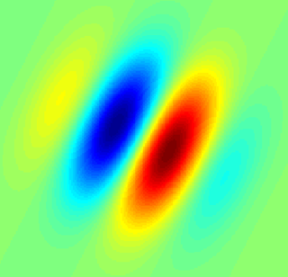
A simple cell in the primary visual cortex is a cell that responds primarily to oriented edges and gratings. These cells were discovered by Torsten Wiesel and David Hubel in the late 1950s.

Complex cells can be found in the primary visual cortex (V1), the secondary visual cortex (V2), and Brodmann area 19 (V3).

<span class="mw-page-title-main">Colour centre</span> Brain region responsible for colour processing

The colour centre is a region in the brain primarily responsible for visual perception and cortical processing of colour signals received by the eye, which ultimately results in colour vision. The colour centre in humans is thought to be located in the ventral occipital lobe as part of the visual system, in addition to other areas responsible for recognizing and processing specific visual stimuli, such as faces, words, and objects. Many functional magnetic resonance imaging (fMRI) studies in both humans and macaque monkeys have shown colour stimuli to activate multiple areas in the brain, including the fusiform gyrus and the lingual gyrus. These areas, as well as others identified as having a role in colour vision processing, are collectively labelled visual area 4 (V4). The exact mechanisms, location, and function of V4 are still being investigated.

<span class="mw-page-title-main">Hypercomplex cell</span>

A hypercomplex cell is a type of visual processing neuron in the mammalian cerebral cortex. Initially discovered by David Hubel and Torsten Wiesel in 1965, hypercomplex cells are defined by the property of end-stopping, which is a decrease in firing strength with increasingly larger stimuli. The sensitivity to stimulus length is accompanied by selectivity for the specific orientation, motion, and direction of stimuli. For example, a hypercomplex cell may only respond to a line at 45˚ that travels upward. Elongating the line would result in a proportionately weaker response. Ultimately, hypercomplex cells can provide a means for the brain to visually perceive corners and curves in the environment by identifying the ends of a given stimulus.

Globs are millimeter-sized color modules found beyond the visual area V2 in the brain's color processing ventral pathway. They are scattered throughout the posterior inferior temporal cortex in an area called the V4 complex. They are clustered by color preference, and organized as color columns. They are the first part of the brain in which color is processed in terms of the full range of hues found in color space.

Feature detection is a process by which the nervous system sorts or filters complex natural stimuli in order to extract behaviorally relevant cues that have a high probability of being associated with important objects or organisms in their environment, as opposed to irrelevant background or noise.

<span class="mw-page-title-main">Parasol cell</span>

A parasol cell, sometimes called an M cell or M ganglion cell, is one type of retinal ganglion cell (RGC) located in the ganglion cell layer of the retina. These cells project to magnocellular cells in the lateral geniculate nucleus (LGN) as part of the magnocellular pathway in the visual system. They have large cell bodies as well as extensive branching dendrite networks and as such have large receptive fields. Relative to other RGCs, they have fast conduction velocities. While they do show clear center-surround antagonism, they receive no information about color. Parasol ganglion cells contribute information about the motion and depth of objects to the visual system.

Surround suppression is where the relative firing rate of a neuron may under certain conditions decrease when a particular stimulus is enlarged. It has been observed in electrophysiology studies of the brain and has been noted in many sensory neurons, most notably in the early visual system. Surround suppression is defined as a reduction in the activity of a neuron in response to a stimulus outside its classical receptive field.

References

  1. 1 2 3 4 5 6 7 8 9 10 11 12 13 14 15 16 17 18 19 20 21 22 23 24 25 Qian, Ning (1997). "Binocular Disparity and the Perception of Depth". Neuron. 18 (3): 359–368. doi: 10.1016/s0896-6273(00)81238-6 . PMID   9115731.
  2. Scholl, B; Burge J; Priebe NJ (2013). "Binocular integration and disparity selectivity in mouse primary visual cortex". Journal of Neurophysiology. 109 (12): 3013–24. doi:10.1152/jn.01021.2012. PMC   3680810 . PMID   23515794.
  3. 1 2 Hubel, David; Torsten Wiesel (1962). "Receptive fields, binocular interaction and functional architecture in the cat's visual cortex". J. Physiol. 160 (1): 106–154. doi:10.1113/jphysiol.1962.sp006837. PMC   1359523 . PMID   14449617.
  4. Bishop, Peter; John Pettigrew (1986). "Neural mechanisms of binocular vision". Vision Research. 26 (9): 1587–1600. doi:10.1016/0042-6989(86)90177-x. PMID   3303676. S2CID   7664762.
  5. Poggio, G; B. Motter; S. Squatrito; Y. Trotter (1985). "Responses of neurons in visual cortex (V1 and V2) of the alert mecaque to dynamic random-dot stereograms". Vision Research. 25 (3): 397–406. doi:10.1016/0042-6989(85)90065-3. PMID   4024459. S2CID   43335583.
  6. Cottereau, Benoit; Suzanne McKee; Justin Ales; Anthony Norcia (2011). "Disparity-Tuned Population Responses from Human Visual Cortex". The Journal of Neuroscience. 31 (3): 954–965. doi:10.1523/jneurosci.3795-10.2011. PMC   3298090 . PMID   21248120.
  7. 1 2 3 4 5 6 7 8 9 10 11 12 13 14 Parker, Andrew (2007). "Binocular depth perception and the cerebral cortex". Nature. 8 (5): 379–391. doi:10.1038/nrn2131. PMID   17453018. S2CID   6234144.
  8. 1 2 Purves, Dale (2012). Neuroscience. Sunderland, MA: Sinauer Associates, Inc.
  9. Sasaki, KS; Tabuchi Y; Ohzawa I (2013). "Complex cells in the cat striate cortex have multiple disparity detectors in the three-dimensional binocular receptive fields". Journal of Neuroscience. 30 (41): 13826–37. doi:10.1523/JNEUROSCI.1135-10.2010. PMC   6633723 . PMID   20943923.
  10. Grunewald, Alexander; Stephen Grossberg (1998). "Self-Organization of Binocular Disparity Tuning by Reciprocal Corticogeniculate Interactions". Journal of Cognitive Neuroscience. 10 (2): 199–215. doi:10.1162/089892998562654. hdl: 2144/2326 . PMID   9555107. S2CID   7200376.
  11. Wardle, SG; Cass J; Brooks KR; Alais D (2010). "Breaking camouflage: binocular disparity reduces contrast masking in natural images". Journal of Vision. 10 (14): 38. doi: 10.1167/10.14.38 . PMID   21196512.
  12. Cao, Y; Grossberg S (2012). "Stereopsis and 3D surface perception by spiking neurons in laminar cortical circuits: a method for converting neural rate models into spiking models". Neural Networks. 26: 75–98. doi:10.1016/j.neunet.2011.10.010. PMID   22119530.
  13. 1 2 3 4 5 6 7 8 Ohzawa, I; G. DeAngelis; R. Freeman (1990). "Stereoscopic depth discrimination in the visual cortex: neurons ideally suited as disparity detectors". Science. 249 (4972): 1037–1041. Bibcode:1990Sci...249.1037O. CiteSeerX   10.1.1.473.8284 . doi:10.1126/science.2396096. PMID   2396096.
  14. 1 2 3 4 5 6 7 8 9 10 11 Fleet, David; Hermann Wagner; David Heeger (1996). "Neural Encoding of Binocular Disparity: Energy Models, Position Shifts and Phase Shifts". Vision Research. 36 (12): 1839–1857. doi: 10.1016/0042-6989(95)00313-4 . PMID   8759452.
  15. Read, Jenny; Andrew Parker; Bruce Cumming (2002). "A simple model accounts for the response of disparity-tuned V1 neurons to anticorrelated images". Visual Neuroscience. 19 (6): 735–753. doi:10.1017/s0952523802196052. PMID   12688669. S2CID   7851440.
  16. 1 2 Chen, Yuzhi; Yunjiu Wang; Ning Qian (2001). "Modeling V1 Disparity Tuning to Time-Varying Stimuli". Journal of Neurophysiology. 86 (1): 143–155. doi:10.1152/jn.2001.86.1.143. PMID   11431496.
  17. Zhu, Y; N. Qian (1996). "Binocular receptive field models, disparity tuning, and characteristic disparity". Neural Comput. 8 (8): 1647–1677. doi:10.1162/neco.1996.8.8.1611. PMID   8888610. S2CID   38166972.
  18. Menz, Michael; Ralph Freeman (2003). "Functional Connectivity of Disparity-Tuned Neurons in the Visual Cortex". Journal of Neurophysiology. 91 (4): 1794–1807. doi:10.1152/jn.00574.2003. PMID   14668293. S2CID   9655002.