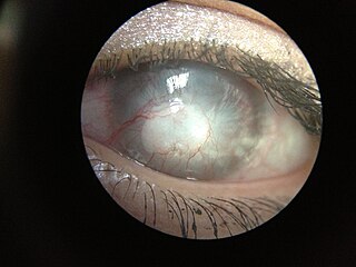
Related Research Articles

The cornea is the transparent front part of the eye that covers the iris, pupil, and anterior chamber. Along with the anterior chamber and lens, the cornea refracts light, accounting for approximately two-thirds of the eye's total optical power. In humans, the refractive power of the cornea is approximately 43 dioptres. The cornea can be reshaped by surgical procedures such as LASIK.

Keratitis is a condition in which the eye's cornea, the clear dome on the front surface of the eye, becomes inflamed. The condition is often marked by moderate to intense pain and usually involves any of the following symptoms: pain, impaired eyesight, photophobia, red eye and a 'gritty' sensation. Diagnosis of infectious keratitis is usually made clinically based on the signs and symptoms as well as eye examination, but corneal scrapings may be obtained and evaluated using microbiological culture or other testing to identify the causative pathogen.
This is a partial list of human eye diseases and disorders.

A red eye is an eye that appears red due to illness or injury. It is usually injection and prominence of the superficial blood vessels of the conjunctiva, which may be caused by disorders of these or adjacent structures. Conjunctivitis and subconjunctival hemorrhage are two of the less serious but more common causes.

Corneal abrasion is a scratch to the surface of the cornea of the eye. Symptoms include pain, redness, light sensitivity, and a feeling like a foreign body is in the eye. Most people recover completely within three days.

Phlyctenular keratoconjunctivitis is an inflammatory syndrome caused by a delayed hypersensitivity reaction to one or more antigens. The triggering antigen is usually a bacterial protein, but may also be a virus, fungus, or nematode.

A corneal ulcer, or ulcerative keratitis, is an inflammatory condition of the cornea involving loss of its outer layer. It is very common in dogs and is sometimes seen in cats. In veterinary medicine, the term corneal ulcer is a generic name for any condition involving the loss of the outer layer of the cornea, and as such is used to describe conditions with both inflammatory and traumatic causes.
Cogan syndrome is a rare disorder characterized by recurrent inflammation of the front of the eye and often fever, fatigue, and weight loss, episodes of vertigo (dizziness), tinnitus and hearing loss. It can lead to deafness or blindness if untreated. The classic form of the disease was first described by D. G. Cogan in 1945.

Acanthamoeba keratitis (AK) is a rare disease in which amoebae of the genus Acanthamoeba invade the clear portion of the front (cornea) of the eye. It affects roughly 100 people in the United States each year. Acanthamoeba are protozoa found nearly ubiquitously in soil and water and can cause infections of the skin, eyes, and central nervous system.

Corneal neovascularization (CNV) is the in-growth of new blood vessels from the pericorneal plexus into avascular corneal tissue as a result of oxygen deprivation. Maintaining avascularity of the corneal stroma is an important aspect of healthy corneal physiology as it is required for corneal transparency and optimal vision. A decrease in corneal transparency causes visual acuity deterioration. Corneal tissue is avascular in nature and the presence of vascularization, which can be deep or superficial, is always pathologically related.

Corneal ulcer, also called keratitis, is an inflammatory or, more seriously, infective condition of the cornea involving disruption of its epithelial layer with involvement of the corneal stroma. It is a common condition in humans particularly in the tropics and in farming. In developing countries, children afflicted by vitamin A deficiency are at high risk for corneal ulcer and may become blind in both eyes persisting throughout life. In ophthalmology, a corneal ulcer usually refers to having an infection, while the term corneal abrasion refers more to a scratch injury.

Chronic superficial keratitis (CSK), also known as pannus or Uberreiter's disease, is an inflammatory condition of the cornea in dogs, particularly seen in the German Shepherd. Both eyes are usually affected. The corneas gradually become pigmented and infiltrated by blood vessels, and the dog may eventually become blind.

Band keratopathy is a corneal disease derived from the appearance of calcium on the central cornea. This is an example of metastatic calcification, which by definition, occurs in the presence of hypercalcemia.
Diffuse lamellar keratitis (DLK) is a sterile inflammation of the cornea which may occur after refractive surgery, such as LASIK. Its incidence has been estimated to be 1 in 500 patients, though this may be as high as 32% in some cases.

Herpetic simplex keratitis is a form of keratitis caused by recurrent herpes simplex virus (HSV) infection in the cornea.
Christmas Eye refers to a seasonal epidemic of corneal ulceration which predominantly occurs in a particular region of Australia, caused by chemicals released upon death by small native beetles in the area.
Neurotrophic keratitis (NK) is a degenerative disease of the cornea caused by damage of the trigeminal nerve, which results in impairment of corneal sensitivity, spontaneous corneal epithelium breakdown, poor corneal healing and development of corneal ulceration, melting and perforation. This is because, in addition to the primary sensory role, the nerve also plays a role maintaining the integrity of the cornea by supplying it with trophic factors and regulating tissue metabolism.
Keratoendotheliitis fugax hereditaria is an autosomal dominantly inherited disease of the cornea, caused by a point mutation in cryopyrin that in humans is encoded by the NLRP3 gene located on the long arm of chromosome 1.
Exposure keratopathy is medical condition affecting the cornea of eyes. It can lead to corneal ulceration and permanent loss of vision due to corneal opacity.
Peripheral Ulcerative Keratitis (PUK) is a group of destructive inflammatory diseases involving the peripheral cornea in human eyes. The symptoms of PUK include pain, redness of the eyeball, photophobia, and decreased vision accompanied by distinctive signs of crescent-shaped damage of the cornea. The causes of this disease are broad, ranging from injuries, contamination of contact lenses, to association with other systemic conditions. PUK is associated with different ocular and systemic diseases. Mooren's ulcer is a common form of PUK. The majority of PUK is mediated by local or systemic immunological processes, which can lead to inflammation and eventually tissue damage. Standard PUK diagnostic test involves reviewing the medical history and a completing physical examinations. Two major treatments are the use of medications such as corticosteroids or other immunosuppressive agents and surgical resection of the conjunctiva. The prognosis of PUK is unclear with one study providing potential complications. PUK is a rare condition with an estimated incidence of 3 per million annually.
References
- ↑ Ramachandran, Tarakad. "Cogan Syndrome". Medlink. MedLink Corporation. Archived from the original on 9 March 2016. Retrieved 11 January 2012.
- ↑ Majmudar PA. "Keratitis, Interstitial" emedicine Dec 2007
- ↑ Dr Khairul Nazri Mohammad (Articles' Author), Waterford General Hospital, IRELAND
- ↑ Kanski JJ. "Clinical Ophthalmology 5th ed"