
Microscopy is the technical field of using microscopes to view objects and areas of objects that cannot be seen with the naked eye. There are three well-known branches of microscopy: optical, electron, and scanning probe microscopy, along with the emerging field of X-ray microscopy.

A microscope is a laboratory instrument used to examine objects that are too small to be seen by the naked eye. Microscopy is the science of investigating small objects and structures using a microscope. Microscopic means being invisible to the eye unless aided by a microscope.

A scanning electron microscope (SEM) is a type of electron microscope that produces images of a sample by scanning the surface with a focused beam of electrons. The electrons interact with atoms in the sample, producing various signals that contain information about the surface topography and composition of the sample. The electron beam is scanned in a raster scan pattern, and the position of the beam is combined with the intensity of the detected signal to produce an image. In the most common SEM mode, secondary electrons emitted by atoms excited by the electron beam are detected using a secondary electron detector. The number of secondary electrons that can be detected, and thus the signal intensity, depends, among other things, on specimen topography. Some SEMs can achieve resolutions better than 1 nanometer.

The optical microscope, also referred to as a light microscope, is a type of microscope that commonly uses visible light and a system of lenses to generate magnified images of small objects. Optical microscopes are the oldest design of microscope and were possibly invented in their present compound form in the 17th century. Basic optical microscopes can be very simple, although many complex designs aim to improve resolution and sample contrast.

A monocular is a compact refracting telescope used to magnify images of distant objects, typically using an optical prism to ensure an erect image, instead of using relay lenses like most telescopic sights. The volume and weight of a monocular are typically less than half of a pair of binoculars with similar optical properties, making it more portable and also less expensive. This is because binoculars are essentially a pair of monoculars packed together — one for each eye. As a result, monoculars only produce two-dimensional images, while binoculars can use two parallaxed images to produce binocular vision, which allows stereopsis and depth perception.

Transmission electron microscopy (TEM) is a microscopy technique in which a beam of electrons is transmitted through a specimen to form an image. The specimen is most often an ultrathin section less than 100 nm thick or a suspension on a grid. An image is formed from the interaction of the electrons with the sample as the beam is transmitted through the specimen. The image is then magnified and focused onto an imaging device, such as a fluorescent screen, a layer of photographic film, or a detector such as a scintillator attached to a charge-coupled device or a direct electron detector.

Angular resolution describes the ability of any image-forming device such as an optical or radio telescope, a microscope, a camera, or an eye, to distinguish small details of an object, thereby making it a major determinant of image resolution. It is used in optics applied to light waves, in antenna theory applied to radio waves, and in acoustics applied to sound waves. The colloquial use of the term "resolution" sometimes causes confusion; when an optical system is said to have a high resolution or high angular resolution, it means that the perceived distance, or actual angular distance, between resolved neighboring objects is small. The value that quantifies this property, θ, which is given by the Rayleigh criterion, is low for a system with a high resolution. The closely related term spatial resolution refers to the precision of a measurement with respect to space, which is directly connected to angular resolution in imaging instruments. The Rayleigh criterion shows that the minimum angular spread that can be resolved by an image-forming system is limited by diffraction to the ratio of the wavelength of the waves to the aperture width. For this reason, high-resolution imaging systems such as astronomical telescopes, long distance telephoto camera lenses and radio telescopes have large apertures.

In optical engineering, an objective is an optical element that gathers light from an object being observed and focuses the light rays from it to produce a real image of the object. Objectives can be a single lens or mirror, or combinations of several optical elements. They are used in microscopes, binoculars, telescopes, cameras, slide projectors, CD players and many other optical instruments. Objectives are also called object lenses, object glasses, or objective glasses.

Magnification is the process of enlarging the apparent size, not physical size, of something. This enlargement is quantified by a size ratio called optical magnification. When this number is less than one, it refers to a reduction in size, sometimes called de-magnification.
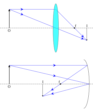
In optics, an image is defined as the collection of focus points of light rays coming from an object. A real image is the collection of focus points actually made by converging/diverging rays, while a virtual image is the collection of focus points made by extensions of diverging or converging rays. In other words, a real image is an image which is located in the plane of convergence for the light rays that originate from a given object. Examples of real images include the image produced on a detector in the rear of a camera, and the image produced on an eyeball retina.

A telescopic sight, commonly called a scope informally, is an optical sighting device based on a refracting telescope. It is equipped with some form of a referencing pattern – known as a reticle – mounted in a focally appropriate position in its optical system to provide an accurate point of aim. Telescopic sights are used with all types of systems that require magnification in addition to reliable visual aiming, as opposed to non-magnifying iron sights, reflector (reflex) sights, holographic sights or laser sights, and are most commonly found on long-barrel firearms, particularly rifles, usually via a scope mount. Similar devices are also found on other platforms such as artillery, tanks and even aircraft. The optical components may be combined with optoelectronics to add night vision or smart device features.
A total internal reflection fluorescence microscope (TIRFM) is a type of microscope with which a thin region of a specimen, usually less than 200 nanometers can be observed.

Confocal microscopy, most frequently confocal laser scanning microscopy (CLSM) or laser scanning confocal microscopy (LSCM), is an optical imaging technique for increasing optical resolution and contrast of a micrograph by means of using a spatial pinhole to block out-of-focus light in image formation. Capturing multiple two-dimensional images at different depths in a sample enables the reconstruction of three-dimensional structures within an object. This technique is used extensively in the scientific and industrial communities and typical applications are in life sciences, semiconductor inspection and materials science.

Dark-field microscopy describes microscopy methods, in both light and electron microscopy, which exclude the unscattered beam from the image. Consequently, the field around the specimen is generally dark.
Köhler illumination is a method of specimen illumination used for transmitted and reflected light optical microscopy. Köhler illumination acts to generate an even illumination of the sample and ensures that an image of the illumination source is not visible in the resulting image. Köhler illumination is the predominant technique for sample illumination in modern scientific light microscopy. It requires additional optical elements which are more expensive and may not be present in more basic light microscopes.

In light microscopy, oil immersion is a technique used to increase the resolving power of a microscope. This is achieved by immersing both the objective lens and the specimen in a transparent oil of high refractive index, thereby increasing the numerical aperture of the objective lens.

The stereo, stereoscopic or dissecting microscope is an optical microscope variant designed for low magnification observation of a sample, typically using light reflected from the surface of an object rather than transmitted through it. The instrument uses two separate optical paths with two objectives and eyepieces to provide slightly different viewing angles to the left and right eyes. This arrangement produces a three-dimensional visualization of the sample being examined. Stereomicroscopy overlaps macrophotography for recording and examining solid samples with complex surface topography, where a three-dimensional view is needed for analyzing the detail.

A condenser is an optical lens that renders a divergent light beam from a point light source into a parallel or converging beam to illuminate an object to be imaged.

Multifocal plane microscopy (MUM), also known as multiplane microscopy or multifocus microscopy, is a form of light microscopy that allows the tracking of the 3D dynamics in live cells at high temporal and spatial resolution by simultaneously imaging different focal planes within the specimen. In this methodology, the light collected from the sample by an infinity-corrected objective lens is split into two paths. In each path the split light is focused onto a detector which is placed at a specific calibrated distance from the tube lens. In this way, each detector images a distinct plane within the sample. The first developed MUM setup was capable of imaging two distinct planes within the sample. However, the setup can be modified to image more than two planes by further splitting the light in each light path and focusing it onto detectors placed at specific calibrated distances. It has later been improved for imaging up to four distinct planes. To image a greater number of focal planes, simpler techniques based on image splitting optics have been developed. One example is by using a customized image splitting prism, which is capable of capturing up to 8 focal planes using only two cameras. Better yet, standard off-the-shelf partial beamsplitters can be used to construct a so-called z-splitter prism that allows simultaneous imaging of 9 individual focal planes using a single camera. Another technique called multifocus microscopy (MFM) uses diffractive Fourier optics to image up to 25 focal planes.

A Lieberkühn reflector (also known as Lieberkühn mirror or simply Lieberkühn) is an illumination device for incident light illumination (epi-illumination) in light microscopes. It encircles the objective, with the mirrored surface facing towards the specimen. This allows illuminating an opaque object from the side of the objective, with the light source positioned behind the specimen as in a transmission microscope.
