Methylene blue, also known as methylthioninium chloride, is a medication and dye. As a medication, it is mainly used to treat methemoglobinemia. Specifically, it is used to treat methemoglobin levels that are greater than 30% or in which there are symptoms despite oxygen therapy. It has previously been used for cyanide poisoning and urinary tract infections, but this use is no longer recommended. It is typically given by injection into a vein.

Methyl violet is a family of organic compounds that are mainly used as dyes. Depending on the number of attached methyl groups, the color of the dye can be altered. Its main use is as a purple dye for textiles and to give deep violet colors in paint and ink, it is also used as a hydration indicator for silica gel. Methyl violet 10B is also known as crystal violet and has medical uses.

Staining is a technique used to enhance contrast in samples, generally at the microscopic level. Stains and dyes are frequently used in histology and in the medical fields of histopathology, hematology, and cytopathology that focus on the study and diagnoses disease at a microscopic level. Stains may be used to define biological tissues, cell populations, or organelles within individual cells.

Bromothymol blue is a pH indicator. It is mostly used in applications that require measuring substances that would have a relatively neutral pH. A common use is for measuring the presence of carbonic acid in a liquid. It is typically sold in solid form as the sodium salt of the acid indicator.
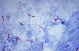
Ziehl-Neelsen staining is a type of Acid-fast stain, first introduced by Paul Ehrlich. Ziehl–Neelsen staining is a bacteriological stain used to identify acid-fast organisms, mainly Mycobacteria. It is named for two German doctors who modified the stain: the bacteriologist Franz Ziehl (1859–1926) and the pathologist Friedrich Neelsen (1854–1898).
Methyl blue is a chemical compound with the molecular formula C37H27N3Na2O9S3. It is used as a stain in histology, and stains collagen blue in tissue sections. It can be used in some differential staining techniques such as Mallory's connective tissue stain and Gömöri trichrome stain, and can be used to mediate electron transfer in microbial fuel cells. Fungal cell walls are also stained by methyl blue.

Crystal violet or gentian violet is a triarylmethane dye used as a histological stain and in Gram's method of classifying bacteria. Crystal violet has antibacterial, antifungal, and anthelmintic properties and was formerly important as a topical antiseptic. The medical use of the dye has been largely superseded by more modern drugs, although it is still listed by the World Health Organization.
Trichrome staining is a histological staining method that uses two or more acid dyes in conjunction with a polyacid. Staining differentiates tissues by tinting them in contrasting colours. It increases the contrast of microscopic features in cells and tissues, which makes them easier to see when viewed through a microscope.

Periodic acid–Schiff (PAS) is a staining method used to detect polysaccharides such as glycogen, and mucosubstances such as glycoproteins, glycolipids and mucins in tissues. The reaction of periodic acid oxidizes the vicinal diols in these sugars, usually breaking up the bond between two adjacent carbons not involved in the glycosidic linkage or ring closure in the ring of the monosaccharide units that are parts of the long polysaccharides, and creating a pair of aldehydes at the two free tips of each broken monosaccharide ring. The oxidation condition has to be sufficiently regulated so as to not oxidize the aldehydes further. These aldehydes then react with the Schiff reagent to give a purple-magenta color. A suitable basic stain is often used as a counterstain.
Trypan blue is an azo dye. It is a direct dye for cotton textiles. In biosciences, it is used as a vital stain to selectively colour dead tissues or cells blue.

Masson's trichrome is a three-colour staining protocol used in histology. The recipes evolved from Claude L. Pierre Masson's (1880–1959) original formulation have different specific applications, but all are suited for distinguishing cells from surrounding connective tissue.
Aniline Blue WS, also called aniline blue, China blue, or Soluble blue, is a mixture of methyl blue and water blue. It may also be either one of them. It is a soluble dye used as a biological dye, in fluorescence microscopy, appearing a yellow-green colour after excitation with violet light. It is a mixture of the trisulfonates of triphenyl rosaniline and of diphenyl rosaniline.

Water blue, also known as aniline blue, Acid blue 22, Soluble Blue 3M, Marine Blue V, or C.I. 42755, is a chemical compound used as a stain in histology. Water blue stains collagen blue in tissue sections. It is soluble in water and slightly soluble in ethanol.

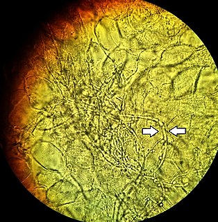
Kerion is the result of the host's response to a fungal ringworm infection of the hair follicles of the scalp that can be accompanied by secondary bacterial infection(s). It usually appears as raised, spongy lesions, and typically occurs in children. This honeycomb is a painful inflammatory reaction with deep suppurative lesions on the scalp. Follicles may be seen discharging pus. There may be sinus formation and rarely mycetoma-like grains are produced. It is usually caused by dermatophytes such as Trichophyton verrucosum, T. mentagrophytes, and Microsporum canis. Treatment with oral griseofulvin common.
A fungal keratitis is an 'inflammation of the eye's cornea' that results from infection by a fungal organism. Keratomycosis is the Greek terminology equivalent of fungal keratitis - it is the fungal infection of the cornea, the anterior part of the eye which covers the pupil. Those experiencing these symptoms are typically advised to immediately visit the appropriate eyecare professional.
The KOH Test for Candida albicans, also known as a potassium hydroxide preparation or KOH prep, is a quick, inexpensive fungal test to differentiate dermatophytes and Candida albicans symptoms from other skin disorders like psoriasis and eczema.

Acid fuchsin or fuchsine acid, (also called Acid Violet 19 and C.I. 42685) is an acidic magenta dye with the chemical formula C20H17N3Na2O9S3. Acid fuchsin has wide use in histology, and is one of the dyes used in Masson's trichrome stain. This method is commonly used to stain cytoplasm and nuclei of tissue sections in the histology laboratory in order to distinguish muscle from collagen. The muscle stains red with the acid fuchsin, and the collagen is stained green or blue with light green SF yellowish or methyl blue. It can also be used to identify growing bacteria.

Calcofluor-white or CFW is a fluorescent blue dye that is used to bind to the polysaccharide polymers of amebic cysts. It functions by being able to bind to 1-3 beta and 1-4 beta polysaccharides on chitin and cellulose that is present in cell walls on fungi, plants, and algae.
Trulla is a fungal genus in the family Steccherinaceae containing six species of polypores. It was circumscribed by mycologists Otto Miettinen and Leif Ryvarden in 2016, as a continuation of prior work that outlined a revised framework for the Steccherinaceae based on molecular phylogenetics. Its closest relative in the Steccherinaceae is the genus Nigroporus, from which it differs in its light-coloured fruit bodies and monomitic context.
