Lead shielding

Last updated
Lead bricks are commonly used as radiation shielding. Lead shielding.jpg
Lead bricks are commonly used as radiation shielding.

Lead shielding refers to the use of lead as a form of radiation protection to shield people or objects from radiation so as to reduce the effective dose. Lead can effectively attenuate certain kinds of radiation because of its high density and high atomic number; principally, it is effective at stopping gamma rays and x-rays.

Contents

Operation

Fluoroscopy room with control space, separated by lead glass. Fluoroscopy room with control space.jpg
Fluoroscopy room with control space, separated by lead glass.

Lead's high density is caused by the combination of its high atomic number and the relatively short bond lengths and atomic radius. The high atomic number means that more electrons are needed to maintain a neutral charge and the short bond length and a small atomic radius means that many atoms can be packed into a particular lead structure.

Because of lead's density and large number of electrons, it is well suited to scattering x-rays and gamma-rays. These rays form photons, a type of boson, which impart energy onto electrons when they come into contact. Without a lead shield, the electrons within a person's body would be affected, which could damage their DNA. When the radiation attempts to pass through lead, its electrons absorb and scatter the energy. Eventually though, the lead will degrade from the energy to which it is exposed. However, lead is not effective against all types of radiation. High energy electrons (including beta radiation) incident on lead may create bremsstrahlung radiation, which is potentially more dangerous to tissue than the original radiation. Furthermore, lead is not a particularly effective absorber of neutron radiation.

Types

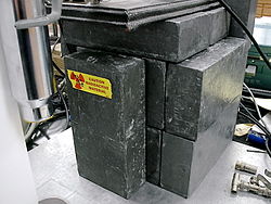
Lead is used for shielding in x-ray machines, nuclear power plants, labs, medical facilities, military equipment, and other places where radiation may be encountered. There is great variety in the types of shielding available both to protect people and to shield equipment and experiments. In gamma-spectroscopy for example, lead castles are constructed to shield the probe from environmental radiation. Personal shielding includes lead aprons (such as the familiar garment used during dental x-rays), thyroid shields, and lead gloves. There are also a variety of shielding devices available for laboratory equipment, including lead castles, structures composed of lead bricks, and lead pigs, made of solid lead or lead-lined containers for storing and transporting radioactive samples. In many facilities where radiation is produced, regulations require construction with lead-lined plywood or drywall to protect adjoining rooms from scatter radiation. [1]

Wear

A lead apron or leaded apron is a type of protective clothing that acts as a radiation shield. It is constructed of a thin rubber exterior and an interior of lead in the shape of a hospital apron. The purpose of the lead apron is to reduce exposure of a hospital patient to x-rays to vital organs that are potentially exposed to ionizing radiation during medical imaging that uses x-rays (radiography, fluoroscopy, computed tomography).

Protection of the reproductive organs with a lead rubber apron is considered important because DNA changes to sperm or egg cells of the patient may pass on genetic defects to the offspring of the patient, causing serious and unnecessary hardship for child and parents.[ citation needed ]

The thyroid gland is especially vulnerable to x-ray exposure. Care should be taken to place a lead apron over the thyroid gland before taking dental radiographs. [2] [3] Aprons used for dental imaging should include thyroid collars. However, in poorer or loosely regulated [4] countries, possibly due to the cost of such equipment (approx. 40 USD), [5] no such lead protection is given to the patients themselves, [6] though the operators do get out of the x-ray room for their own safety.

The correct thickness of lead-equivalent (Pbeq) wear will depend on how long and how often the person is working in an exposed environment. The minimum requirement is to wear 0.25 mm Pbeq when not behind lead shielding. In a theatre using fluoroscopy (e.g. orthopaedics, cardiology or interventional radiology) 0.35 or 0.5 mm lead may be appropriate because of the higher KV employed, and on proximity to the primary beam. [7]

See also

Related Research Articles

Background radiation is a measure of the level of ionizing radiation present in the environment at a particular location which is not due to deliberate introduction of radiation sources.

<span class="mw-page-title-main">X-ray</span> Form of short-wavelength electromagnetic radiation

An X-ray, or, much less commonly, X-radiation, is a penetrating form of high-energy electromagnetic radiation. Most X-rays have a wavelength ranging from 10 picometers to 10 nanometers, corresponding to frequencies in the range 30 petahertz to 30 exahertz (3×10¹⁶ Hz to 3×10¹⁹ Hz) and energies in the range 145 eV to 124 keV. X-ray wavelengths are shorter than those of UV rays and typically longer than those of gamma rays. In many languages, X-radiation is referred to as Röntgen radiation, after the German scientist Wilhelm Conrad Röntgen, who discovered it on November 8, 1895. He named it X-radiation to signify an unknown type of radiation. Spellings of X-ray(s) in English include the variants x-ray(s), xray(s), and X ray(s). The most familiar use of X-rays is checking for fractures (broken bones), but X-rays are also used in other ways. For example, chest X-rays can spot pneumonia. Mammograms use X-rays to look for breast cancer.

<span class="mw-page-title-main">Radiography</span> Imaging technique using ionizing and non-ionizing radiation

Radiography is an imaging technique using X-rays, gamma rays, or similar ionizing radiation and non-ionizing radiation to view the internal form of an object. Applications of radiography include medical radiography and industrial radiography. Similar techniques are used in airport security. To create an image in conventional radiography, a beam of X-rays is produced by an X-ray generator and is projected toward the object. A certain amount of the X-rays or other radiation is absorbed by the object, dependent on the object's density and structural composition. The X-rays that pass through the object are captured behind the object by a detector. The generation of flat two dimensional images by this technique is called projectional radiography. In computed tomography an X-ray source and its associated detectors rotate around the subject which itself moves through the conical X-ray beam produced. Any given point within the subject is crossed from many directions by many different beams at different times. Information regarding attenuation of these beams is collated and subjected to computation to generate two dimensional images in three planes which can be further processed to produce a three dimensional image.

<span class="mw-page-title-main">Acute radiation syndrome</span> Health problems caused by exposure to high levels of ionizing radiation

Acute radiation syndrome (ARS), also known as radiation sickness or radiation poisoning, is a collection of health effects that are caused by being exposed to high amounts of ionizing radiation in a short period of time. Symptoms can start within an hour of exposure, and can last for several months. Early symptoms are usually nausea, vomiting and loss of appetite. In the following hours or weeks, initial symptoms may appear to improve, before the development of additional symptoms, after which either recovery or death follow.

Ionizing radiation, including nuclear radiation, consists of subatomic particles or electromagnetic waves that have sufficient energy to ionize atoms or molecules by detaching electrons from them. Some particles can travel up to 99% of the speed of light, and the electromagnetic waves are on the high-energy portion of the electromagnetic spectrum.

Medical physics deals with the application of the concepts and methods of physics to the prevention, diagnosis and treatment of human diseases with a specific goal of improving human health and well-being. Since 2008, medical physics has been included as a health profession according to International Standard Classification of Occupation of the International Labour Organization.

<span class="mw-page-title-main">External beam radiotherapy</span> Treatment of cancer with ionized radiation

External beam radiotherapy (EBRT) is the most common form of radiotherapy. The patient sits or lies on a couch and an external source of ionizing radiation is pointed at a particular part of the body. In contrast to brachytherapy and unsealed source radiotherapy, in which the radiation source is inside the body, external beam radiotherapy directs the radiation at the tumour from outside the body. Orthovoltage ("superficial") X-rays are used for treating skin cancer and superficial structures. Megavoltage X-rays are used to treat deep-seated tumours, whereas megavoltage electron beams are typically used to treat superficial lesions extending to a depth of approximately 5 cm. X-rays and electron beams are by far the most widely used sources for external beam radiotherapy. A small number of centers operate experimental and pilot programs employing beams of heavier particles, particularly protons, owing to the rapid dropoff in absorbed dose beneath the depth of the target.

Radiation protection, also known as radiological protection, is defined by the International Atomic Energy Agency (IAEA) as "The protection of people from harmful effects of exposure to ionizing radiation, and the means for achieving this". Exposure can be from a source of radiation external to the human body or due to internal irradiation caused by the ingestion of radioactive contamination.

<span class="mw-page-title-main">Nuclear medicine</span> Medical specialty

Nuclear medicine or nucleology is a medical specialty involving the application of radioactive substances in the diagnosis and treatment of disease. Nuclear imaging, in a sense, is "radiology done inside out" because it records radiation emitting from within the body rather than radiation that is generated by external sources like X-rays. In addition, nuclear medicine scans differ from radiology, as the emphasis is not on imaging anatomy, but on the function. For such reason, it is called a physiological imaging modality. Single photon emission computed tomography (SPECT) and positron emission tomography (PET) scans are the two most common imaging modalities in nuclear medicine.

<span class="mw-page-title-main">Fluoroscopy</span> Production of an image when X-rays strike a fluorescent screen

Fluoroscopy is an imaging technique that uses X-rays to obtain real-time moving images of the interior of an object. There are two main sub-category of Fluoroscopy. Larger, typically Floor, Wall or Ceiling mounted device often called Cath Lab, and Smaller Mobile C-Arm. In its primary application of medical imaging, a fluoroscope allows a surgeon to see the internal structure and function of a patient mainly during surgery so that the pumping action of the heart or the motion of swallowing, for example, can be watched. This is useful for both diagnosis and therapy and occurs in general radiology, interventional radiology, and image-guided surgery.

<span class="mw-page-title-main">Radioactive contamination</span> Undesirable radioactive elements on surfaces or in gases, liquids, or solids

Radioactive contamination, also called radiological pollution, is the deposition of, or presence of radioactive substances on surfaces or within solids, liquids, or gases, where their presence is unintended or undesirable.

An X-ray image intensifier (XRII) is an image intensifier that converts X-rays into visible light at higher intensity than the more traditional fluorescent screens can. Such intensifiers are used in X-ray imaging systems to allow low-intensity X-rays to be converted to a conveniently bright visible light output. The device contains a low absorbency/scatter input window, typically aluminum, input fluorescent screen, photocathode, electron optics, output fluorescent screen and output window. These parts are all mounted in a high vacuum environment within glass or, more recently, metal/ceramic. By its intensifying effect, It allows the viewer to more easily see the structure of the object being imaged than fluorescent screens alone, whose images are dim. The XRII requires lower absorbed doses due to more efficient conversion of X-ray quanta to visible light. This device was originally introduced in 1948.

<span class="mw-page-title-main">Isotopes of iodine</span> Nuclides with atomic number of 53 but with different mass numbers

There are 37 known isotopes of iodine (53I) from 108I to 144I; all undergo radioactive decay except 127I, which is stable. Iodine is thus a monoisotopic element.

<span class="mw-page-title-main">Radiation burn</span> Damage to skin or biological tissue from radiation exposure

A radiation burn is a damage to the skin or other biological tissue and organs as an effect of radiation. The radiation types of greatest concern are thermal radiation, radio frequency energy, ultraviolet light and ionizing radiation.

<span class="mw-page-title-main">Industrial radiography</span> Type of non-destructive testing

Industrial radiography is a modality of non-destructive testing that uses ionizing radiation to inspect materials and components with the objective of locating and quantifying defects and degradation in material properties that would lead to the failure of engineering structures. It plays an important role in the science and technology needed to ensure product quality and reliability. In Australia, industrial radiographic non-destructive testing is colloquially referred to as "bombing" a component with a "bomb".

<span class="mw-page-title-main">Dental radiography</span> X-ray imaging in dentistry

Dental radiographs, commonly known as X-rays, are radiographs used to diagnose hidden dental structures, malignant or benign masses, bone loss, and cavities.

<span class="mw-page-title-main">Projectional radiography</span> Formation of 2D images using X-rays

Projectional radiography, also known as conventional radiography, is a form of radiography and medical imaging that produces two-dimensional images by x-ray radiation. The image acquisition is generally performed by radiographers, and the images are often examined by radiologists. Both the procedure and any resultant images are often simply called "X-ray". Plain radiography or roentgenography generally refers to projectional radiography. Plain radiography can also refer to radiography without a radiocontrast agent or radiography that generates single static images, as contrasted to fluoroscopy, which are technically also projectional.

<span class="mw-page-title-main">Gamma ray</span> Energetic electromagnetic radiation arising from radioactive decay of atomic nuclei

A gamma ray, also known as gamma radiation (symbol γ or ), is a penetrating form of electromagnetic radiation arising from the radioactive decay of atomic nuclei. It consists of the shortest wavelength electromagnetic waves, typically shorter than those of X-rays. With frequencies above 30 exahertz (3×10¹⁹ Hz), it imparts the highest photon energy. Paul Villard, a French chemist and physicist, discovered gamma radiation in 1900 while studying radiation emitted by radium. In 1903, Ernest Rutherford named this radiation gamma rays based on their relatively strong penetration of matter; in 1900 he had already named two less penetrating types of decay radiation (discovered by Henri Becquerel) alpha rays and beta rays in ascending order of penetrating power.

<span class="mw-page-title-main">X-ray detector</span> Instrument that can measure properties of X-rays

X-ray detectors are devices used to measure the flux, spatial distribution, spectrum, and/or other properties of X-rays.

StemRad is an Israeli-American start-up company that develops and manufactures personal protective equipment (PPE) against ionizing radiation. Its first product was the 360 Gamma, a device that protects the user's pelvic bone marrow from gamma radiation. StemRad has also developed the StemRad MD, a protective suit designed to provide whole-body radiation protection to physicians, and the AstroRad vest for radiation protection in space, which is currently being tested on the International Space Station and is one of the primary payloads onboard NASA's Artemis 1 lunar mission.

References

  1. NCRP. (2004). Structural shielding design for medical X-ray imaging facilities. National Council on Radiation Protection and Measurement. ISBN   1-60119-854-X. OCLC   1058579305.
  2. A Patient's Guide to Medical Imaging By Ronald Eisenberg, JD, MD, FACR, Alexander Margulis, MD
  3. Current oral and maxillofacial imaging,Thomas F. Razmus, Gail F. Williamson, Page 107
  4. "YouTube". www.youtube.com. Archived from the original on 2021-12-21. Retrieved 2020-07-06.
  5. "Quickship Standard Thyroid Collar". www.universalmedicalinc.com. Retrieved 2020-07-06.
  6. Orach, C. G. (October 2009). "Health equity: challenges in low income countries". African Health Sciences. 9 (Suppl 2): S49–S51. ISSN   1680-6905. PMC   2877288 . PMID   20589106.
  7. Livingstone RS, Varghese A, Keshava SN. A Study on the Use of Radiation-Protective Apron among Interventionists in Radiology. J Clin Imaging Sci 2018;8:34