Lectins are carbohydrate-binding proteins that are highly specific for sugar groups that are part of other molecules, so cause agglutination of particular cells or precipitation of glycoconjugates and polysaccharides. Lectins have a role in recognition at the cellular and molecular level and play numerous roles in biological recognition phenomena involving cells, carbohydrates, and proteins. Lectins also mediate attachment and binding of bacteria, viruses, and fungi to their intended targets.
An oligosaccharide is a saccharide polymer containing a small number of monosaccharides. Oligosaccharides can have many functions including cell recognition and cell binding. For example, glycolipids have an important role in the immune response.

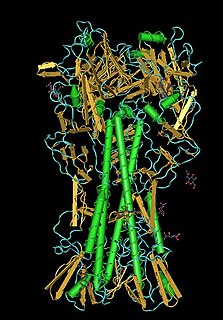
Concanavalin A (ConA) is a lectin originally extracted from the jack-bean. It is a member of the legume lectin family. It binds specifically to certain structures found in various sugars, glycoproteins, and glycolipids, mainly internal and nonreducing terminal α-D-mannosyl and α-D-glucosyl groups. ConA is a plant mitogen, and is known for its ability to stimulate mouse T-cell subsets giving rise to four functionally distinct T cell populations, including precursors to regulatory T cells; a subset of human suppressor T-cells is also sensitive to ConA. ConA was the first lectin to be available on a commercial basis, and is widely used in biology and biochemistry to characterize glycoproteins and other sugar-containing entities on the surface of various cells. It is also used to purify glycosylated macromolecules in lectin affinity chromatography, as well as to study immune regulation by various immune cells.

Eosinophil major basic protein, often shortened to major basic protein is encoded in humans by the PRG2 gene.

EF-Tu is a prokaryotic elongation factor responsible for catalyzing the binding of an aminoacyl-tRNA (aa-tRNA) to the ribosome. It is a G-protein, and facilitates the selection and binding of an aa-tRNA to the A-site of the ribosome. As a reflection of its crucial role in translation, EF-Tu is one of the most abundant and highly conserved proteins in prokaryotes. It is found in eukaryotic mitochrondria as TUFM.
Mannose-binding lectin (MBL), also called mannan-binding lectin or mannan-binding protein (MBP), is a lectin that is instrumental in innate immunity as an opsonin and via the lectin pathway.

Thermolysin is a thermostable neutral metalloproteinase enzyme produced by the Gram-positive bacteria Bacillus thermoproteolyticus. It requires one zinc ion for enzyme activity and four calcium ions for structural stability. Thermolysin specifically catalyzes the hydrolysis of peptide bonds containing hydrophobic amino acids. However thermolysin is also widely used for peptide bond formation through the reverse reaction of hydrolysis. Thermolysin is the most stable member of a family of metalloproteinases produced by various Bacillus species. These enzymes are also termed 'neutral' proteinases or thermolysin -like proteinases (TLPs).

Galectin-1 is a protein that in humans is encoded by the LGALS1 gene.

Platelet glycoprotein Ib alpha chain also known as glycoprotein Ib (platelet), alpha polypeptide or CD42b, is a protein that in humans is encoded by the GP1BA gene.

The EGF-like domain is an evolutionary conserved protein domain, which derives its name from the epidermal growth factor where it was first described. It comprises about 30 to 40 amino-acid residues and has been found in a large number of mostly animal proteins. Most occurrences of the EGF-like domain are found in the extracellular domain of membrane-bound proteins or in proteins known to be secreted. An exception to this is the prostaglandin-endoperoxide synthase. The EGF-like domain includes 6 cysteine residues which in the epidermal growth factor have been shown to form 3 disulfide bonds. The structures of 4-disulfide EGF-domains have been solved from the laminin and integrin proteins. The main structure of EGF-like domains is a two-stranded β-sheet followed by a loop to a short C-terminal, two-stranded β-sheet. These two β-sheets are usually denoted as the major (N-terminal) and minor (C-terminal) sheets. EGF-like domains frequently occur in numerous tandem copies in proteins: these repeats typically fold together to form a single, linear solenoid domain block as a functional unit.

Glycoprotein Ib (platelet), beta polypeptide (GP1BB) also known as CD42c, is a protein that in humans is encoded by the GP1BB gene.

Fibulin-2 is a protein that in humans is encoded by the FBLN2 gene.

The legume lectins are a family of sugar-binding proteins or lectins found in the seeds and, in smaller amounts, in the roots, stems, leaves and bark of plants of the family Fabaceae. The exact function of the legume lectins in vivo is unknown but they are probably involved in the defense of plants against predators. Related proteins in other plant families and in animals have also been found. They have been used for decades as a model system for the study of protein-carbohydrate interactions, because they show an amazing variety of binding specificities and are easy to obtain and purify. Over the years, a quite impressive amount of structural data has been gathered. Well-studied members of this protein family include phytohemagglutinin and concanavalin A.
The Walker A and Walker B motifs are protein sequence motifs, known to have highly conserved three-dimensional structures. These were first reported in ATP-binding proteins by Walker and co-workers in 1982.
Mamannamana Vijayan is an Indian structural biologist. His main area of research is protein structures. His contributions have been towards the structure and carbohydrate specificity of lectins and protein hydration. He has also contributed towards the area of structure and interactions of mycobacterial proteins and supramolecular association with reference to chemical evolution and origin of life. Vijayan did biological macromolecular crystallography in India.

A circular permutation is a relationship between proteins whereby the proteins have a changed order of amino acids in their peptide sequence. The result is a protein structure with different connectivity, but overall similar three-dimensional (3D) shape. In 1979, the first pair of circularly permuted proteins – concanavalin A and lectin – were discovered; over 2000 such proteins are now known.

In molecular biology, a carbohydrate-binding module (CBM) is a protein domain found in carbohydrate-active enzymes. The majority of these domains have carbohydrate-binding activity. Some of these domains are found on cellulosomal scaffoldin proteins. CBMs were previously known as cellulose-binding domains. CBMs are classified into numerous families, based on amino acid sequence similarity. There are currently 64 families of CBM in the CAZy database.
In molecular biology, the galactose binding lectin domain is a protein domain. It is found in many proteins including the lectin purified from sea urchin eggs, SUEL. This lectin exists as a disulfide-linked homodimer of two subunits; the dimeric form is essential for hemagglutination activity. The sea urchin egg lectin (SUEL) forms a new class of lectins. Although SUEL was first isolated as a D-galactoside binding lectin, it was later shown that it binds to L-rhamnose preferentially. L-rhamnose and D-galactose share the same hydroxyl group orientation at C2 and C4 of the pyranose ring structure.

In molecular biology the L-like lectin domain is a protein domain found in lectins which are similar to the leguminous plant lectins.

Intelectins are lectins expressed in humans and other chordates. Humans express two types of intelectins encoded by ITLN1 and ITLN2 genes respectively. Several intelectins bind microbe-specific carbohydrate residues. Therefore, intelectins have been proposed to function as immune lectins. Even though intelectins contain fibrinogen-like domain found in the ficolins family of immune lectins, there is significant structural divergence. Thus, intelectins may not function through the same lectin-complement pathway. Most intelectins are still poorly characterized and they may have diverse biological roles. Human intelectin-1 (hIntL-1) has also been shown to bind lactoferrin, but the functional consequence has yet to be elucidated. Additionally, hIntL-1 is a major component of asthmatic mucus and may be involved in insulin physiology as well.
