Related Research Articles

The atom probe was introduced at the 14th Field Emission Symposium in 1967 by Erwin Wilhelm Müller and J. A. Panitz. It combined a field ion microscope with a mass spectrometer having a single particle detection capability and, for the first time, an instrument could “... determine the nature of one single atom seen on a metal surface and selected from neighboring atoms at the discretion of the observer”.
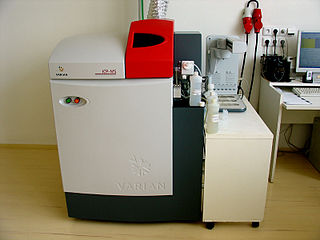
Inductively coupled plasma mass spectrometry (ICP-MS) is a type of mass spectrometry that uses an inductively coupled plasma to ionize the sample. It atomizes the sample and creates atomic and small polyatomic ions, which are then detected. It is known and used for its ability to detect metals and several non-metals in liquid samples at very low concentrations. It can detect different isotopes of the same element, which makes it a versatile tool in isotopic labeling.
Field-emission electric propulsion (FEEP) is an advanced electrostatic space propulsion concept, a form of ion thruster, that uses a liquid metal as a propellant – usually either caesium, indium, or mercury.

Transmission electron microscopy (TEM) is a microscopy technique in which a beam of electrons is transmitted through a specimen to form an image. The specimen is most often an ultrathin section less than 100 nm thick or a suspension on a grid. An image is formed from the interaction of the electrons with the sample as the beam is transmitted through the specimen. The image is then magnified and focused onto an imaging device, such as a fluorescent screen, a layer of photographic film, or a sensor such as a scintillator attached to a charge-coupled device.
Particle-induced X-ray emission or proton-induced X-ray emission (PIXE) is a technique used for determining the elemental composition of a material or a sample. When a material is exposed to an ion beam, atomic interactions occur that give off EM radiation of wavelengths in the x-ray part of the electromagnetic spectrum specific to an element. PIXE is a powerful yet non-destructive elemental analysis technique now used routinely by geologists, archaeologists, art conservators and others to help answer questions of provenance, dating and authenticity.

An ion source is a device that creates atomic and molecular ions. Ion sources are used to form ions for mass spectrometers, optical emission spectrometers, particle accelerators, ion implanters and ion engines.
Electrospray ionization (ESI) is a technique used in mass spectrometry to produce ions using an electrospray in which a high voltage is applied to a liquid to create an aerosol. It is especially useful in producing ions from macromolecules because it overcomes the propensity of these molecules to fragment when ionized. ESI is different from other ionization processes since it may produce multiple-charged ions, effectively extending the mass range of the analyser to accommodate the kDa-MDa orders of magnitude observed in proteins and their associated polypeptide fragments.

Secondary-ion mass spectrometry (SIMS) is a technique used to analyze the composition of solid surfaces and thin films by sputtering the surface of the specimen with a focused primary ion beam and collecting and analyzing ejected secondary ions. The mass/charge ratios of these secondary ions are measured with a mass spectrometer to determine the elemental, isotopic, or molecular composition of the surface to a depth of 1 to 2 nm. Due to the large variation in ionization probabilities among elements sputtered from different materials, comparison against well-calibrated standards is necessary to achieve accurate quantitative results. SIMS is the most sensitive surface analysis technique, with elemental detection limits ranging from parts per million to parts per billion.

An X-ray tube is a vacuum tube that converts electrical input power into X-rays. The availability of this controllable source of X-rays created the field of radiography, the imaging of partly opaque objects with penetrating radiation. In contrast to other sources of ionizing radiation, X-rays are only produced as long as the X-ray tube is energized. X-ray tubes are also used in CT scanners, airport luggage scanners, X-ray crystallography, material and structure analysis, and for industrial inspection.
Pyroelectric fusion refers to the technique of using pyroelectric crystals to generate high strength electrostatic fields to accelerate deuterium ions (tritium might also be used someday) into a metal hydride target also containing deuterium (or tritium) with sufficient kinetic energy to cause these ions to undergo nuclear fusion. It was reported in April 2005 by a team at UCLA. The scientists used a pyroelectric crystal heated from −34 to 7 °C (−29 to 45 °F), combined with a tungsten needle to produce an electric field of about 25 gigavolts per meter to ionize and accelerate deuterium nuclei into an erbium deuteride target. Though the energy of the deuterium ions generated by the crystal has not been directly measured, the authors used 100 keV (a temperature of about 109 K) as an estimate in their modeling. At these energy levels, two deuterium nuclei can fuse to produce a helium-3 nucleus, a 2.45 MeV neutron and bremsstrahlung. Although it makes a useful neutron generator, the apparatus is not intended for power generation since it requires far more energy than it produces.
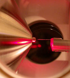
The name electrospray is used for an apparatus that employs electricity to disperse a liquid or for the fine aerosol resulting from this process. High voltage is applied to a liquid supplied through an emitter. Ideally the liquid reaching the emitter tip forms a Taylor cone, which emits a liquid jet through its apex. Varicose waves on the surface of the jet lead to the formation of small and highly charged liquid droplets, which are radially dispersed due to Coulomb repulsion.

Liquid chromatography–mass spectrometry (LC–MS) is an analytical chemistry technique that combines the physical separation capabilities of liquid chromatography with the mass analysis capabilities of mass spectrometry (MS). Coupled chromatography – MS systems are popular in chemical analysis because the individual capabilities of each technique are enhanced synergistically. While liquid chromatography separates mixtures with multiple components, mass spectrometry provides spectral information that may help to identify each separated component. MS is not only sensitive, but provides selective detection, relieving the need for complete chromatographic separation. LC–MS is also appropriate for metabolomics because of its good coverage of a wide range of chemicals. This tandem technique can be used to analyze biochemical, organic, and inorganic compounds commonly found in complex samples of environmental and biological origin. Therefore, LC–MS may be applied in a wide range of sectors including biotechnology, environment monitoring, food processing, and pharmaceutical, agrochemical, and cosmetic industries. Since the early 2000s, LC–MS has also begun to be used in clinical applications.

Focused ion beam, also known as FIB, is a technique used particularly in the semiconductor industry, materials science and increasingly in the biological field for site-specific analysis, deposition, and ablation of materials. A FIB setup is a scientific instrument that resembles a scanning electron microscope (SEM). However, while the SEM uses a focused beam of electrons to image the sample in the chamber, a FIB setup uses a focused beam of ions instead. FIB can also be incorporated in a system with both electron and ion beam columns, allowing the same feature to be investigated using either of the beams. FIB should not be confused with using a beam of focused ions for direct write lithography. These are generally quite different systems where the material is modified by other mechanisms.

Field desorption (FD) is a method of ion formation used in mass spectrometry (MS) in which a high-potential electric field is applied to an emitter with a sharp surface, such as a razor blade, or more commonly, a filament from which tiny "whiskers" have formed. This results in a high electric field which can result in ionization of gaseous molecules of the analyte. Mass spectra produced by FD have little or no fragmentation because FD is a soft ionization method. They are dominated by molecular radical cations M+. and less often, protonated molecules . The technique was first reported by Beckey in 1969. It is also the first ionization method to ionize nonvolatile and thermally labile compounds. One major difference of FD with other ionization methods is that it does not need a primary beam to bombard a sample.
Static secondary-ion mass spectrometry, or static SIMS is a secondary ion mass spectrometry technique for chemical analysis including elemental composition and chemical structure of the uppermost atomic or molecular layer of a solid which may be a metal, semiconductor or plastic with insignificant disturbance to its composition and structure. It is one of the two principal modes of operation of SIMS, which is the mass spectrometry of ionized particles emitted by a solid surface upon bombardment by energetic primary particles.

Instrumental analysis is a field of analytical chemistry that investigates analytes using scientific instruments.
Field-emission microscopy (FEM) is an analytical technique that is used in materials science to study the surfaces of needle apexes. The FEM was invented by Erwin Wilhelm Müller in 1936, and it was one of the first surface-analysis instruments that could approach near-atomic resolution.
Thermal ionization, also known as surface ionization or contact ionization, is a physical process whereby the atoms are desorbed from a hot surface, and in the process are ionized.

Aerosol mass spectrometry is the application of mass spectrometry to the analysis of the composition of aerosol particles. Aerosol particles are defined as solid and liquid particles suspended in a gas (air), with size range of 3 nm to 100 μm in diameter and are produced from natural and anthropogenic sources, through a variety of different processes that include wind-blown suspension and combustion of fossil fuels and biomass. Analysis of these particles is important owing to their major impacts on global climate change, visibility, regional air pollution and human health. Aerosols are very complex in structure, can contain thousands of different chemical compounds within a single particle, and need to be analysed for both size and chemical composition, in real-time or off-line applications.
A probe tip is an instrument used in scanning probe microscopes (SPMs) to scan the surface of a sample and make nano-scale images of surfaces and structures. The probe tip is mounted on the end of a cantilever and can be as sharp as a single atom. In microscopy, probe tip geometry and the composition of both the tip and the surface being probed directly affect resolution and imaging quality. Tip size and shape are extremely important in monitoring and detecting interactions between surfaces. SPMs can precisely measure electrostatic forces, magnetic forces, chemical bonding, Van der Waals forces, and capillary forces. SPMs can also reveal the morphology and topography of a surface.
References
- ↑ Swanson, L.W. (1983). "Liquid metal ion sources: Mechanism and applications". Nuclear Instruments and Methods in Physics Research. 218 (1–3): 347–353. doi:10.1016/0167-5087(83)91005-0. ISSN 0167-5087.
- ↑ Clampitt, R. (1981). "Advances in molten metal field ion sources". Nuclear Instruments and Methods in Physics Research. 189 (1): 111–116. doi:10.1016/0029-554X(81)90132-4. ISSN 0167-5087.
- ↑ Jon Orloff (24 October 2008). Handbook of Charged Particle Optics, Second Edition. CRC Press. p. 32. ISBN 978-1-4200-4555-0.