In medicine, a pulse represents the tactile arterial palpation of the cardiac cycle (heartbeat) by fingertips. The pulse may be palpated in any place that allows an artery to be compressed near the surface of the body, such as at the neck, wrist, at the groin, behind the knee, near the ankle joint, and on foot. The radial pulse is commonly measured using three fingers. This has a reason: the finger closest to the heart is used to occlude the pulse pressure, the middle finger is used get a crude estimate of the blood pressure, and the finger most distal to the heart is used to nullify the effect of the ulnar pulse as the two arteries are connected via the palmar arches. The study of the pulse is known as sphygmology.

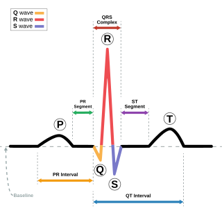
Electrocardiography is the process of producing an electrocardiogram, a recording of the heart's electrical activity through repeated cardiac cycles. It is an electrogram of the heart which is a graph of voltage versus time of the electrical activity of the heart using electrodes placed on the skin. These electrodes detect the small electrical changes that are a consequence of cardiac muscle depolarization followed by repolarization during each cardiac cycle (heartbeat). Changes in the normal ECG pattern occur in numerous cardiac abnormalities, including:

Palpitations are perceived abnormalities of the heartbeat characterized by awareness of cardiac muscle contractions in the chest, which is further characterized by the hard, fast and/or irregular beatings of the heart.

Dextrocardia is a rare congenital condition in which the apex of the heart is located on the right side of the body, rather than the more typical placement towards the left. There are two main types of dextrocardia: dextrocardia of embryonic arrest and dextrocardia situs inversus. Dextrocardia situs inversus is further divided.

A cardiac stress test is a cardiological examination that evaluates the cardiovascular system's response to external stress within a controlled clinical setting. This stress response can be induced through physical exercise or intravenous pharmacological stimulation of heart rate.

Atrioventricular block is a type of heart block that occurs when the electrical signal traveling from the atria, or the upper chambers of the heart, to ventricles, or the lower chambers of the heart, is impaired. Normally, the sinoatrial node produces an electrical signal to control the heart rate. The signal travels from the SA node to the ventricles through the atrioventricular node. In an AV block, this electrical signal is either delayed or completely blocked. When the signal is completely blocked, the ventricles produce their own electrical signal to control the heart rate. The heart rate produced by the ventricles is much slower than that produced by the SA node.
Impedance cardiography (ICG) is a non-invasive technology measuring total electrical conductivity of the thorax and its changes in time to process continuously a number of cardiodynamic parameters, such as stroke volume (SV), heart rate (HR), cardiac output (CO), ventricular ejection time (VET), pre-ejection period and used to detect the impedance changes caused by a high-frequency, low magnitude current flowing through the thorax between additional two pairs of electrodes located outside of the measured segment. The sensing electrodes also detect the ECG signal, which is used as a timing clock of the system.

A biosignal is any signal in living beings that can be continually measured and monitored. The term biosignal is often used to refer to bioelectrical signals, but it may refer to both electrical and non-electrical signals. The usual understanding is to refer only to time-varying signals, although spatial parameter variations are sometimes subsumed as well.

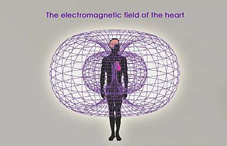
Magnetocardiography (MCG) is a technique to measure the magnetic fields produced by electrical currents in the heart using extremely sensitive devices such as the superconducting quantum interference device (SQUID). If the magnetic field is measured using a multichannel device, a map of the magnetic field is obtained over the chest; from such a map, using mathematical algorithms that take into account the conductivity structure of the torso, it is possible to locate the source of the activity. For example, sources of abnormal rhythms or arrhythmia may be located using MCG.

Cardiac amyloidosis is a subcategory of amyloidosis where there is depositing of the protein amyloid in the cardiac muscle and surrounding tissues. Amyloid, a misfolded and insoluble protein, can become a deposit in the heart's atria, valves, or ventricles. These deposits can cause thickening of different sections of the heart, leading to decreased cardiac function. The overall decrease in cardiac function leads to a plethora of symptoms. This multisystem disease was often misdiagnosed, with a corrected analysis only during autopsy. Advancements of technologies have increased earlier accuracy of diagnosis. Cardiac amyloidosis has multiple sub-types including light chain, familial, and senile. One of the most studied types is light chain cardiac amyloidosis. Prognosis depends on the extent of the deposits in the body and the type of amyloidosis. New treatment methods are actively being researched in regards to the treatment of heart failure and specific cardiac amyloidosis problems.

Alcoholic cardiomyopathy (ACM) is a disease in which the long-term consumption of alcohol leads to heart failure. ACM is a type of dilated cardiomyopathy. The heart is unable to pump blood efficiently, leading to heart failure. It can affect other parts of the body if the heart failure is severe. It is most common in males between the ages of 35 and 50.

Cardiac magnetic resonance imaging, also known as cardiovascular MRI, is a magnetic resonance imaging (MRI) technology used for non-invasive assessment of the function and structure of the cardiovascular system. Conditions in which it is performed include congenital heart disease, cardiomyopathies and valvular heart disease, diseases of the aorta such as dissection, aneurysm and coarctation, coronary heart disease. It can also be used to look at pulmonary veins.

Coronary ischemia, myocardial ischemia, or cardiac ischemia, is a medical term for abnormally reduced blood flow in the coronary circulation through the coronary arteries. Coronary ischemia is linked to heart disease, and heart attacks. Coronary arteries deliver oxygen-rich blood to the heart muscle. Reduced blood flow to the heart associated with coronary ischemia can result in inadequate oxygen supply to the heart muscle. When oxygen supply to the heart is unable to keep up with oxygen demand from the muscle, the result is the characteristic symptoms of coronary ischemia, the most common of which is chest pain. Chest pain due to coronary ischemia commonly radiates to the arm or neck. Certain individuals such as women, diabetics, and the elderly may present with more varied symptoms. If blood flow through the coronary arteries is stopped completely, cardiac muscle cells may die, known as a myocardial infarction, or heart attack.

Hypertrophic cardiomyopathy screening is an assessment and testing to detect hypertrophic cardiomyopathy (HCM).
John Peter Wikswo, Jr. is a biological physicist at Vanderbilt University. He was born in Lynchburg, Virginia, United States.
A diagnosis of myocardial infarction is created by integrating the history of the presenting illness and physical examination with electrocardiogram findings and cardiac markers. A coronary angiogram allows visualization of narrowings or obstructions on the heart vessels, and therapeutic measures can follow immediately. At autopsy, a pathologist can diagnose a myocardial infarction based on anatomopathological findings.

Magnetomyography (MMG) is a technique for mapping muscle activity by recording magnetic fields produced by electrical currents occurring naturally in the muscles, using arrays of SQUIDs. It has a better capability than electromyography for detecting slow or direct currents. The magnitude of the MMG signal is in the scale of pico (10−12) to femto (10−15) Tesla (T). Miniaturizing MMG offers a prospect to modernize the bulky SQUID to wearable miniaturized magnetic sensors.

In medicine, monitoring is the observation of a disease, condition or one or several medical parameters over time.
Bioinstrumentation or biomedical instrumentation is an application of biomedical engineering which focuses on development of devices and mechanics used to measure, evaluate, and treat biological systems. The goal of biomedical instrumentation focuses on the use of multiple sensors to monitor physiological characteristics of a human or animal for diagnostic and disease treatment purposes. Such instrumentation originated as a necessity to constantly monitor vital signs of Astronauts during NASA's Mercury, Gemini, and Apollo missions.

Jaakko A. Malmivuo is a Finnish engineer, academic, author, and opera singer. He was a professor of Bioelectromagnetism at Tampere University of Technology (TUT) from 1976 to 2010, an adjunct professor in the Faculty of Medicine at the University of Tampere as well as a visiting professor in the Faculty of Electrical Engineering and Computer Science, Electronics, and Medical Signal Processing at Technische Universität Berlin. Moreover, he was a director of the Ragnar Granit Institute at TUT from 1992.


